
 Worksheets
Chapter 13: Lower Gastrointestinal System
Total questions: 75
Worksheet time: 1hrs 15mins
Which part of the small intestine has a feathery appearance when filled with barium?
Ileum
Jejunum
Cecum
Duodenum
Which part of the small intestine has the largest diameter?
Duodenum
Ileum
Jejunum
Cecum
Which part of the small intestine is the shortest?
Duodenum
Ileum
Jejunum
Pylorus
Which part of the small intestine makes up three fifths of its entirety?
Duodenum
Jejunum
Ileum
Ilium
Which aspect of the large intestine is located highest, or most superior, in the abdomen?
Right colic flexure
Left colic flexure
Transverse colon
Ascending colon
Which of the following structures is not considered part of the colon?
Transverse colon
Right and left colic flexures
Rectum
All of the above are part of the colon.
Which part of the large intestine is located between the rectum and the descending colon?
Left colic flexure
Right colic flexure
Sigmoid colon
Cecum
Which part of the large intestine has the widest diameter?
Descending colon
Transverse colon
Cecum
Ascending colon
Which part of the colon has the greatest amount of potential movement?
Descending colon
Transverse colon
Sigmoid colon
Ascending colon
What is the term for the three bands of muscle that pull the large intestine into pouches?
Haustra
Valvulae conniventes
Suspensory ligaments
Taenia coli
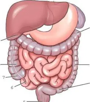
Which structure is labeled 6?
6 is the appendix.
6 is the ileum.
6 is the cecum.
6 is the sigmoid colon.
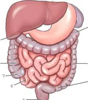
Which structure is labeled 1?
Ascending colon
Left colic flexure
Right colic flexure
Sigmoid colon
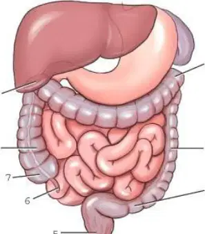
Which structure is labeled as number 7?
Descending colon
Sigmoid colon
Cecum
Rectum
The ascending colon and upper rectum are intraperitoneal structures.
True
False
Which sections of the large intestine will most likely be filled with air with the patient in the prone position during a double-contrast barium enema (BE)?
Ascending colon, descending colon, and rectum
Transverse and sigmoid colon
Rectum only
Right and left colic flexure and sigmoid colon
The part of the gastrointestinal (GI) tract that synthesizes and absorbs vitamins B and K is:
Ileum
Duodenum
Jejunum
Large intestine
The circular staircase, or herringbone pattern, is a common radiographic sign for a mechanical ileus.
True
False
Meckel diverticulum is best diagnosed with a radionuclide (nuclear medicine) scan.
True
False
Which of the following conditions may lead to an adynamic ileus?
Small bowel tumor
Scar tissue within the jejunum
Peritonitis
Stricture of ileum due to an inguinal hernia
Which of the following conditions may produce the “cobblestone” or “string” sign?
Whipple disease
Regional enteritis (Crohn’s disease)
Giardiasis
Ileus
Which of the following procedures is considered as a functional study?
Barium enema
Enteroclysis
Air-contrast BE
Small bowel series
When are small bowel series deemed to be complete?
Two hours after the ingestion of barium
Once the contrast media passes the ileocecal valve
Once the contrast media reaches the rectum
Once the contrast media passes the duodenojejunal flexure
The term describing a double-contrast small bowel procedure is:
two-stage small bowel procedure.
diagnostic intubation.
enteroclysis.
none of the above.
The enteroclysis procedure is indicated for patients with regional enteritis.
True
False
For an average adult, the amount of barium ingested is one 16-oz cup for a small-bowel-only series.
True
False
The patient must be NPO a minimum of hours before the small bowel series.
4
6
8
24
The tip of the catheter is advanced to the during an enteroclysis.
duodenojejunal junction (ligament of Treitz)
C-loop of the duodenum
pyloric sphincter
ileocecal sphincter
A twisting of the intestine on its own mesentery is termed:
intussusception.
volvulus.
diverticulosis.
enteritis.
A telescoping, or invagination, of one part of the intestine into another is termed:
diverticulosis.
volvulus.
intussusception.
colitis.
The “stovepipe” radiographic sign is often seen with:
volvulus.
intussusception.
neoplasm.
chronic ulcerative colitis.
The “tapered,” or “corkscrew,” radiographic sign is often seen with:
diverticulosis.
neoplasm.
volvulus.
intussusception.
Which radiographic sign is frequently seen with adenocarcinoma of the large intestine?
“Sail” sign
Diverticula
“Napkin ring” or “apple core” sign
Thickened mucosa
Ultrasound, with graded compression, can be used in diagnosing acute appendicitis.
True
False
CT may be performed to diagnose acute appendicitis.
True
False
Which of the following is classified as an irritant laxative?
Magnesium citrate
Magnesium sulfate
Castor oil
None of the above
Which of the following conditions would contraindicate the use of a cathartic before a barium enema?
Colitis
Diverticulosis
Obstruction
Diverticulitis
Synthetic latex enema tips are safe to use for latex-sensitive patients.
True
False
Rectal retention enema tips should be fully inflated by the technologist before beginning a barium enema.
True
False
At what stage of respiration should the enema tip be inserted into the rectum?
During deep breaths
During shallow breaths
Suspended inspiration
Suspended expiration
In what position is the patient placed for the enema tip insertion?
Sims’
Lithotomy
Modified lithotomy
Prone
During the initial enema tip insertion, the tip is aimed:
toward the coccyx.
toward the umbilicus.
directly posterior.
directly superior.
Which of the following pathologic conditions is best demonstrated with evacuative proctography?
Intussusception
Volvulus
Rectal prolapse
Diverticulosis
Which specific aspect of the large intestine must be demonstrated during evacuative proctography?
Sigmoid colon
Haustra
Anorectal angle
Rectal ligament
The ideal kV range for a double-contrast barium enema is:
70 to 80.
90 to 100.
100 to 125.
130 to 140.
Evacuative proctography is most commonly performed on geriatric patients.
True
False
During a colostomy barium enema, a double-contrast retention enema tip is used.
True
False
The opening leading into the intestine for the patient with a colostomy is termed the stoma.
True
False
The height of the enema bag should not exceed 36 inches (92 cm) above the radiographic table at the beginning of the study.
True
False
Which imaging modality can demonstrate abscesses in the retroperitoneum?
MRI
Nuclear medicine
Sonography
Conventional radiography
Which one of the following imaging modalities and/or procedures is very effective in detecting the Meckel diverticulum?
CT
Double-contrast barium enema
Sonography
Nuclear medicine
Which of the following statements is NOT true in regard to a pediatric small bowel series?
The transit time for barium through the small intestine is longer than that of an adult.
Barium sulfate is the contrast medium of choice.
The small bowel series should be scheduled early in the morning.
A gonadal shield often cannot be used during the later stages of the study.
What type of enema tip is recommended for a barium enema on an infant?
Rectal retention
Plastic disposable
10 Fr, flexible silicone catheter
Foley catheter
Central ray and image receptor centering for a 1-hour small bowel radiograph should be:
2 inches (5 cm) above level of the iliac crest.
at the level of the iliac crest.
1 inch (2.5 cm) below the level of the iliac crest.
at the level of the ASIS.
Which projection and/or position is most commonly performed during an evacuative proctogram?
Anteroposterior (AP) erect
Lateral
Right posterior oblique (RPO) and left posterior oblique (LPO)
AP axial
Why is the posteroanterior (PA) rather than the AP projection recommended for a small bowel series?
Less gonadal dose for female patients
More comfortable for patient
Places small intestine closer to image receptor (IR)
Better separation of loops of small intestine
Which of the following positions best demonstrates the left colic flexure?
LPO
Left lateral decubitus
Left lateral
Left anterior oblique (LAO)
During a double-contrast barium enema (BE) procedure, the radiologist suspects a polyp in the descending colon. Which position would best demonstrate this?
Supine for AP projection
45° erect PA projection
Right lateral decubitus
Left lateral decubitus
A radiograph of an AP barium enema (BE) projection reveals poor visualization of the sigmoid due to excessive superimposition of the sigmoid colon and rectum. How can this area be better visualized on the repeat exposure?
Angle the central ray (CR) 30° to 40° cephalad with AP projection.
Increase kV.
Take a PA projection with the patient in the left lateral decubitus position.
Take a PA projection with a 30° to 40° cephalad CR angle.
A patient comes to radiology for a barium enema (BE). He has a possible fistula extending from the rectum to the urinary bladder. Which one of the following projections and/or positions would best demonstrate the fistula?
Lateral rectum position
AP erect projection
LPO axial projection
LPO and RPO positions
A patient comes to radiology with possible diverticulosis. Which of the following studies is most diagnostic for detecting this condition?
Single-contrast barium enema
Double-contrast barium enema
Evacuative proctogram
Small bowel series
A patient comes to radiology for a double-contrast barium enema. The patient cannot lie on her side during the study. Which of the following projections could replace the lateral rectum projection?
AP axial
LPO axial projection
Ventral decubitus
Dorsal decubitus
During a single-contrast barium enema, the radiologist detects a possible defect within the right colic flexure. Which of the following projections and/or positions best demonstrates this region of the colon?
RPO
LAO
AP axial
LPO
A patient comes to radiology with a history of rectocele. The procedure that best demonstrates this condition is:
Barium enema
IV pyelogram
Hysterosalpingography
Mammography
An infant is brought to the ED with a possible intussusception. Which of the following procedures may actually correct this condition?
Small bowel enema
Small bowel series
Defecography
Barium or air enema
Which of the following barium enema projections and/or positions provides the greatest amount of gonadal dosage to both male and female patients?
AP/PA
Lateral rectum
Left lateral decubitus
Left posterior oblique
What is another term for the AP axial projection taken during a barium enema procedure?
Sims’ position
Butterfly position
Chassard-Lapine position
Smith position
How much CR angulation is required for the AP axial projection?
10 to 15
20 to 25
30 to 40
45
While attempting to insert an enema tip into the rectum, the technologist experiences resistance. What should be the next step taken by the technologist?
Retry the insertion using more lubrication.
Ask the patient to try to insert it himself.
Have the radiologist insert it using fluoroscopic guidance.
Cancel the procedure.
Why is it important for the technologist to review the patient’s chart and inform the radiologist before beginning the barium enema examination if a biopsy was performed as part of a prior sigmoidoscopy or colonoscopy procedure?
A sigmoidoscopy or colonoscopy would make the BE examination unnecessary.
The biopsy of the colon may weaken that portion of the colon, which could lead to a perforation during the BE examination.
The radiologist would want to confer with the referring physician to see whether the biopsy revealed a malignancy.
None of the above; the radiologist does not need to know this information before the BE examination.
Overhead, radiographic projections are often not taken when using digital fluoroscopy.
True
False
A single-stage, double-contrast barium enema involves instilling both the negative and positive contrast media at the same time.
True
False
During barium enema fluoroscopy, the radiologist detects a possible defect in the right colic flexure. He asks the technologist to produce a radiograph that will best demonstrate this region of the large intestine. Which one of the following positions will accomplish this goal?
LAO
AP axial projection
AP recumbent
LPO
Computed tomography colonography (CTC) is considered as an effective alternative to endoscopic colonoscopy.
True
False
Why is oral contrast media sometimes given during computed tomography colonography?
To determine if a fistula is present
To mark or “tag” possible fecal matter
To demonstrate possible diverticula
To prevent spasm of the large intestine
The average time to scan the large intestine during a computed tomography colonography is:
1 Hour
20 Minutes
30 Minutes
10 Minutes

