
 Worksheets
Chapter 14 Hypersensitivity_MEDL 201
Total questions: 40
Worksheet time: 20mins
Which statement best defines hypersensitivity in immunology?
A diminished immune response to a pathogenic antigen
An exaggerated immune response to a typically harmless antigen that can cause tissue injury and disease
A normal immune response that clears infectious agents without inflammation
A genetic inability to recognize foreign antigens
According to the material, which outcome is directly associated with hypersensitivity?
Enhanced resistance to pathogens
Autoimmune tolerance maintenance
Tissue injury and disease
Rapid wound healing without inflammation
Match each hypersensitivity category with its typical reaction timing based on the instructional graphic.
Type I
Immediate: minutes to hours after antigen exposure
Type II
Immediate: minutes to hours after antigen exposure
Type III
Immediate: minutes to hours after antigen exposure
Type IV
Delayed: develops 24 to 48 hours after antigen exposure
Which statement best defines Type I hypersensitivity based on the lecture notes?
A delayed T-cell–mediated response occurring days after exposure
An anaphylactic hypersensitivity typically thought of as allergies
A complement-dependent cytotoxic reaction against self-antigens
A serum sickness–type response driven by immune complexes
Identify the component that binds to FcεRI receptors on mast cells and basophils during the sensitization phase.
IgG
IgM
IgE
Complement C3b
Match each step of the sensitization phase to its correct description.
APC function
Processes allergens and presents them to Th cells
Th2 role
Induces production of allergen-specific IgE
IgE interaction
Binds to FcεRI receptors on mast cells and basophils
During the activation phase of Type I hypersensitivity, which event initiates mast cell and basophil degranulation?
Allergen cross-linking of adjacent cell-bound IgEs
Th2 secretion of IL-2 and IFN-γ
APC presentation of peptide on MHC I
Formation of immune complexes depositing in tissues
Select all statements that accurately describe the timing and key components associated with Type I hypersensitivity.
Commonly occur within minutes after exposure to an allergen
Primarily involve IgE, mast cells, and basophils
Depend mainly on neutrophils and IgM
Typically thought of as allergies
Which sequence correctly outlines the sensitization phase?
Mast cells release mediators → IgE produced → APC presents allergen
APC presents allergen to Th cells → Th2 induces allergen-specific IgE → IgE binds FcεRI on mast cells/basophils
Allergen cross-links IgE → Th2 activation → APC phagocytosis
Complement activation → IgM binding → eosinophil degranulation
In the activation phase, what is the immediate consequence of allergen cross-linking adjacent cell-bound IgEs?
APCs increase antigen presentation to CD8+ T cells
Mast cells and basophils degranulate, releasing chemical mediators
IgE production ceases and tolerance is induced
Immune complexes deposit and trigger complement
Match each activation-phase event to its outcome.
Allergen cross-linking of IgE
Triggers degranulation
Mast cell/basophil degranulation
Produces allergy symptoms
Mediator release and binding
Acts on target organs
Based on the listed common allergens, which option is a typical trigger for Type I hypersensitivity reactions?
Pollen
Sodium chloride
Distilled water
Vitamin C supplements
Select all items that are recognized as common allergens implicated in Type I hypersensitivity.
Mold spores
Animal dander
Dust mites
Insect venom
Granulated sugar
Match each clinical manifestation of Type I hypersensitivity to its concise description.
Rhinitis
Seasonal nasal symptoms often called hay fever
Allergic asthma
Lower airway hyperreactivity with bronchoconstriction
Urticaria
Wheal-and-flare lesions on the skin known as hives
Systemic anaphylaxis
A potentially fatal, whole-body reaction
Which clinical feature of urticaria is specifically highlighted in Type I hypersensitivity?
Wheal and flare
Productive cough
Purulent exudate
Petechiae
A patient rapidly develops hypotension, airway swelling, and diffuse rash after exposure to an allergen. Which Type I hypersensitivity manifestation best fits this presentation?
Systemic anaphylaxis
Eczema
Rhinitis
Contact dermatitis
Which intervention is correctly categorized as drug therapy for Type I hypersensitivity?
Antihistamines
Elimination diet only
Cold compresses
Surgical excision of the allergen source
Select all pharmacologic agents that are included in drug therapy for Type I hypersensitivity.
Bronchodilators
Mast cell stabilizers
Corticosteroids
Epinephrine
Oral antibiotics for viral infections
Which statement best describes allergy immunotherapy (AIT) in the management of Type I hypersensitivity?
Administer gradually increasing doses of the allergen
Provide a single high dose of allergen to induce tolerance
Avoidance is the only principle; AIT is contraindicated
Replace IgE with IgM using plasma exchange
In vivo testing for Type I hypersensitivity can be performed using which approaches to introduce allergens into the skin?
Percutaneous or intradermal methods
Oral ingestion challenge
Intravenous allergen infusion
Nasal provocation only
During a standard skin test for Type I hypersensitivity, what procedural sequence is followed before interpreting results?
Apply a panel of allergens to separate skin sites, then wait 15–20 minutes
Apply a single allergen to one site, wait 2 hours
Draw blood immediately after allergen application
Wash the skin with alcohol and interpret immediately
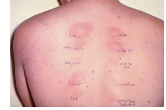
Which finding indicates a positive result in in vivo skin testing for Type I hypersensitivity?
Development of a wheal and flare at the application site
A painless blanching without swelling
No visible change after 30 minutes
Diffuse erythema over the entire back
Allergen-specific IgE testing is used for what primary purpose in evaluating Type I hypersensitivity?
Detect IgE directed to a specific allergen in patient serum
Quantify mast cell numbers in tissue biopsies
Measure total immunoglobulin concentrations across all isotypes
Confirm complement activation by the classical pathway
Match each assay type with what it detects.
Percutaneous/intradermal skin test
Wheal and flare response at the application site
Allergen-specific IgE assay
IgE directed against a defined allergen in serum
Total IgE assay
Overall concentration of IgE in serum
Which statements correctly describe total IgE testing?
Its known as the RadioImmunoSorbent Test
Its known as the Radioallergosorbent Test
It distinguishes which allergen the IgE targets by including antigen in the assay.
It is conceptually different from allergen-specific testing because no defined antigen is required.
A clinician wants to avoid potential systemic reactions from skin testing. Which alternative test provides evidence of sensitization without applying allergens to the skin?
Serum allergen-specific IgE assay
Percutaneous skin prick panel
Intradermal injection testing
Patch testing for contact dermatitis
Which statement best defines Type II hypersensitivity in immunology?
A T-cell–mediated delayed inflammatory response
An antibody-mediated cytotoxic hypersensitivity targeting cell surface antigens
An IgE-driven immediate hypersensitivity against soluble allergens
A complement-independent reaction that only affects viral particles
Identify the key antibody components involved in Type II hypersensitivity.
IgA and IgE
IgG and IgM
IgD and IgE
IgA and IgG
In Type II hypersensitivity, antibodies bound to cell surface antigens can lead to which effects on the target cell? Select all that apply.
Cell destruction through cytotoxic mechanisms
Inhibition of target-cell function
Increase in target-cell function
Induction of granuloma formation
Match each mechanism of cell damage in Type II hypersensitivity with its description.
Complement activation
Classical pathway triggers lysis of the bound cell
Opsonization
Antibody coats the cell to enhance phagocytosis by macrophages
Antibody-dependent cell-mediated cytotoxicity (ADCC)
Fc receptor–bearing NK cells or macrophages kill the antibody-tagged cell
Which process is correctly paired with its immune effector in Type II hypersensitivity?
Opsonization — neutrophil Fc receptors recognizing IgE
ADCC — NK cell Fc receptors engaging antibody-coated cells
Complement activation — alternative pathway triggered by IgA
Cell destruction — eosinophil degranulation via IL-5
Which clinical scenario is most consistent with Type II hypersensitivity?
Serum sickness due to immune complex deposition
Contact dermatitis from poison ivy
Hemolytic disease of the newborn (HDN) due to maternal antibodies
Anaphylaxis after peanut exposure mediated by IgE
Which description best characterizes Type III hypersensitivity?
Cell-mediated destruction by cytotoxic T cells
Antibody-dependent cell cytotoxicity against membrane-bound antigens
Immune complex–mediated reaction where antigen–antibody complexes deposit in tissues
Immediate mast cell degranulation triggered by IgE binding to allergens
In Type III hypersensitivity, which immunoglobulins are the key components directed against a soluble antigen?
IgA and IgE
IgG and IgM
IgD and IgA
IgE and IgM
Select all events that occur after small antigen–antibody complexes precipitate and deposit in tissues during Type III hypersensitivity.
Complement binds to the complexes
Vasodilation increases
Vasopermeability decreases
Neutrophils migrate to the affected area
Lysosomal enzymes released cause tissue damage
Which antigen form is typically targeted in Type III hypersensitivity reactions?
Cell-associated antigen fixed on membranes
Soluble antigen circulating in fluid
Hapten bound to carrier protein on cell surface
Allogeneic MHC molecules on donor cells
Match each clinical entity with its classification related to Type III hypersensitivity.
Arthus reaction
Local immune complex–mediated reaction
Serum sickness
Immune complex disease following exposure to foreign serum proteins
Systemic lupus erythematosus (SLE)
Autoimmune disease with immune complex deposition
Rheumatoid arthritis (RA)
Autoimmune disease with immune complexes in joints
Which step most directly explains why tissue damage occurs in Type III hypersensitivity?
IgE-mediated mast cell degranulation releases histamine
Cytotoxic T lymphocytes perforate target cells
Activated macrophages phagocytose RBCs causing hemolysis
Neutrophils and macrophages release lysosomal enzymes at sites of immune complex deposition
A patient develops a painful, localized reaction after repeated antigen injection at the same site. Which mechanism best explains this Arthus reaction?
Delayed-type hypersensitivity mediated by Th1 cells forming granulomas
Immune complex precipitation in local tissues with complement activation
Direct antibody binding to cell-surface antigens causing ADCC
Immediate IgE-mediated mast cell degranulation in the skin
Which sequence correctly orders key events in Type III hypersensitivity from initiation to tissue injury?
Complement activation → immune complex deposition → neutrophil migration → enzyme release
Soluble antigen binds IgG/IgM → small complexes precipitate in tissues → complement binds → vasodilation/vasopermeability increase → neutrophils migrate → lysosomal enzymes released
Neutrophil migration → antigen recognition by TCR → cytokine release → complement activation
IgE binding to FcεRI → allergen cross-linking → immediate degranulation → tissue damage

