
 Worksheets
Imaging I: Chpt 9-Radiographic Image Quality
Total questions: 61
Worksheet time: 31mins
Which pair of factors directly contributes to the visibility of anatomic structures in a quality radiographic image as defined in this lesson?
Brightness and contrast
Spatial resolution and distortion
Exposure time and patient motion
Tube current and source-to-image distance
A radiograph is judged by both visibility and accuracy. Which combination best represents accuracy of structural lines recorded?
Sharpness characterized by spatial resolution and distortion
Brightness characterized by exposure and window level
Contrast characterized by kVp and subject density
Noise characterized by quantum mottle and scatter
During latent image creation, x-ray attenuation varies across tissues. Which term describes this process and explains why some structural lines are recorded more distinctly than others?
Differential absorption
Uniform transmission
Constant scatter
Isotropic emission
Radiographic image quality is defined by two overarching aspects. Which pair best represents these aspects?
Brightness and contrast alone
Visibility of anatomic structures and accuracy of structural lines
Exposure factors and patient positioning
Pixel size and bit depth
Which statement best distinguishes brightness from contrast in radiographic imaging?
Brightness determines the difference between adjacent structures, while contrast sets the overall lightness of the image.
Brightness controls overall image lightness or darkness on the display, while contrast governs how different structures appear relative to each other.
Brightness and contrast are identical measures with no functional difference.
Contrast is adjusted only on the monitor, whereas brightness is fixed during acquisition.
Which image quality component primarily deals with the accuracy of structural lines rather than their visibility?
Brightness
Contrast
Sharpness (spatial resolution)
Distortion of size or shape
According to the instructional text, what does density represent in a processed radiographic image?
The sharpness of edges throughout the image
The amount of overall blackness on the processed image
The visibility of soft tissue structures only
The uniformity of pixel size across the detector
Which exposure or processing factor primarily controls density in film screen and computed radiography (CR)?
Kilovoltage peak (kVp) exclusively
Milliampere-seconds (mAs) and processing chemicals
Focal spot size and source-to-image distance (SID)
Collimation width and grid ratio
In image formation, what is identified as the main factor affecting radiographic density at the receptor?
The quantity of x-ray photons reaching the image receptor
The duration of post-processing algorithms
The number of pixels displayed on the monitor
The brand of the imaging system
Which outcome is correctly matched with the exposure error described for digital imaging?
Underexposure leads to saturation (severely degraded image).
Extreme overexposure increases quantum noise, producing a grainy image.
Underexposure may increase quantum noise, causing a grainy image.
Moderate overexposure is fully corrected by film chemicals without any degradation.
Which statement best describes the exposure indicator in digital imaging?
It is a numeric value indicating the level of radiation exposure to the digital image receptor.
It is a color scale that adjusts contrast after the image is captured.
It is a physical knob on the x-ray tube that sets patient dose directly.
It is a histogram that displays pixel frequencies without any exposure information.
According to the visual, what should radiographers do before deciding whether to repeat an exposure?
Increase the mAs until the image appears darker.
Review the exposure indicator value along with the image quality.
Ignore the exposure indicator and rely on patient feedback.
Rely solely on standardized numbers without considering the anatomy imaged.
Which control on the display monitor sets how light or dark the image appears overall by determining the midpoint of the visible gray values?
Window width
Window level
Exposure indicator
Bit depth
Displayed image brightness should be optimized primarily for which purpose in clinical practice?
Reducing file size
Matching the x-ray technique chart
Human perception so the radiologist can see needed structures
Standardizing monitor calibration across departments
When the window level is moved up to a higher pixel value, what is the expected effect on visibility?
Decreases overall brightness and reveals brighter regions
Increases overall brightness and improves visibility of darker regions
Has no effect on overall brightness, only contrast
Increases noise and reduces detail in all regions
In a chest radiograph, why might increasing window level be useful?
It enhances visualization of structures inside the lungs that otherwise appear too dark
It reduces motion blur caused by breathing
It increases the exposure indicator value to match protocol
It prevents image saturation in bright bone areas
Lowering the window level to a low pixel value primarily helps with which imaging goal?
Seeing more detail in bright or overexposed regions
Highlighting darker soft-tissue regions
Eliminating quantum noise
Correcting geometric distortion
What happens when an image lacks sufficient contrast between brightness levels or densities?
It becomes sharper but noisier
It appears homogeneous, making different tissues hard to differentiate
It shows increased saturation in dark regions
It displays more anatomic detail due to wider dynamic range
Why are differences in brightness levels (contrast) necessary in radiographic images?
To reduce radiation dose to the patient
To differentiate among different anatomic tissues by showing adjacent structures
To calibrate the exposure indicator automatically
To prevent saturation in the detector at high exposures
Human perception in imaging is optimized by controlling which of the following?
The range of gray values mapped for display using window level.
The mechanical movement of the x-ray tube during exposure.
The total number of pixels collected during acquisition.
The compression ratio used when saving the file.
Which statement best defines subject contrast in radiography?
The visible range of grays on the final image
The natural differences in how body tissues absorb x‑rays
The amount of scatter produced by the x‑ray beam
The brightness setting selected on the monitor
Subject contrast primarily depends on which combination of factors?
Thickness, density, and composition of the body part, plus x‑ray beam energy (kVp)
Tube current (mA) and exposure time only
Monitor brightness and ambient room light
Image receptor brand and pixel pitch
In a chest radiograph, bone appears lighter and lungs appear darker. What principle explains this difference?
Radiographic post‑processing enhances bones only
Bone absorbs more x‑rays than air, producing subject contrast
Air scatters less radiation, reducing image brightness
Lungs produce inherent gray levels unrelated to absorption
Which statement best defines radiographic contrast?
Differences in patient anatomy thickness
The visible difference in shades of gray on the final image
The energy output of the x‑ray tube (kVp)
The amount of radiation dose delivered to the patient
Radiographic contrast depends on subject contrast and which additional set of factors?
Only patient positioning
Technical and processing factors such as kVp, scatter radiation, image receptor response, and digital post‑processing
Ambient light and display brand
Collimation and grid ratio exclusively
A digitally processed image shows clear differences between bone, soft tissue, and air as a good range of grays. What does this indicate?
Low radiographic contrast
High radiographic contrast
No subject contrast
Overexposure with reduced brightness
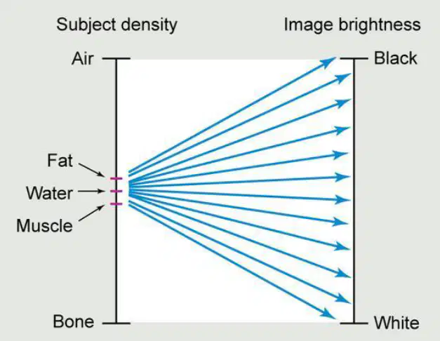
Contrast resolution refers to the imaging system’s ability to do what?
Distinguish very small positional changes
See small differences between tissues that absorb x‑rays almost the same way
Produce maximum spatial resolution regardless of dose
Eliminate all scatter radiation
Why is window width adjustment used in digital radiography displays?
To increase patient dose for better clarity
Because the human eye cannot detect very small differences in gray shades, so the computer changes how many shades of gray are visible
To correct geometric distortion caused by magnification
To remove motion blur created during exposure
Which statement best defines subject contrast in radiography?
The visible difference between light and dark areas on the final image
The variation in X-ray absorption by different tissues within the patient
The difference in pixel values produced by digital windowing controls
The sharpness of structural lines determined by pixel pitch
Radiographic contrast is primarily described as which of the following?
The inherent absorption differences among tissues before imaging
The visible difference in densities on the final image influenced by technique and processing
The number of pixels and their size that determine sharpness
The total brightness of the monitor set by window level
Refer to the diagram of radiographic images labeled “Higher contrast” and “Lower contrast.” Which statement best describes higher contrast in terms of tissue absorption differences and image appearance?
Large differences in tissue absorption produce images with pronounced black‑and‑white separation and fewer visible gray shades.
Small differences in tissue absorption produce images with subtle tonal changes and many gray shades.
Uniform tissue absorption produces uniformly gray images with minimal structure visibility.
Higher contrast occurs when soft tissues absorb more than bone, creating mostly light tones.
In the diagram comparing contrast levels, what scenario most likely yields lower contrast?
Tissues with similar composition and closely matched attenuation coefficients.
A mix of air and dense bone with markedly different attenuation.
Use of a technique that increases absorption differences between soft tissue and bone.
Presence of metal implants next to soft tissue causing extreme brightness differences.
Which statement best defines contrast resolution in medical imaging?
The ability to see small tissue differences by distinguishing subtle shades of gray
The maximum number of line pairs per millimeter an imaging system can display
The capacity to enlarge images without pixelation
The process of reducing motion blur using shorter exposure times
A radiograph appears to have greater contrast (more pronounced differences between light and dark regions). Which window parameter adjustment most directly produces this effect?
Narrowing the window width to include fewer shades of gray
Widening the window width to include more shades of gray
Increasing window level to shift the image brightness upward
Decreasing spatial frequency to lower image sharpness
Why is computer adjustment of window width important for tissue differentiation on a radiograph?
It controls the number of gray levels displayed, enhancing visibility of small tissue differences
It changes the physical resolution limit of the detector to reveal smaller objects
It compensates for patient motion by re-aligning pixels
It replaces the need for proper exposure by automatically setting mAs and kVp
In digital image windowing, what is the primary effect of adjusting the window level on the displayed image?
It alters overall brightness by choosing which pixel value range is centered
It sharpens edges by increasing spatial resolution
It changes detector efficiency, reducing patient dose
It modifies the number of pixels in the matrix
A technologist wants to visualize fine lung markings that are close in pixel values to surrounding tissue. Based on windowing principles shown in the diagram, which strategy is most appropriate?
Use a narrow window width to enhance contrast between similar pixel values
Use a wide window width to include more pixel values, reducing contrast
Lower the window level to darken the entire image uniformly
Raise the window level to brighten the entire image without affecting contrast
Recall: Which statement best defines spatial resolution in digital imaging?
The range of brightness values captured by a sensor
The sharpness and accuracy of detail displayed in an image
The amount of compression applied during file saving
The color gamut a monitor can reproduce
Which statement best describes the relationship between field of view (FOV) and pixel size when matrix size is held constant?
Increasing FOV increases pixel size
Increasing FOV decreases pixel size
FOV and pixel size are unrelated
Decreasing FOV increases pixel size
When matrix size increases while FOV remains the same, what happens to pixel size and image detail?
Pixel size decreases and image detail improves
Pixel size increases and image detail worsens
Pixel size remains the same and detail is unchanged
Pixel size decreases but detail worsens
Which statement best defines spatial resolution in medical imaging?
The amount of radiation hitting the image receptor
The ability of an imaging system to distinguish small or closely spaced structures as separate
The difference in brightness between two areas on an image
The degree of magnification caused by geometric factors
Recorded detail is most directly assessed using which measurement approach?
Counting pixels per inch on a display
Evaluating line pairs per millimeter as an indicator of image sharpness
Measuring exposure time in milliseconds
Comparing grayscale values across regions of interest
A system with higher spatial resolution will most likely show which outcome when imaging fine anatomical structures?
Increased unsharpness and blur at edges
Reduced ability to distinguish adjacent structures
Enhanced visibility of smaller detectable objects and sharper edges
Greater graininess due to noise
Consider two imaging systems: System A resolves 5 line pairs per millimeter, and System B resolves 10 line pairs per millimeter. Which inference is most reasonable?
System A provides better recorded detail because fewer line pairs reduce noise
System B provides higher recorded detail, indicating greater image sharpness
Both systems have equal spatial resolution because millimeters are the same unit
System A will better distinguish adjacent structures due to lower frequency
Which statement best defines spatial frequency in imaging systems?
The rate at which image brightness changes over time
The number of line pairs per millimeter that the system can distinguish
The total number of pixels in the detector array
The wavelength of the x‑ray beam used during exposure
MTF describes how well an imaging system can
Control radiation dose
Transfer detail from object to image
Enlarge small structures for clarity
Filter our scatter
A system with an MTF of 1.0 indicates
Some loss of detail
No detail transferred
Perfect reproduction of detail
Excessive quantum noise
DQE measures how efficiently the receptor
Reduces scatter
Converts x-rays into a useful image
Compresses digital data
Changes magnification
Shape distortion occurs when
kVp is too low
Beam, part, or IR are misaligned
Exposure time is too short
Collimation is opened too wide
Elongation means the image appears
Shorter than it really is
Sharper than expected
More magnified with no cause
Longer than it really is
Scatter primarily affects image
Brightness
Contrast
Spatial Resolution only
Pixel depth
Scatter creates
Useful anatomical detail
Increased patient dose without image change
Fog that reduces visibility of structures
Higher MTF values
Quantum noise appears most when
Too many photons reach the IR
Too few photons reach the IR
The collimation is too tight and cuts off
The mAs is set too high
Quantum noise makes an image look
Sharper and more detailed
Grainy and lacking detail
More magnified
Overexposed
Higher SNR means
Noise is stronger than signal
Signal is stronger than noise
Image will be grainier
The image cannot be used
Increasing SNR results in
Poorer detail
Clearer image quality
Lower contrast visibility
More scatter production
High CNR makes it easier to
See small contrast differences
Increase elongation
Create more digital artifacts
Reduce spatial resolution
Digital systems have a wide dynamic range, meaning
They require perfect exposure
They can display both dark and light areas well
They cannot handle underexposure
They always reduce patient dose
An artifact refers to
Any intentional enhancement feature
Any unwanted image appearance
A type of pixel used in digital imaging
A standard test tool
A minus-density artifact appears
Lighter than expected
Darker than expected
A plus-density artifact appears
Lighter than expected
Darker than expected

