
NEW
Font size
 Worksheets
Intracranial Regulation Part B Introduction
Total questions: 63
Worksheet time: 32mins
Which of the following best describes the concept of intracranial regulation?
Processes that impact intracranial compensation and adaptive neurological function.
Processes that only affect blood flow in the brain.
Processes that regulate hormone levels in the body.
Processes that control muscle movement.
Which condition is listed as an exemplar of issues affecting intracranial regulation?
Traumatic brain injury
Diabetes mellitus
Hypertension
Asthma
Select the condition that is NOT mentioned as affecting intracranial regulation.
Cerebral edema
Meningitis
Seizures/status epilepticus
Diabetes mellitus
Which of the following best describes a head injury?
A broad classification that includes injury to the scalp, skull, or brain
A specific injury to the brain only, excluding the scalp and skull
An injury that only affects the skull and not the brain or scalp
A minor injury that does not require medical attention
Approximately how many people experience head injuries every year in the United States?
2.5 million people
500,000 people
10 million people
1 million people
What is the most common cause of death from trauma?
Head injury
Spinal cord injury with paralysis and respiratory failure
Severe bleeding from limb injuries
Multiple organ failure
What is the most common cause of brain trauma?
Falls
Motor vehicle accidents
Sports injuries
Gunshot wounds
Which group is at the highest risk for brain trauma?
Males age 15-19, children 0-4 years, and adults 65 and older
Females age 20-30
Adults age 40-50
Teenagers age 13-15 only
Refer to the image showing a patient after a bilateral decompressive craniectomy. What is the main purpose of this surgical procedure?
To relieve pressure on the brain as a last ditch effort
To repair a fractured skull and restore its shape
To remove a blood clot from the brain
To treat a scalp laceration
Which of the following best describes a primary brain injury?
Damage that occurs due to the initial trauma, such as contusions, lacerations, or penetration by a foreign object
Damage that develops over days due to lack of oxygen
Swelling of the brain caused by infection
A psychological response to trauma
Which of the following is a mechanism of secondary brain injury?
Inadequate delivery of nutrients and oxygen to the cells
Immediate laceration of blood vessels
Penetration of the skull by a foreign object
Direct contusion of the brain tissue
What is the main difference between primary and secondary brain injury?
Primary injury occurs at the time of trauma, while secondary injury evolves after the initial insult over hours to days
Primary injury is always less severe than secondary injury
Secondary injury is caused by external objects only, while primary injury is not
Primary injury only affects children, while secondary injury only affects adults
Which event directly follows an increase in intracranial volume after a traumatic brain injury?
Intracranial pressure increases due to the rigid cranium
Cerebral blood flow ceases immediately
Brain herniates before pressure rises
Cerebral hypoxia and ischemia occur first
What is the main consequence of increased intracranial pressure on cerebral blood vessels after a traumatic brain injury?
Blood flow to the brain slows down
Brain tissue immediately regenerates
Oxygen supply to the brain increases
Intracranial volume decreases
Based on the CT scan images labeled Day 1, Day 2, and Day 3, what trend is most likely observed in the progression of traumatic brain injury?
Increasing damage and swelling over time
Immediate recovery of brain tissue
No change in brain structure
Decreasing intracranial pressure each day
Arrange the following steps in the correct order as they occur in the pathophysiology of traumatic brain injury: 1) Cerebral hypoxia and ischemia occur, 2) Brain suffers traumatic injury, 3) Intracranial pressure increases, 4) Cerebral blood flow ceases.
2, 3, 1, 4
3, 2, 1, 4
2, 1, 3, 4
1, 2, 3, 4
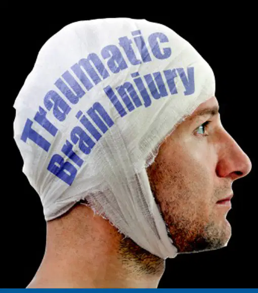
Which of the following is a common manifestation of brain injury?
Altered level of consciousness (LOC)
Increased appetite
Improved vision
Rapid weight gain

Which type of brain injury is defined as a temporary loss of neurologic function with no apparent structural damage to the brain?
Concussion
Contusion
Closed brain injury
Open brain injury
Which of the following is NOT listed as a neurologic change associated with brain injury?
Hearing loss
Sensation
Movement
Reflexes
Brain injuries may exhibit fast or slow bleeding. Which type of blood vessel bleeds faster in brain injuries?
Arteries
Veins
Capillaries
Lymphatic vessels
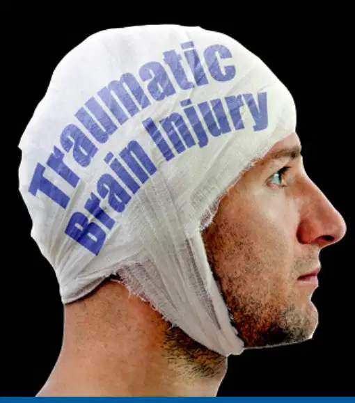
A patient presents with abnormal pupils and seizures after a head injury. Which other symptom would most likely indicate a brain injury?
Altered level of consciousness (LOC)
Improved digestion
Increased hair growth
Decreased thirst
Which statement best explains the difference between a concussion and a contusion?
A concussion involves temporary loss of neurologic function without structural damage, while a contusion involves actual structural damage to the brain.
A concussion is always more severe than a contusion and requires surgery.
A contusion is a temporary loss of consciousness, while a concussion is permanent.
A concussion and a contusion are identical in symptoms and treatment.
Which of the following best describes a concussion?
A temporary loss of neurological function with no structural damage
A permanent loss of consciousness due to brain injury
A temporary loss of memory with structural brain damage
A chronic condition affecting only physical abilities
What is the main difference between a grade 1 (mild) and grade 2 (moderate) concussion?
Grade 1 symptoms last less than 15 minutes, while grade 2 symptoms last more than 15 minutes
Grade 1 involves loss of consciousness, while grade 2 does not
Grade 2 symptoms are always physical, while grade 1 symptoms are emotional
Grade 1 requires hospitalization, while grade 2 does not
Which grade of concussion is characterized by loss of consciousness?
Grade 1 (Mild)
Grade 2 (Moderate)
Grade 3 (Severe)
None of the above
Repeated concussions can lead to which syndrome?
Chronic Traumatic Encephalopathy
Post-Traumatic Stress Disorder
Epilepsy
Parkinson's Disease
According to the table, which of the following is a physical symptom of concussion? (Refer to the table showing concussion symptoms in four categories.)
Headache
Difficulty thinking clearly
Sadness
Trouble falling asleep
Which of the following is NOT a category of concussion symptoms listed in the table? (Refer to the table showing concussion symptoms in four categories.)
Digestive
Thinking/Remembering
Physical
Sleep
What is the recommended action if a patient is sent home after a concussion?
Teach the family long-term signs and symptoms to watch for
Advise complete bed rest for one week
Schedule immediate surgery
Prescribe strong painkillers
Which symptom should be reported immediately after head trauma?
Severe headache
Mild thirst
Slight fatigue
Temporary happiness
Which of the following is a sign that should be observed in a patient after head trauma?
Difficulty speaking or movement
Increased appetite
Improved memory
Rapid weight loss
Why should a patient be aroused and assessed frequently after a concussion, unless the concussion is mild?
To monitor for worsening symptoms and ensure patient safety
To prevent the patient from sleeping too much
To increase the patient's alertness artificially
To reduce the risk of dehydration
Which of the following is a possible emotional/mood symptom of concussion according to the table? (Refer to the table showing concussion symptoms in four categories.)
Irritability
Headache
Sleeping more than usual
Difficulty remembering new information
Which of the following best describes a contusion in the context of traumatic brain injury (TBI)?
A more severe TBI with structural damage and possible surface hemorrhage, similar to a bruise.
A mild TBI with no structural damage and no bleeding.
A brain injury that only affects the spinal cord.
A type of injury that only occurs outside the skull.
What factor most influences the symptoms and recovery of a person with a traumatic brain injury (TBI)?
The amount of damage and associated cerebral edema.
The age of the patient only.
The time of day the injury occurred.
The color of the bruise on the head.
Which of the following is a likely symptom of a more severe TBI with structural damage?
Longer period of unconsciousness with more symptoms of neurologic deficits and changes in vital signs.
Immediate full recovery with no symptoms.
Only mild headache and no other symptoms.
Short period of unconsciousness with no neurologic deficits.
What is the primary cause of a closed brain injury?
Blunt trauma to the head.
Exposure to loud noises.
Infection in the brain tissue.
Lack of oxygen to the brain.
How does an acceleration/deceleration injury damage brain tissue?
When the head accelerates and then rapidly decelerates, causing the brain to move within the skull and damage tissue.
By a slow and steady movement of the head.
By exposure to chemicals in the environment.
By a gradual loss of memory over time.
Refer to the diagram showing coup and contrecoup injuries. What does the coup injury represent?
The initial impact site where the brain hits the inside of the skull.
The secondary impact site on the opposite side of the brain.
A bruise on the scalp only.
A tear in the spinal cord.
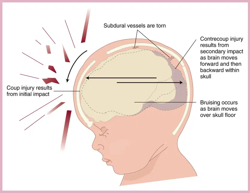
According to the diagram of closed brain injury, what happens to subdural vessels during acceleration-deceleration injury?
They are torn as the brain moves within the skull.
They become stronger and more flexible.
They remain completely unaffected.
They move outside the skull.
A patient experiences a longer period of unconsciousness and more symptoms of neurologic deficits after a head injury. Based on the information provided, what type of brain injury is most likely?
A more severe TBI with structural damage and possible surface hemorrhage (contusion).
A mild concussion with no structural damage.
A spinal cord injury only.
A simple scalp laceration.
What is the medical term for the deformity of the ear shown in the lower left image, which is commonly caused by trauma and results in swelling and a lumpy appearance?
Cauliflower ear
Auricular hematoma with infection and hearing loss
Simple ear bruise
External otitis
The lower right image shows bruising behind the ear. What serious condition is this sign most commonly associated with?
Basilar skull fracture
Middle ear infection with fever
Simple contusion from minor trauma
Allergic reaction causing skin discoloration
What is diffuse axonal injury most commonly associated with?
Widespread axon damage in the brain seen with head trauma
Localized bleeding in the brain tissue after a stroke
Loss of memory due to aging
Infection of the meninges causing inflammation
Which of the following best describes the clinical consequence of diffuse axonal injury?
Immediate coma following head trauma
Gradual loss of motor function over several months
Sudden onset of seizures without trauma
Temporary confusion that resolves within hours
Why is diffuse axonal injury considered one of the most devastating types of traumatic brain injury?
Because it causes damage over a widespread area and is a major cause of unconsciousness and persistent vegetative state after head trauma
Because it only affects a small, localized region of the brain
Because it is always accompanied by skull fractures
Because it results in mild symptoms that resolve quickly

Examine the images showing axonal damage highlighted in yellow. What do these images represent in the context of traumatic brain injury?
They show widespread axon damage characteristic of diffuse axonal injury
They show localized bleeding in the brain tissue
They show normal, healthy axons without injury
They show infection spreading along nerve fibers
Which type of intracranial bleeding involves blood collecting between the skull and the dura mater?
Epidural hematoma
Subdural hematoma with chronic symptoms
Intracerebral hemorrhage
Subarachnoid hemorrhage
What is the main difference between a subdural hematoma and an epidural hematoma?
A subdural hematoma involves blood between the dura and the brain, while an epidural hematoma involves blood between the skull and the dura
A subdural hematoma is always acute, while an epidural hematoma is always chronic
A subdural hematoma occurs only in children, while an epidural hematoma occurs only in adults
A subdural hematoma is less serious than an epidural hematoma
Which type of intracranial bleeding is characterized by bleeding within the brain tissue itself?
Intracerebral hemorrhage and hematoma
Epidural hematoma
Subdural hematoma
Subarachnoid hemorrhage
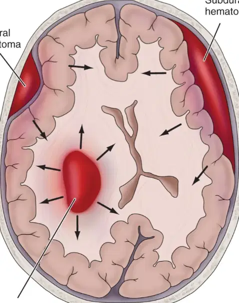
Refer to the diagram of the brain showing labeled areas of bleeding. Which area is labeled as 'subdural hematoma'?
The area between the dura and the brain
The area between the skull and the dura
The area within the brain tissue
The area outside the skull
Where does blood collect in an epidural hematoma?
Between the skull and the dura
Between the brain and the dura
Within the brain tissue
In the ventricles
Which type of blood vessel is most commonly involved in an epidural hematoma?
Artery
Vein with slow bleeding
Capillary
Lymphatic vessel
A patient with an epidural hematoma may initially lose consciousness, then regain it briefly before deteriorating rapidly. What is this period of regained consciousness called?
Lucid interval
Prodromal phase with confusion
Postictal state
Recovery phase
Which of the following is a key emergency treatment for epidural hematoma?
Craniotomy or burr holes to remove the clot and stop bleeding
Administration of antibiotics
Physical therapy for rehabilitation
Steroid injection to reduce inflammation
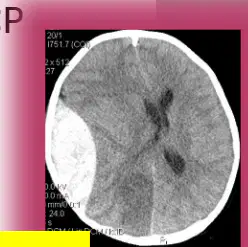
According to the CT scan image provided, which side of the brain shows the epidural hematoma? (Refer to the image showing a white, lens-shaped area between the skull and the dura.)
Right side
Left side with diffuse swelling
Both sides equally
No abnormality visible
Which population is epidural hematoma rare in?
Children
Adults with hypertension
Elderly patients
Athletes
Where does blood collect in a subdural hematoma?
Between the dura and the brain
Between the skull and the dura
Within the brain tissue
In the subarachnoid space
What is the usual origin of bleeding in a subdural hematoma?
Venous
Arterial with rapid bleeding
Capillary
Lymphatic
Which statement best describes the development of symptoms in chronic subdural hematoma?
Symptoms develop over weeks to months and may fluctuate
Symptoms appear suddenly and are always severe
Symptoms are limited to headaches only
Symptoms resolve without treatment
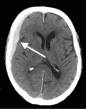
According to the CT scan image provided, what is the typical appearance of a subdural hematoma? (Refer to the image showing a crescent-shaped area between the dura and the brain.)
Crescent-shaped area between the dura and the brain
Lens-shaped area between the skull and dura
Diffuse swelling throughout the brain
No abnormality visible
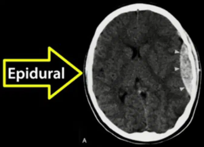
Based on the CT scans shown, which type of hematoma is located between the skull and the dura mater?
Epidural hematoma
Subdural hematoma
Intracerebral hematoma
Subarachnoid hemorrhage

Examine the CT scans labeled 'Epidural' and 'Subdural.' Which feature best distinguishes an epidural hematoma from a subdural hematoma on imaging?
An epidural hematoma typically appears as a lens-shaped (biconvex) area, while a subdural hematoma appears as a crescent-shaped area.
An epidural hematoma is always larger than a subdural hematoma, regardless of the cause.
A subdural hematoma is found only in the elderly, while an epidural hematoma occurs only in children.
Both types of hematomas have identical shapes and locations on CT scans.

