
 Worksheets
First trimester part 3
Total questions: 85
Worksheet time: 1hrs 14mins
Is a pregnancy located outside central/fundal location of uterus within the fallopian tube (ampullary portion)
(a)
Ectopic pregnancies occur in the fallopian tubes __% of the time
5
95
65
3
Ectopic pregancnies occur in ovary, broad ligament, peritoneum, cervix, and cornua __% of the time
5
95
85
1
if EP is in the (a) portion of the fallopian tube near the uterine cornu => increased risk for massive hemorrhage with rupture=> lead to hysterectomy or even death
One of most emergent diagnoses made with US
(a)
(a) % of of maternal deaths are related to EP
1. Rise in incidence of pelvic infections
2. Use of intrauterine contraceptive devices (IUCDs)
3. Fallopian tube surgeries
4. Infertility treatments
5. History of ectopic pregnancy
(a)
Pain is reported in (a) % of patients, may be consistent with appendicitis or PID
Vaginal bleeding, empty uterus, presence of adnexal mass, and a positive pregnancy test is found in (a) % of cases
Correlating clinical findings with US in EP – imperative for DX
True
False
in a normal pregnancy: β-hCG level doubles every (a) hours until it reaches 10,000-20,000 mIU/mL
in a normal pregnancy: β-hCG level doubles every 48 hours until it reaches ___-___ mIU/mL
(a)
in EP: β-HCG levels usually increase (a)
Mean serum β-HCG levels are (a)
EP may have a similar appearance to an early intrauterine pregnancy
True
False
Serial beta-hCG levels are preferred when it comes to (a)
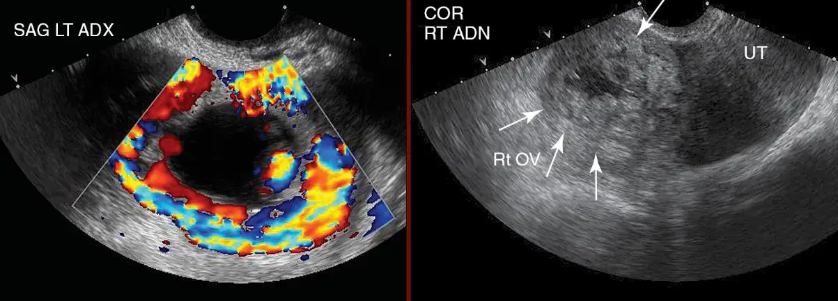
These images both show
(a)
Most important finding when scanning for EP is to determine
If uterine cavity empty and adnexal mass present
If uterine cavity empty and adnexal mass is not present
If normal intrauterine gestation (reducing probability of ectopic
pregnancy)
Non normal intrauterine sac
20% of patients with EP demonstrate intrauterine saclike structure known as
(a)
Do Pseudo GS contain a living embryo or yolk sac?
Yes
No
Pseudo GS is located (a) within the endometrial cavity, normal GS are eccentrically located
(a) level echoes are commonly observed in pseudo GS
Presence of ___ in uterus positively indicates an intrauterine gestation
Amniotic sac
Yolk sac
Gestational sac
embryo
Which pattern is considered normal for an intrauterine pregnancy?
Low resistive, low diastolic
Low resistive, high diastolic
high resistive, high diastolic
high resistive, low diastolic
Peak high velocities in an intrauterine pregnancy measures:
30cm/sec
20cm/sec
40cm/sec
80cm/sec
A pseudogestational sac demonstrates which pattern of flow?
high resisitive, high diastolic
low resistive, low diastolic
high resistive, low diastolic
low resisitive, high diastolic
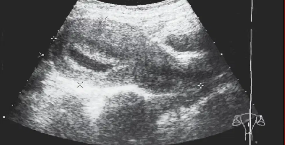
Where is this ectopic pregnancy located?
Endometrium
Fundus
Cervix
Myometrium
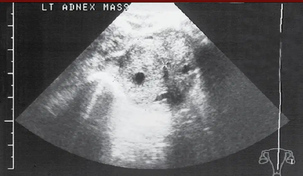
This image shows a (a) in the LT adnexa
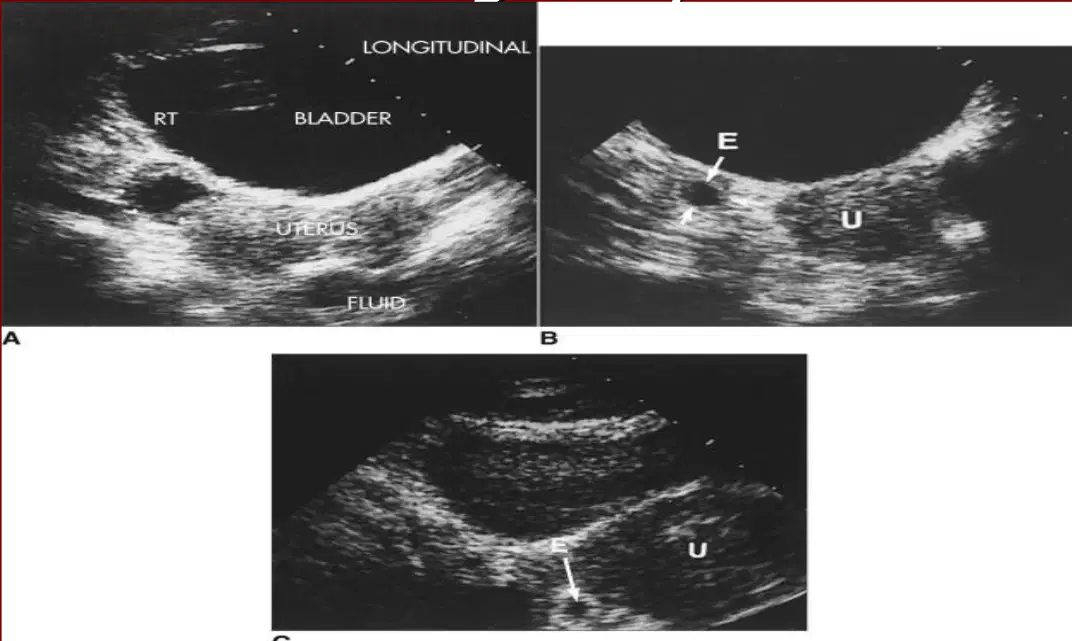
These images all show
(a)
Should there be internal echoes in a normal IUP?
Yes
No
An empty uterus is normal from __ to __ weeks
5-6
3-4
4-5
6-7
A recent spontaneous abortion could indicate a (a) uterus
No peritrophoblastic flow is characteristic of a (a)
Simultaneous intrauterine pregnancy and ectopic pregnancy (see findings above for IUP and ectopic pregnancy)
(a)
Pseudogestational sac
• Extrauterine sac in adnexa with thickened echogenic ring
(a)
•No yolk sac in the uterus
•Possible yolk sac outside uterus
(a)
Located outside the uterine cavity in that part of the fallopian tube that penetrates the muscular layer of the uterus
(a)
• Rupture with hemorrhage
• Eccentric intrauterine gestational sac with incomplete myometrial mantel surrounding sac
(a)
•Pseudogestational sac of ectopic pregnancy
(a)
Examining adnexa sonographically:
• Critical in evaluation of ectopic pregnancy
True
false
Identification of live embryo within adnexa is most specific for
(a) gestation ( occurs ~ 25% of the time)
Only approximately (a) % of live ectopic pregnancies identified using transabdominal sonography
What is one of the most frequent findings of EP?
(a)
Thickened echogenic ring, separate from ovary, which represents trophoblastic tissue or chorionic villi
(a)
Will an embryo or YS be seen in an extrauterine GS within adnexa?
Yes
No
Possibly
Absent intrauterine GS + corresponding adnexal mass => risk of ectopic pregnancy
>90%
>100%
<50%
<70%
Complex adnexal masses, aside from extrauterine GS, often represent (a) in peritoneal cavity
Where are hematomas in the peritoneal cavity usually located?
Within FT or round ligament
Within FT or ovarian ligament
Within FT or broad ligament
Within FT or fornix ligament
Ovarian processes can be differentiated from hematoma based on their location within
the ovary visualization of surrounding ovarian tissue
True
False
80% of patients with EP demonstrate at least (a) ml of blood within the peritoneum (blood escaping from the distal tube - fimbria)
~ (a) % of women with EP demonstrate intraperitoneal fluid by TVS transvaginal sonography
Presence of (a) - highly specific for hemoperitoneum and to be highly correlated with EP
The combination of an __ and __ fluid is the most precise sonographic correlation in the diagnosis of EP
(a)
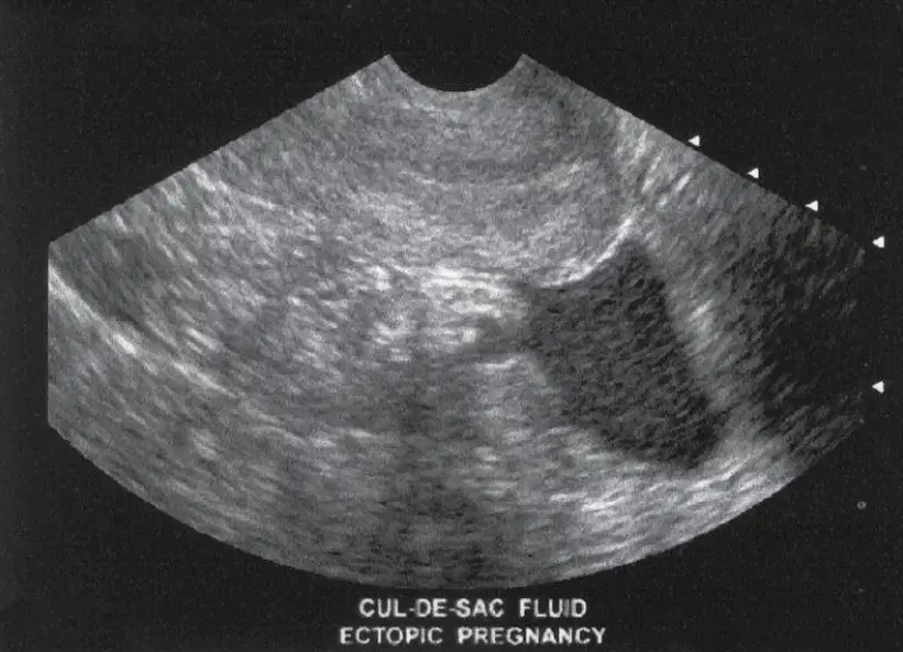
This shows
(a)
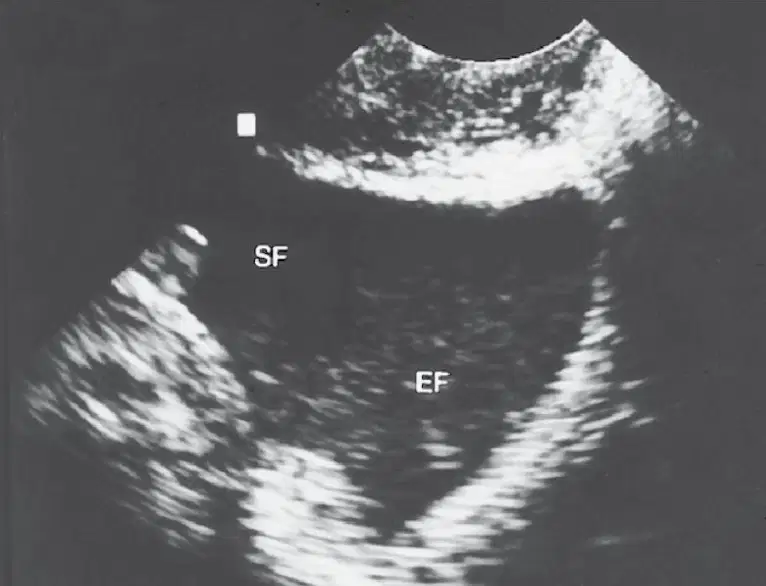
This shows
(a)
Simultaneous intrauterine and extrauterine pregnancies are extremely uncommon, even in patients undergoing an infertility workup
(a)
Ovulation induction and IVF can lead to a higher risk of:
Heterotopic
Corneal pregnancy
Interstitial Pregnancy
Ectopic pregnancies
An Interstitial pregnancy is (a)
What is the most life threatening ectopic gestation?
Interstitial
Ovarian
Round ligament
Broad ligament
in segment of fallopian tube that enters uterus which involves parauterine and myometrial vasculature, creating life- threatening hemorrhage when rupture occurs
(a)
Interstitial pregnancies occur (a) % of all EP
Is an interstitial pregnancy easy to identify on ultrasound?
Yes
No
Described as an eccentrically placed GS within the uterus that has an incomplete myometrial mantle surrounding the sac (some institutions measure the myometrial thickness and follow these implantations carefully for evidence of myometrial thinning and
uterine rupture)
(a)
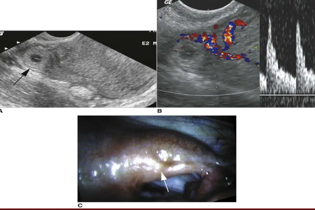
These images show (a)
Cervical pregnancies are extremely (a)
Which ectopic pregnancy is associated with an increased risk of hysterectomy because of uncontrollable bleeding?
Ovarian pregnancy
Interstitial pregnancy
Cervical Pregnancy
Heterotopic pregnancy
Concentric shape with decidual reaction and increased color Doppler flow around the trophoblast.
(a)
misshapen sac, possibly hour-glassing (the hour-glass sign)
through the cervical os, with lack of color flow around the decidual ring
(a)
Ovarian pregnancies are rare, occurring in ____% of ectopic pregnancies
<4
<3
<7
<1
reported cases have demonstrated complex adnexal masses that involve or contain the ovary
(a)
distinguishing ovarian pregnancy from a hemorrhagic ovarian cyst
or from other ovarian processes may be difficult.
(a)
Maximum thickness of subcutaneous lucency at back of neck in embryo at 11 to 14 weeks’ gestation
(a)
An (a) NT measurement is associated with trisomies T 21, T 13 and T 18
and in fetuses with cardiac defects and other genetic syndromes
NT measurement combined with biochemical markers—free beta- hCG and pregnancy-associated plasma protein (PAPP-A)—to assess risk for (a)
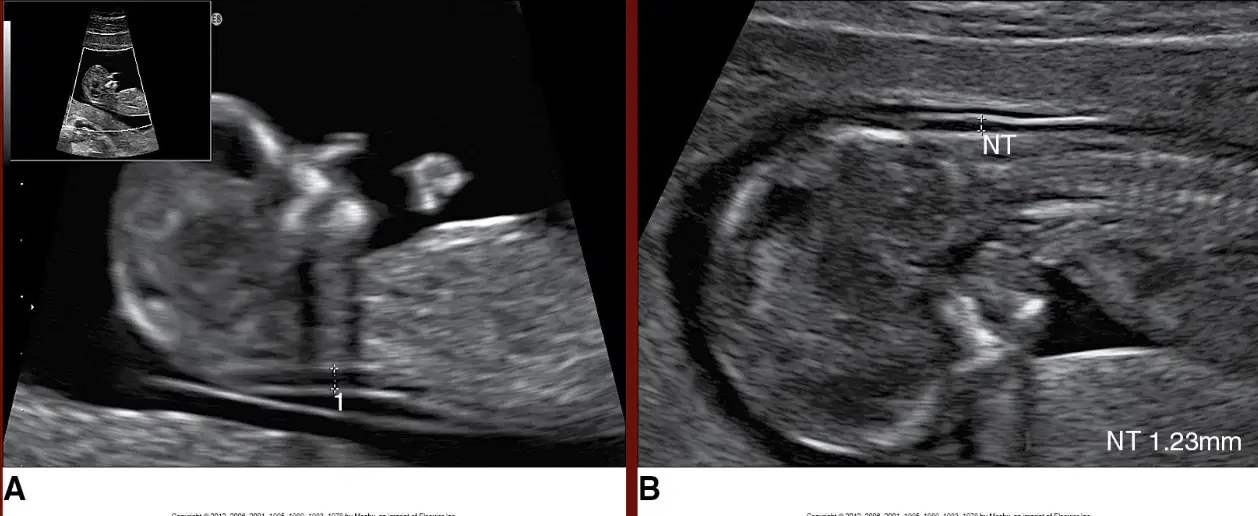
This shows (a)
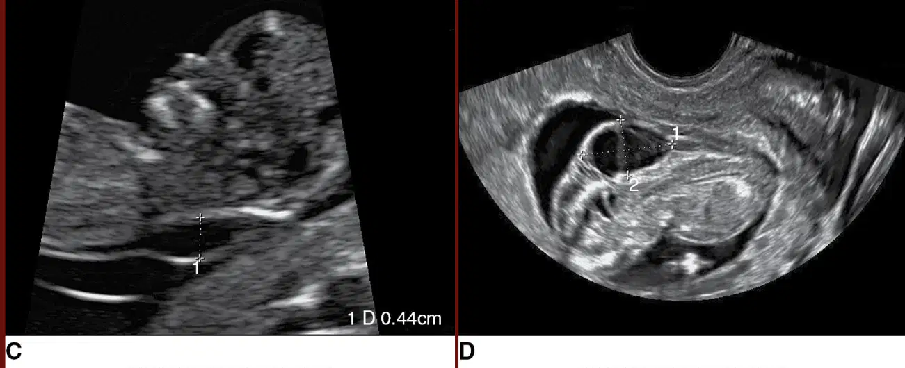
This image shows an (a) nuchal translucency
Fetuses must be between __ and __ 6 days for Nuchal Translucency
(a)
CRL must be __ and __ for nuchal translucency
(a)
Sonographer must obtain optimal image of Nuchal translucency (a)
Embryo must be (a) from amniotic membrane with head in neutral position, no hyperextension or flexion
✓ Nuchal thickening
✓ Presence or absence of nasal bone
✓ Tricuspid regurgitation
✓ Abnormal flow in ductus venosus
✓ Possibly abnormalities of hindbrain
(a)
Strong relationship between increased NT and fetuses with (a)
Ability to detect four-chamber view and great vessels as early as (a)
✓ Increased Nuchal translucency
✓ Tricuspid regurgitation
✓ Reversal (or absence) of flow in Ductus Venosus
(a)
Other cardiac associations that can be detected in the first trimester are Ectopia Cordis and Limb body wall complex
True
False

