
 Worksheets
SLIDE 1: Head and Neck Muscles
Total questions: 95
Worksheet time: 48mins
Which muscle elevates the mandible during forceful chewing?
Temporalis
Platysma
Zygomaticus
Frontalis
Which muscle primarily closes the eyelids?
Orbicularis oculi
Orbicularis oris
Occipitalis
Masseter
Which muscle tenses the skin of the neck and depresses the mandible slightly?
Platysma
Zygomaticus
Sternocleidomastoid
Frontalis
Which paired muscle rotates the head to the opposite side and flexes the neck?
Sternocleidomastoid
Occipitalis
Masseter
Temporalis
Which muscle elevates the corners of the mouth to produce a smile?
Zygomaticus
Orbicularis oris
Platysma
Occipitalis
Which muscle forms a sphincter around the mouth and purses the lips?
Orbicularis oris
Orbicularis oculi
Masseter
Frontalis
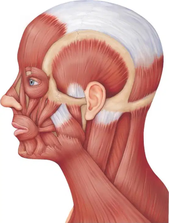
You palpate a patient clenching their teeth and feel a strong bulge over the ramus of the mandible. Which muscle is most responsible?
Masseter
Platysma
Frontalis
Occipitalis
A patient cannot raise their eyebrows to create horizontal forehead wrinkles. Which superficial muscle is impaired?
Frontalis
Temporalis
Orbicularis oris
Sternocleidomastoid
Which muscle retracts the scalp posteriorly and works with the frontal belly during facial expression?
Occipitalis
Zygomaticus
Temporalis
Platysma
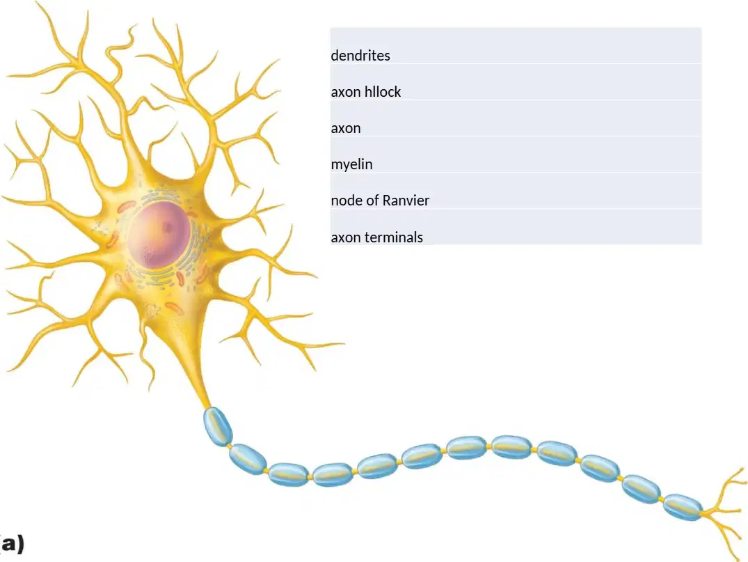
Which neuron region integrates incoming graded potentials and most directly initiates an action potential if threshold is reached?
Dendritic spines receiving signals
Axon hillock at the soma base
Myelin sheath around the axon
Axon terminals at synaptic ends
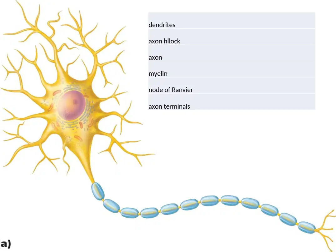
In the diagram, which label corresponds to the regularly spaced gaps between myelin segments that enable saltatory conduction?
Dendrites branching from the soma
Node of Ranvier along the axon
Axon hillock near the soma
Axon terminals forming synapses
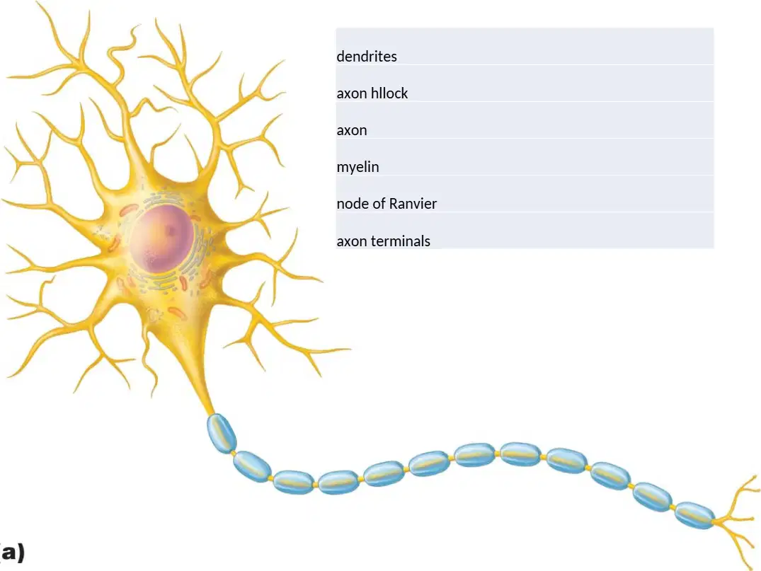
Select the component whose primary function is to receive synaptic input from other neurons.
Dendrites extending from the cell body
Axon propagating action potentials
Myelin insulating the axon fiber
Axon terminals releasing transmitters
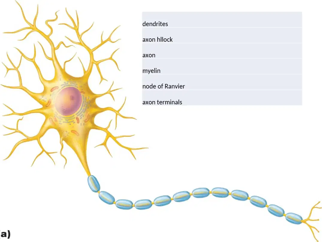
Which statement best matches the role of myelin as depicted on the axon?
It generates neurotransmitters for release
It electrically insulates to speed conduction
It initiates spikes at the axon hillock
It forms synapses with target cells
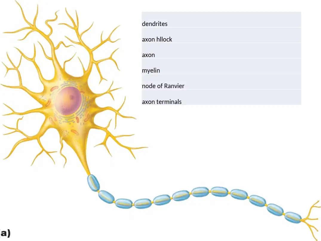
A toxin uniformly removes the myelin sheaths in the pictured neuron while leaving nodes intact. What immediate effect on conduction is most likely?
Faster saltatory conduction between nodes
Failure of rapid conduction and spike decay
Greater spike initiation at the hillock
Enhanced neurotransmitter release distally
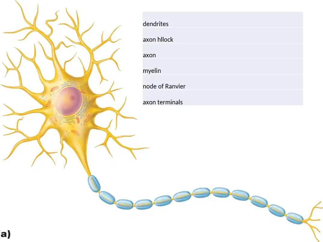
Electrical signals travel from the soma toward the target cell. Which sequence best traces the route in this neuron?
Dendrites → nodes → soma
Hillock → axon → terminals
Myelin → soma → dendrites
Terminals → axon → hillock
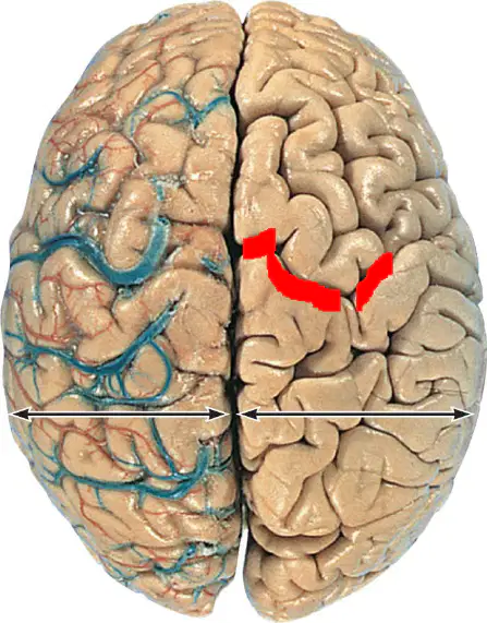
On the superior view shown, the deep groove separating the two cerebral hemispheres along the midline is called the:
longitudinal fissure
central sulcus
lateral fissure
parieto‑occipital sulcus
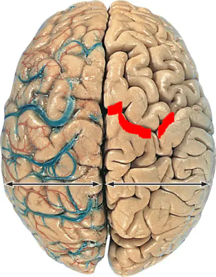
In the diagram, the raised ridges between grooves on the cerebral surface are termed:
gyri
sulci
fissures
lobules
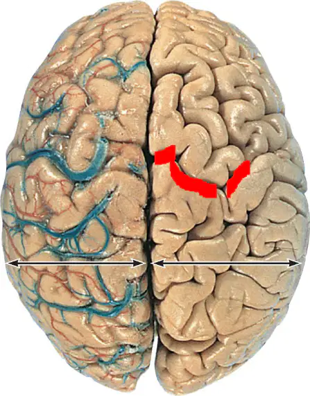
Which statement best distinguishes a sulcus from a gyrus on the cortical surface?
A sulcus is a shallow groove; a gyrus is a raised ridge
A sulcus is a raised ridge; a gyrus is a deep groove
A sulcus is a broad lobe; a gyrus is a thin membrane
A sulcus and gyrus are identical surface features
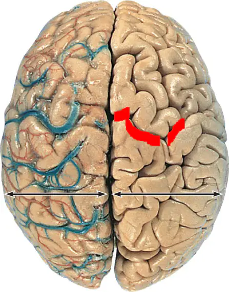
Identify the lobe located most posteriorly in the cerebral hemispheres, primarily involved in visual processing:
occipital lobe
frontal lobe
temporal lobe
parietal lobe
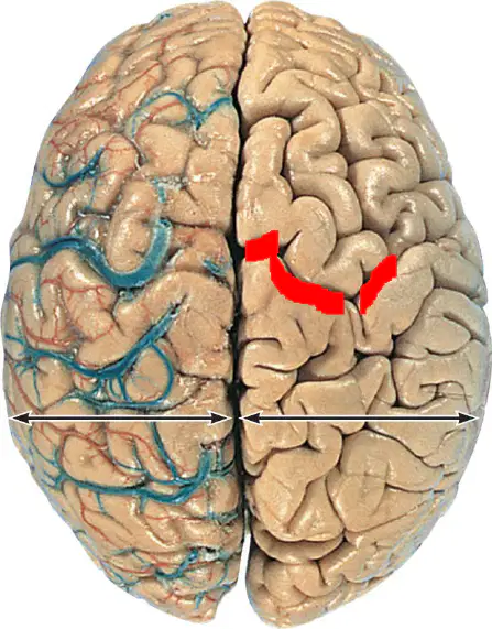
A lesion restricted to the anterior lobe highlighted near the forehead region would MOST likely involve which lobe?
frontal lobe
occipital lobe
parietal lobe
insula lobe
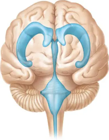
In the left diagram (a), which labeled cavity represents the paired spaces within the cerebral hemispheres that arch into the frontal and occipital lobes?
Lateral ventricles within cerebral hemispheres
Third ventricle around thalamic region
Cerebral aqueduct in midbrain conduit
Fourth ventricle between pons and cerebellum
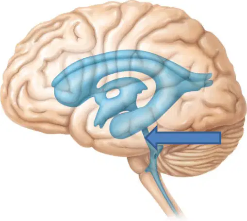
On the right sagittal diagram (b), cerebrospinal fluid flows from the third ventricle to the fourth ventricle through which narrow channel indicated near the midbrain?
Cerebral aqueduct connecting third to fourth
Interventricular foramina near lateral bodies
Central canal continuing into spinal cord
Median aperture toward subarachnoid space
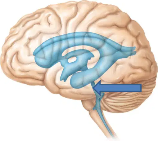
Which structure is best identified as the midline cavity bordered by the thalamus, situated inferior to the lateral ventricles and superior to the cerebral aqueduct in the sagittal view?
Third ventricle between diencephalic walls
Fourth ventricle dorsal to pons region
Lateral ventricle within cerebral lobes
Central canal within lower brainstem
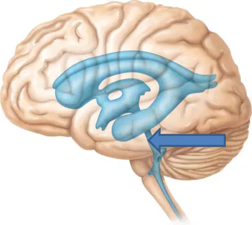
A tumor compresses the channel highlighted by the arrow in diagram (b). Predict the most immediate ventricular dilation pattern that would occur upstream of the blockage.
Enlargement of lateral and third ventricles
Expansion of fourth ventricle only
Dilation of central canal exclusively
Uniform enlargement of all ventricles equally
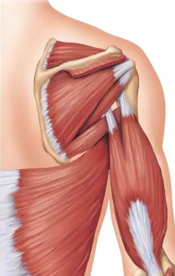
In the left diagram, which muscle forms the rounded contour over the lateral shoulder, capping the glenohumeral joint?
Deltoid muscle over the shoulder cap
Trapezius across the upper back
Latissimus dorsi along the lower back
Pectoralis major over the chest wall
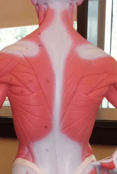
Which structure in the right diagram spans from the cervical region to the mid-back, forming a broad superficial sheet over the upper back?
Trapezius forming a broad superficial sheet
Deltoid covering the lateral shoulder
Biceps brachii on the anterior arm
Pectoralis major spanning the anterior chest
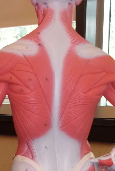
In the central diagram, identify the large muscle that sweeps from the lower thoracic and lumbar region toward the humerus, contributing to shoulder adduction and extension.
Latissimus dorsi sweeping toward the humerus
Pectoralis major crossing the anterior chest
Deltoid inserting on the humeral tuberosity
Biceps brachii running along the anterior arm
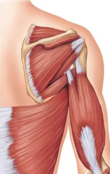
A client struggles to initiate shoulder abduction from neutral in the left diagram. Which primary muscle weakness most directly explains this deficit?
Deltoid initiating shoulder abduction
Latissimus dorsi driving shoulder flexion
Trapezius powering elbow extension
Pectoralis major starting forearm supination
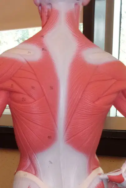
During a pull-up depicted by muscles in the central and right diagrams, which pair primarily produces shoulder extension and scapular depression?
Latissimus dorsi with lower trapezius
Deltoid with anterior fibers
Pectoralis major with clavicular head
Biceps brachii with short head
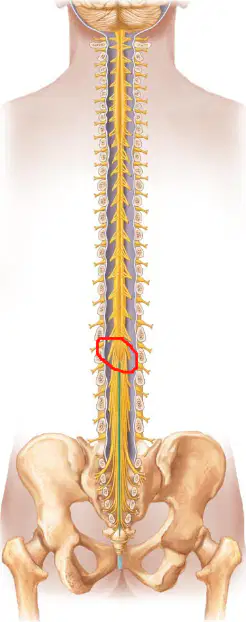
In the diagram of the lower spinal cord, which labeled structure represents the tapered terminal end of the spinal cord proper?
Conus medullaris
Filum terminale
Cauda equina
Lumbar enlargement
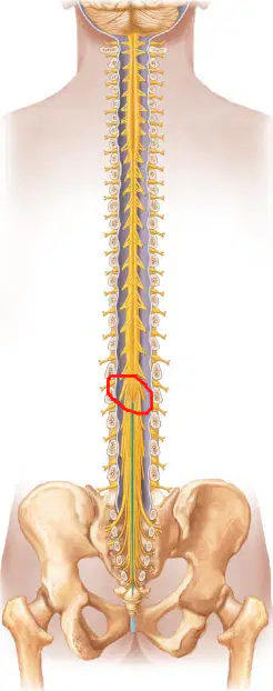
Which option best describes the filum terminale as seen in an inferior spinal cord diagram?
A thin connective tissue strand anchoring the cord
A bundle of lumbar and sacral nerve roots
The cone-shaped termination of spinal cord
A widened region housing more motor neurons
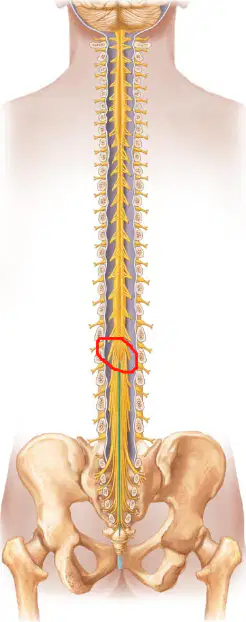
You plan a lumbar puncture at the L4–L5 level shown in the diagram. Which anatomical rationale most justifies this site selection?
The spinal cord has ended, leaving cauda equina roots
The filum terminale fills the dural sac completely
The conus medullaris occupies the vertebral canal here
The lumbar enlargement protects against needle injury
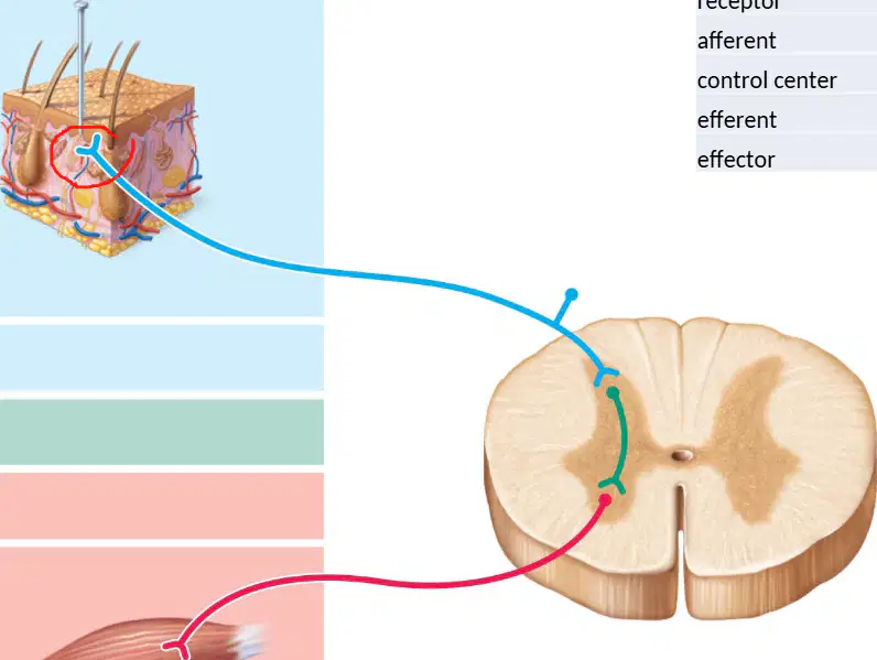
Which sequence best represents the flow of information in a simple reflex arc from initial event to response?
Stimulus → receptor → afferent → control center → efferent → effector
Receptor → stimulus → efferent → control center → afferent → effector
Stimulus → efferent → receptor → control center → afferent → effector
Receptor → afferent → stimulus → efferent → control center → effector
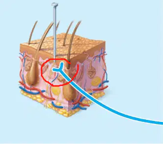
In the diagram, the blue fiber traveling from the skin to the spinal cord represents which component of the reflex arc?
Afferent pathway carrying sensory input
Efferent pathway carrying motor output
Effector producing the physical response
Control center generating the stimulus
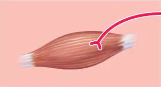
Which element serves as the effector in the depicted reflex arc?
Skeletal muscle contracting to produce movement
Cutaneous receptor detecting tissue damage
Interneurons within the spinal cord gray matter
Afferent neuron transmitting sensory signals
A pinprick activates a cutaneous receptor that sends an impulse to the spinal cord and produces a rapid withdrawal. Which step immediately precedes motor neuron activation?
Signal processing in the control center
Stimulation of the effector organ
Transduction by the skin receptor
Arrival of the external stimulus
If the afferent pathway were severed but all other components remained intact, what outcome would most likely occur?
No reflex because sensory input cannot reach the control center
Exaggerated reflex because efferent neurons fire continuously
Normal reflex because efferent and effector are unaffected
Delayed reflex because stimulus travels a longer route
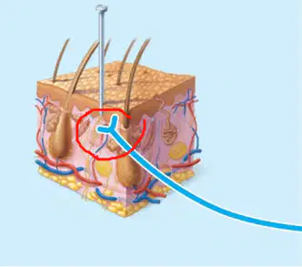
Which pairing correctly matches a component with its functional description in this reflex arc?
Receptor: converts stimulus into neural signal
Effector: detects environmental change at the skin
Afferent: delivers motor commands to muscle
Control center: produces the external stimulus
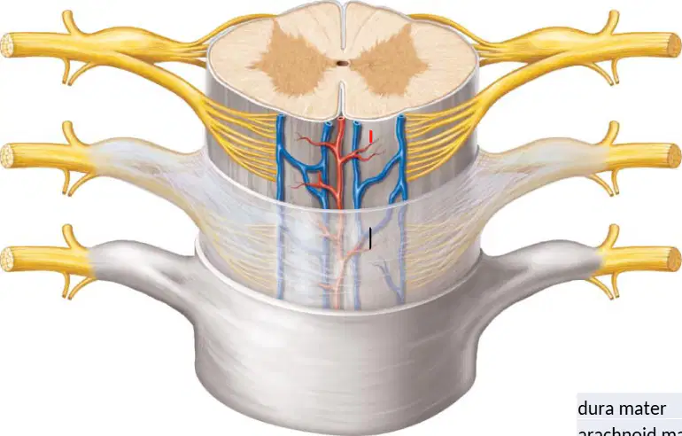
In the diagram of a spinal cord cross-section, which structure is the tough outer meningeal layer surrounding the cord?
Dura mater
Arachnoid mater
Pia mater
Epidural fat
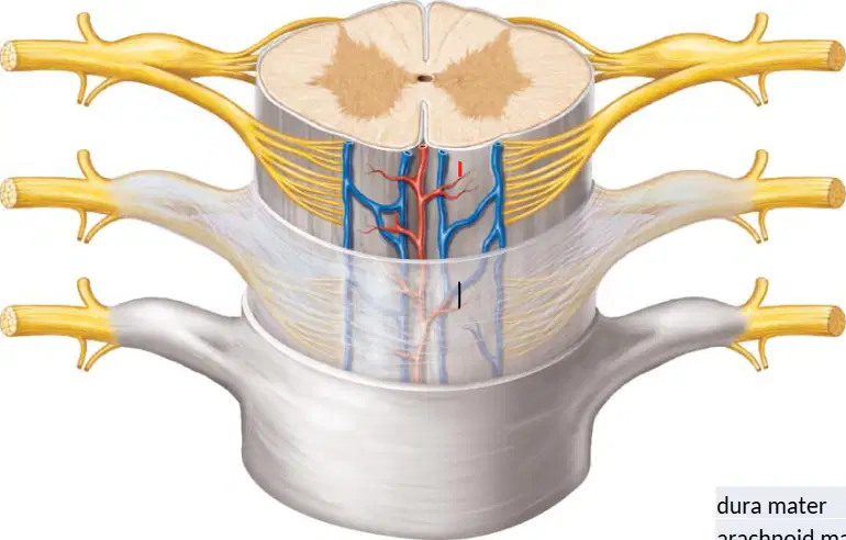
Which labeled feature marks the shallow midline groove on the posterior surface of the spinal cord?
Ventral median fissure
Dorsal median sulcus
Central canal opening
Posterolateral sulcus
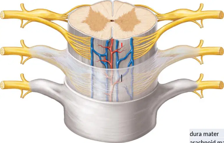
In the diagram, dorsal roots and ventral roots merge to form which mixed structure carrying both sensory and motor fibers?
Dorsal root ganglion
Spinal nerve
Sympathetic chain
Ventral ramus
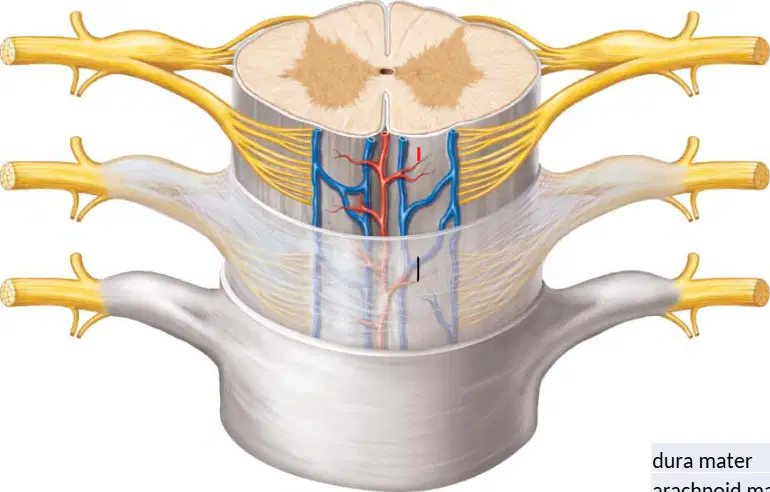
Which component contains the cell bodies of sensory neurons associated with the dorsal roots?
Ventral horn nucleus
Dorsal root ganglion
Lateral horn cluster
Interneuronal commissure
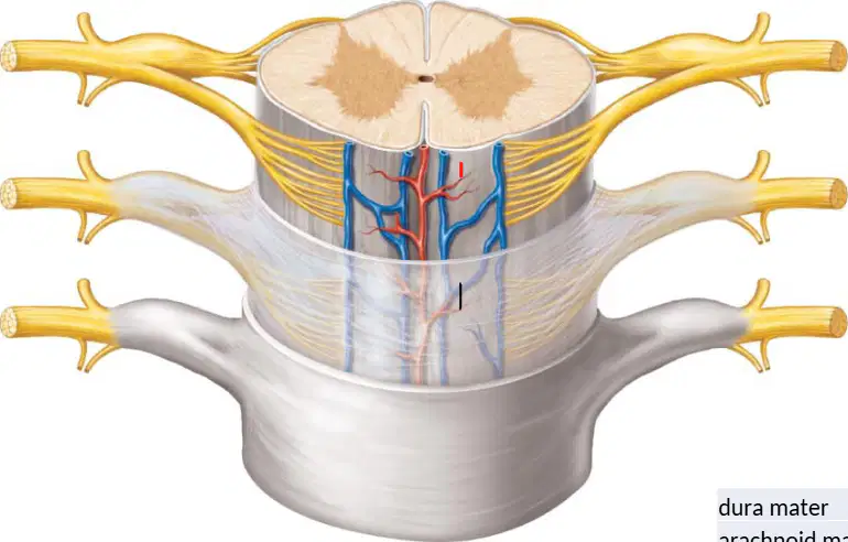
Which option correctly pairs root type with predominant fiber direction at the cord?
Dorsal roots—efferent fibers
Ventral roots—afferent fibers
Dorsal roots—afferent fibers
Ventral roots—interneuronal fibers
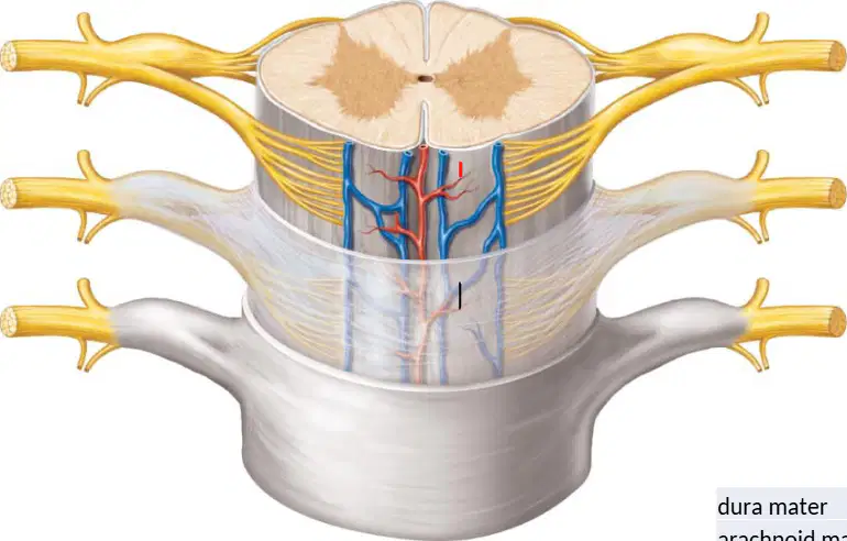
In the midline on the anterior aspect of the cord, which deeper groove partially divides the cord into halves?
Dorsal median sulcus
Ventral median fissure
Anterolateral sulcus
Posterior intermediate sulcus
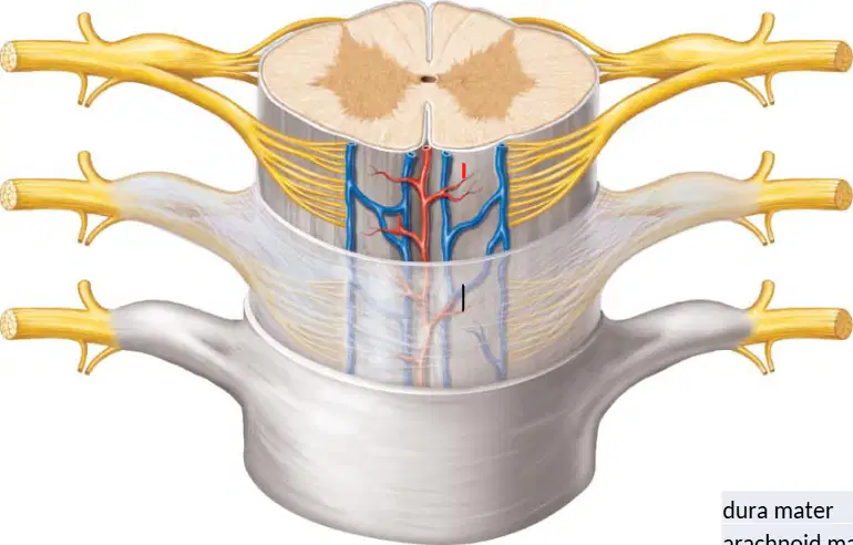
Comparing the meninges shown, which layer lies directly deep to the dura and forms a web-like space above the pia?
Arachnoid mater
Pia mater
Epidural periosteum
Endoneurium
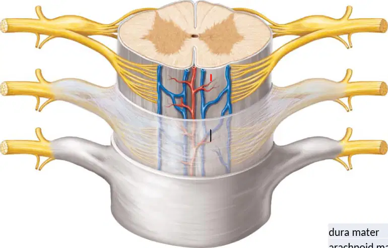
A patient’s dorsal root is compressed proximal to the ganglion in the diagrammed region. Which primary deficit is most expected at that level?
Loss of motor function only
Loss of sensory input only
Loss of autonomic outflow only
Complete loss of mixed conduction
In the arm diagram, which nerve primarily runs along the posterior compartment to the lateral forearm and hand?
Radial nerve along posterior compartment
Ulnar nerve across anterior compartment
Median nerve through central compartment
Musculocutaneous nerve to medial compartment
In the pelvis and anterior thigh diagram, which nerve emerges near the inguinal region to supply the anterior thigh muscles?
Femoral nerve supplying anterior thigh
Obturator nerve to medial thigh
Sciatic nerve for posterior thigh
Gluteal nerve to lateral pelvis
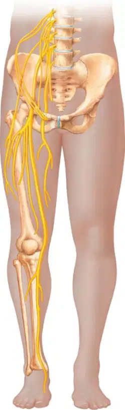
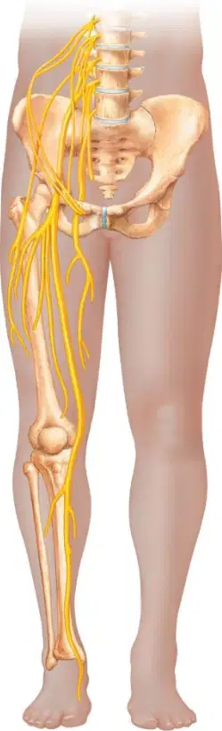
In the posterior thigh and leg diagram, the thick nerve descending from the sacral plexus and splitting near the knee is best identified as which nerve?
Sciatic nerve splitting at popliteal fossa
Femoral nerve bifurcating at knee joint
Obturator nerve dividing in thigh canal
Saphenous nerve branching near fibular head
Which nerve most likely innervates most intrinsic hand muscles on the medial side, as suggested by the medial forearm course in the arm diagram?
Ulnar nerve to medial intrinsic hand
Radial nerve to thenar intrinsic hand
Femoral nerve to medial intrinsic hand
Sciatic nerve to medial intrinsic hand
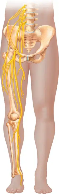
A patient has weakness adducting the thigh; based on the medial thigh pathway seen in the pelvis diagram, which nerve is most implicated?
Obturator nerve for thigh adduction
Femoral nerve for thigh adduction
Sciatic nerve for thigh adduction
Radial nerve for thigh adduction

Which muscle is the vertically oriented, segmented pair running on either side of the midline and enclosed by the rectus sheath in the diagram?
Rectus abdominis
External oblique
Internal oblique
Transversus abdominis
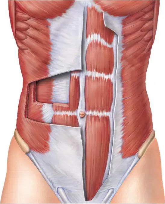
In the illustrated abdominal wall, which muscle has fibers running inferomedially (hands-in-pockets orientation) and forms the most superficial lateral layer?
External oblique
Internal oblique
Transversus abdominis
Rectus abdominis
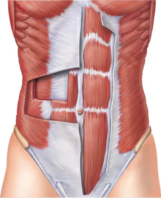
A clinician plans an incision that minimizes damage to muscle fibers by following the predominant fiber direction of the deepest lateral abdominal layer. Based on the diagram, which layer should be considered and what is its fiber direction?
Transversus abdominis; horizontal fibers
Internal oblique; inferomedial fibers
External oblique; superomedial fibers
Rectus abdominis; oblique fibers
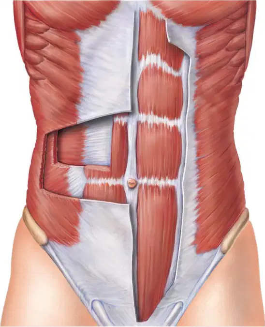
Which statement best distinguishes internal oblique from the other three muscles shown in the diagram?
It has fibers running superomedially between two lateral layers
It forms the midline segmented pair within a sheath
It is the outermost layer with inferomedial fibers
It is the deepest lateral layer with transverse fibers
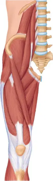
In the lateral thigh diagram, which muscle forms the large superficial mass over the posterior hip and upper thigh?
Gluteus maximus
Rectus femoris
Vastus medialis
Sartorius
Vastus lateralis
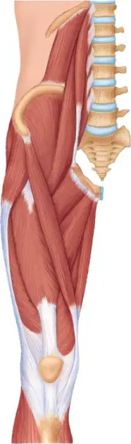
Which muscle runs obliquely from the anterior superior iliac spine across the thigh to the medial knee, visible as a thin strap-like band?
Sartorius
Hamstrings
Vastus lateralis
Vastus medialis
Rectus femoris
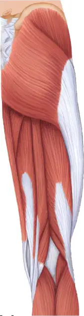
On the anterior thigh, which quadriceps muscle lies centrally and runs straight down the front of the femur?
Rectus femoris
Vastus lateralis
Vastus medialis
Sartorius
Hamstrings
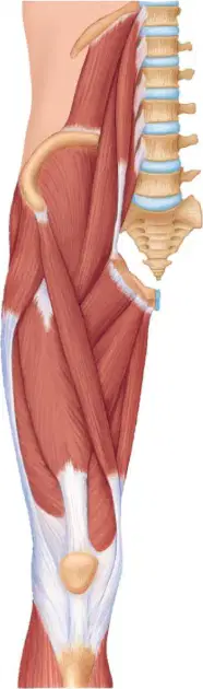
Which pair best distinguishes the medial and lateral components of the quadriceps on the distal anterior thigh?
Vastus medialis medial; vastus lateralis lateral
Vastus lateralis medial; vastus medialis lateral
Vastus medialis lateral; sartorius medial
Rectus femoris medial; hamstrings lateral
Gluteus maximus medial; rectus femoris lateral
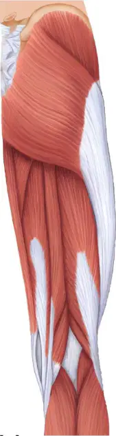
In the posterior thigh region, which group of muscles occupies most of the mass and primarily performs knee flexion?
Hamstrings
Quadriceps
Adductors
Gluteals
Tensor fasciae
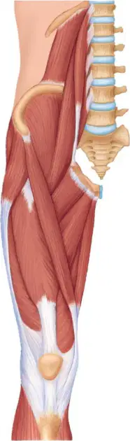
A sprinter reports pain where the strap-like anterior thigh muscle crosses toward the medial knee. Based on the diagram, which muscle is likely strained and why?
Sartorius, because it crosses from hip to medial knee
Rectus femoris, because it lies deep along femur
Vastus lateralis, because it crosses the knee diagonally
Gluteus maximus, because it runs vertically to patella
Hamstrings, because they insert on the anterior tibia
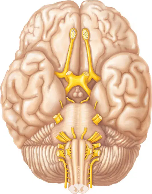
Which structure represents the crossing of optic nerve fibers at the base of the brain in an inferior view?
Olfactory bulbs and tracts crossing midline
Optic chiasm forming an X-shaped junction
Trochlear nerves decussating around pons
Abducens nerves entering the cavernous sinus
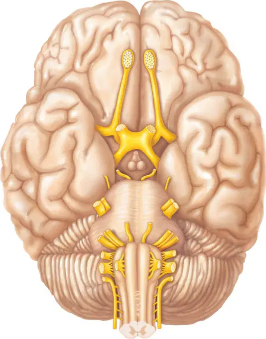
From an inferior view, which cranial nerve emerges between the midbrain and pons and typically appears as paired bundles near the interpeduncular fossa?
Oculomotor nerve exiting between midbrain and pons
Trochlear nerve emerging from dorsal midbrain
Abducens nerve arising at pontomedullary junction
Trigeminal nerve arising from lateral pons
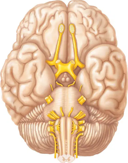
Which nerve is the large sensory-motor root arising from the lateral aspect of the pons in the inferior view?
Trigeminal nerve from lateral pons
Abducens nerve from medial medulla
Olfactory nerve from frontal lobe
Trochlear nerve from dorsal surface
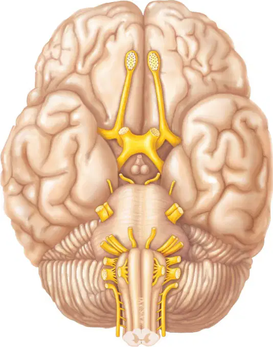
In the inferior view, which cranial nerve is most closely associated with the superior oblique muscle and emerges posteriorly before wrapping around to the ventral aspect?
Oculomotor nerve to superior oblique
Trochlear nerve winding around midbrain
Abducens nerve supplying lateral rectus
Optic nerve exiting the retina
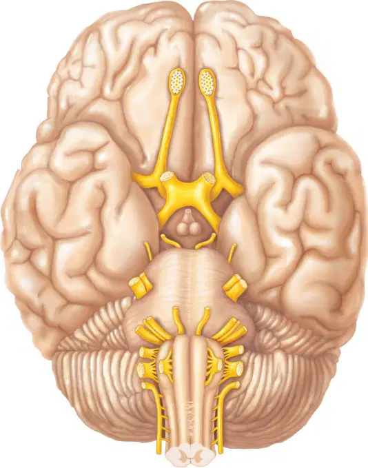
Which pair of structures lies most rostrally on the inferior surface, projecting from the frontal lobes as elongated bulbs?
Olfactory bulbs and tracts anteriorly
Optic nerves from diencephalon
Pons with transverse fibers
Medulla pyramids and olives
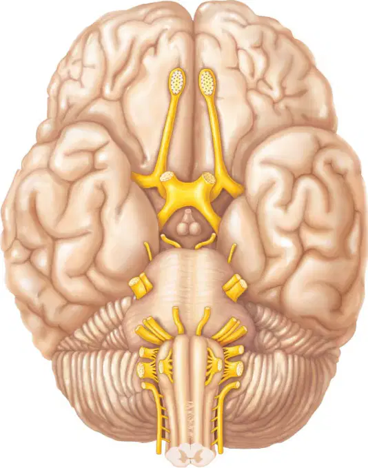
Identify the cranial nerve that emerges at the pontomedullary junction near the midline and courses forward toward the orbit through the cavernous sinus.
Abducens nerve from pontomedullary junction
Trigeminal nerve from lateral mid-pons
Oculomotor nerve from interpeduncular fossa
Optic nerve from eyeball to chiasm
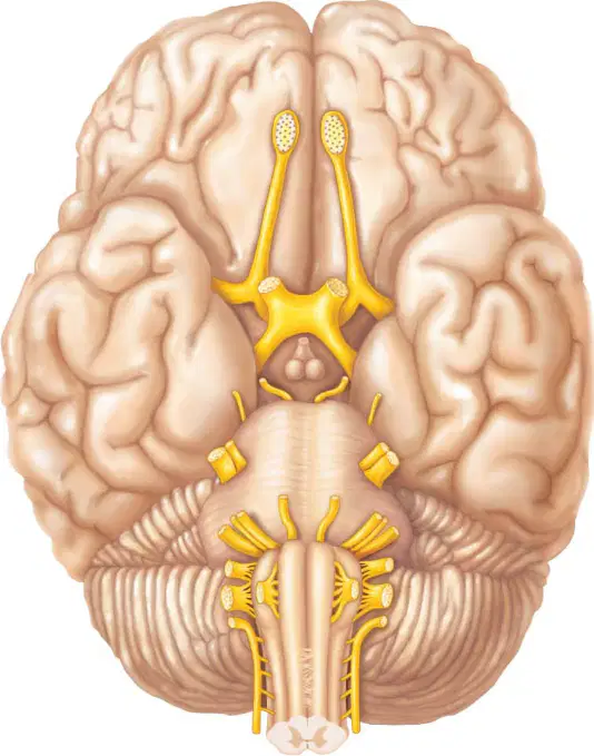
Which brainstem region forms a rounded bulge anterior to the cerebellum and serves as the origin site for several cranial nerve roots in the inferior view?
Pons bulge anterior to cerebellum
Medulla tapering to spinal cord
Midbrain colliculi on dorsal surface
Diencephalon around third ventricle
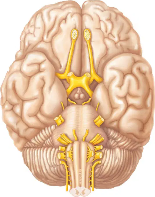
From the inferior perspective, which vertical structure continues caudally from the pons and contains the pyramids flanking the midline?
Medulla oblongata with ventral pyramids
Optic chiasm forming central X
Olfactory tracts along frontal base
Cerebral peduncles of midbrain
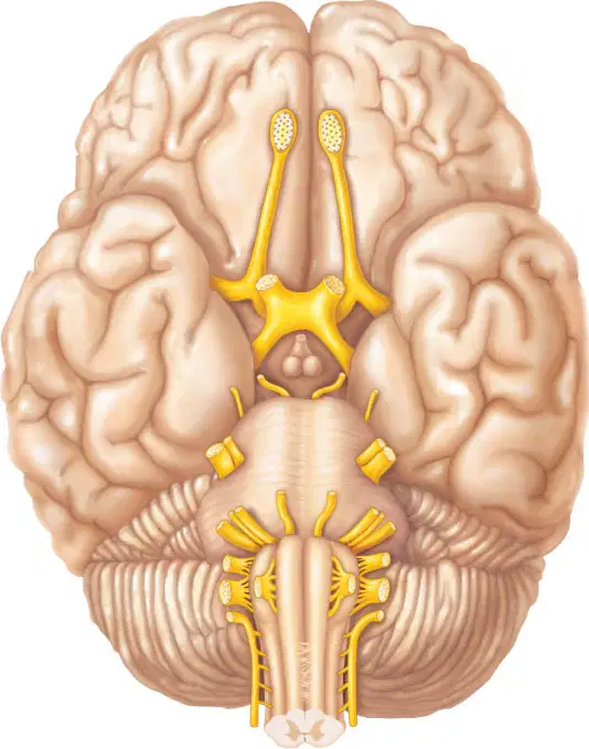
Two round anterior projections are connected to the diencephalon by the optic tracts and converge at an X-shaped junction. What are these paired projections called?
Optic nerves converging from each eye
Olfactory bulbs projecting from cortex
Abducens roots from medulla
Trochlear nerves circling midbrain
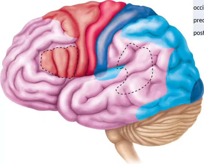
On the lateral surface shown, which groove separates the precentral gyrus from the postcentral gyrus?
Central sulcus
Lateral sulcus
Parieto‑occipital sulcus
Longitudinal fissure
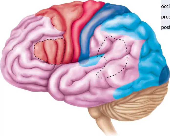
Which labeled region corresponds to the lobe most posterior on the lateral surface?
Occipital lobe
Parietal lobe
Temporal lobe
Frontal lobe
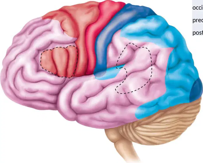
In the diagram, the gyrus immediately anterior to the central sulcus is identified as which structure?
Precentral gyrus
Postcentral gyrus
Superior temporal gyrus
Cingulate gyrus

Which option best locates the temporal lobe on this lateral view?
Inferolateral cerebrum above cerebellum
Superior posterior cortex near midline
Anterior superior cortex behind forehead
Medial surface adjacent to corpus callosum
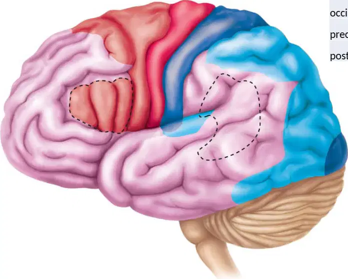
Damage to the postcentral gyrus in the depicted hemisphere would most directly impair which function?
Primary somatosensory processing
Primary motor initiation
Visual field mapping
Auditory language comprehension
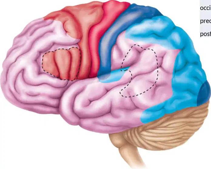
On the image, which structure lies inferior and posterior to the occipital lobe, with tightly folded cortex?
Cerebellum
Parietal operculum
Insular cortex
Pons
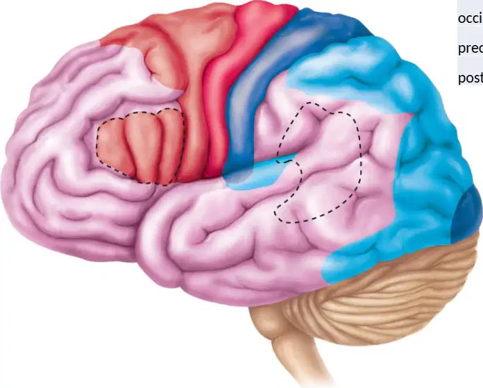
A lesion just posterior to the central sulcus extending superiorly would most likely involve which lobe?
Parietal lobe
Frontal lobe
Temporal lobe
Occipital lobe

In the sagittal brain diagram, which structure forms the C-shaped white matter tract connecting the cerebral hemispheres, arching above the lateral ventricles?
Corpus callosum
Thalamus
Hypothalamus
Midbrain
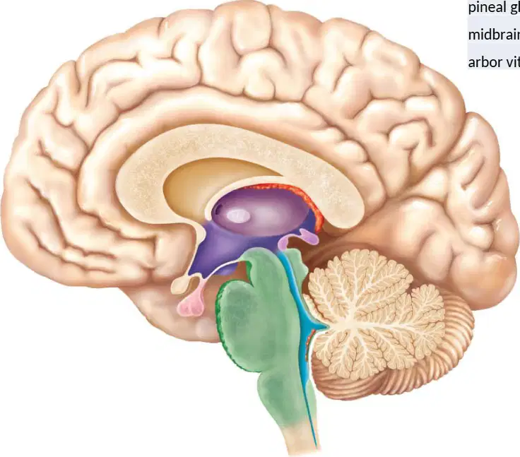
Which midline diencephalic structure in the diagram acts primarily as a relay for sensory information to the cortex?
Thalamus
Hypothalamus
Pineal gland
Arbor vitae
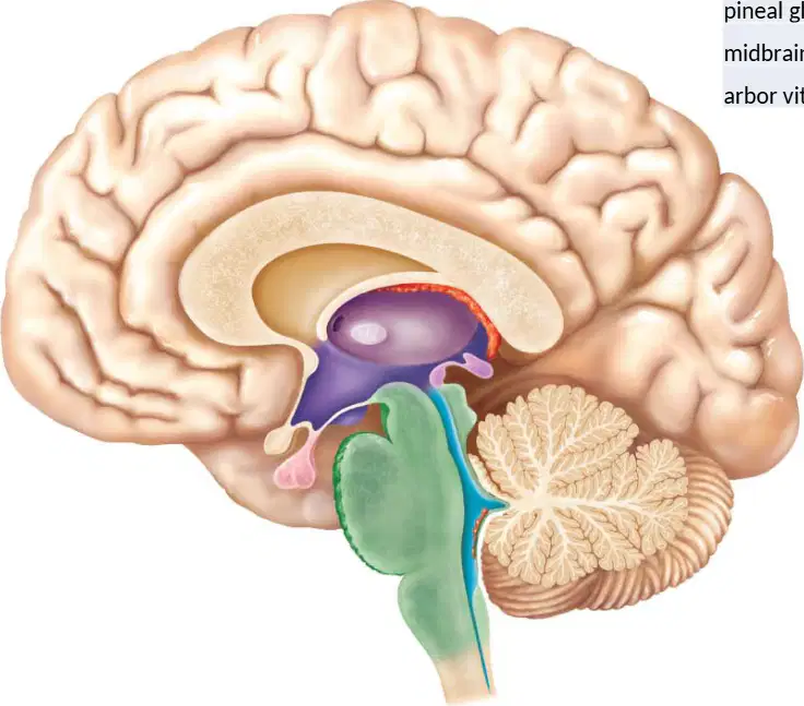
In the image, which small endocrine organ projects posteriorly from the diencephalon and is associated with melatonin secretion?
Pineal gland
Pituitary gland
Amygdala
Caudate nucleus
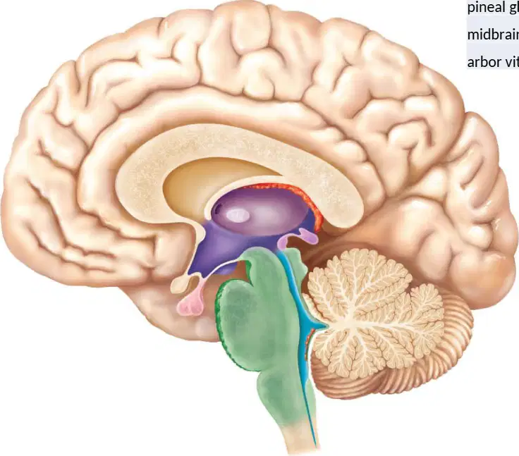
Which structure in the sagittal view lies inferior to the thalamus and is most directly involved in homeostatic regulation such as temperature and hunger?
Hypothalamus
Corpus callosum
Arbor vitae
Pineal gland
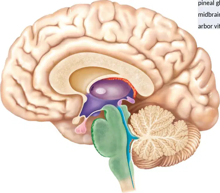
In the sagittal diagram, which region is the segment of the brainstem that connects the pons and forebrain and contains the cerebral aqueduct?
Midbrain
Medulla
Cerebellum
Temporal lobe

Which cerebellar feature, visible as tree-like branching white matter in the sagittal section, is labeled in the diagram?
Arbor vitae
Corpus callosum
Internal capsule
Basal nuclei

In an anterior view of the lower leg, which muscle lies just lateral to the tibia and is a primary dorsiflexor of the ankle?
Tibialis anterior
Fibularis longus
Gastrocnemius
Soleus
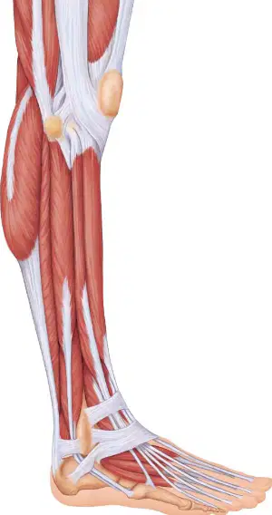
When viewing the lateral side of the lower leg, which muscle forms the prominent belly behind the fibula and contributes to foot eversion?
Fibularis longus
Tibialis anterior
Gastrocnemius
Soleus
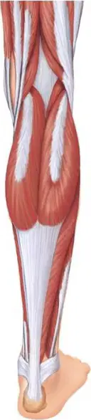
You are asked to plan a taping strategy to limit excessive plantarflexion during rehab. Based on posterior leg anatomy, which superficial muscle would you primarily target to reduce plantarflexor power?
Gastrocnemius
Tibialis anterior
Fibularis longus
Deep toe flexors
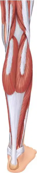
During a calf-raise test with the knee flexed to 90°, plantarflexion strength decreases less than expected. Which muscle most likely maintains force in this position due to its monoarticular design?
Soleus
Gastrocnemius
Tibialis anterior
Fibularis longus
Which plexus is formed by anterior rami from C1–C4 and primarily innervates structures of the neck near the upper spine?
Cervical plexus near the upper spine
Brachial plexus of the shoulder region
Lumbar plexus within the lower back
Sacral plexus of the pelvic cavity
In a diagram highlighting nerves supplying the upper limb emerging between C5 and T1, which plexus is depicted?
Brachial plexus supplying the upper limb
Lumbar plexus serving anterior thigh
Sacral plexus innervating posterior leg
Cervical plexus serving superficial neck
A patient shows sensory loss over the anterior thigh and weakness in knee extension after a lumbar disc issue. Which plexus is most likely affected?
Lumbar plexus involving anterior thigh
Cervical plexus controlling diaphragm
Brachial plexus serving forearm flexors
Sacral plexus innervating plantar foot
Damage to nerves arising from L4–S4 most strongly impairs which functional region shown in a spinal plexus diagram of the lower body?
Posterior thigh and most of the leg
Anterior neck and superficial scalp
Anterior arm and lateral forearm
Anterior thigh and iliacus muscle
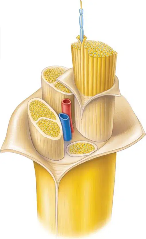
In the peripheral nerve diagram, which term names a bundle of axons grouped together within the nerve?
Fascicle
Endoneurium
Epineurium
Perineurium
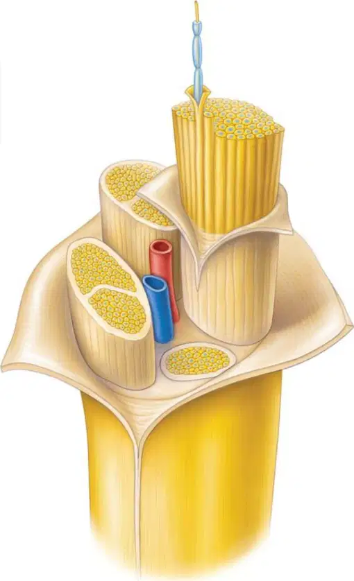
Which connective tissue layer directly surrounds individual nerve fibers within a fascicle in the diagram?
Endoneurium
Perineurium
Epineurium
Myelin sheath
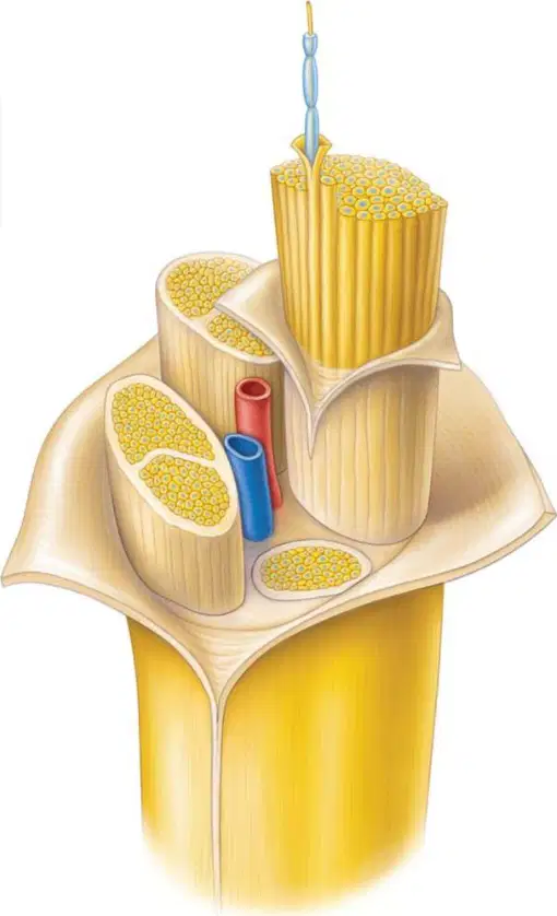
Looking at the whole-nerve cross-section, which layer encloses the entire nerve and includes blood vessels?
Epineurium
Perineurium
Endoneurium
Neurilemma
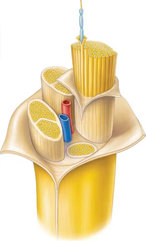
A fascicle is wrapped by a protective sheath forming the blood–nerve barrier. Based on the diagram, which layer provides this function around each fascicle?
Perineurium
Epineurium
Endoneurium
Axolemma

