
 Worksheets
Pelvic Sonogram Examination Quiz
Total questions: 60
Worksheet time: 30mins
What are the two approaches used in a pelvic sonogram examination?
Transabdominal and Transvaginal approaches
Transrectal and Transvaginal approaches
Transabdominal and Transrectal approaches
Transvaginal and Transurethral approaches
What is applied to the lower abdomen before scanning in the transabdominal approach of a pelvic sonogram?
Cold gel
Warm gel
Ultrasound dye
Sterile water
What condition must be met before performing the transvaginal approach in a pelvic sonogram?
Full bladder
Empty bladder
Application of warm gel
Use of a transrectal probe
The transvaginal approach is used to evaluate which of the following?
Abdominal examination
Pelvic examination
Rectal examination
Thoracic examination
With transvaginal imaging, oblique angulation is often necessary to visualize what part of the uterus?
Cervix and uterus
Ovaries and adnexal
Myometrium and cervix
Uterine size and shape
Which of the following should be documented during a pelvic ultrasound examination?
Uterine size, shape, orientation, endometrium, myometrium, cervix
Uterine size, shape, orientation, ovaries, fallopian tubes
Endometrium, ovaries, fallopian tubes, cervix
Myometrium, ovaries, uterine size, fallopian tubes
What role does the vagina serve in a pelvic ultrasound examination?
It serves as a landmark for cervix and lower uterine segment
It serves as a landmark for ovaries and fallopian tubes
It serves as a landmark for uterine size and shape
It serves as a landmark for endometrium and myometrium
What is the purpose of a distended bladder during a pelvic ultrasound examination?
To serve as an acoustic window for viewing pelvic anatomy and pathology.
To reduce the size of the uterus for better imaging.
To eliminate the need for transverse imaging.
To increase the resolution of the ultrasound machine.
When is the bladder considered optimally full during a pelvic ultrasound examination?
When it covers the fundus of the normal-sized uterus.
When it is completely empty.
When it is partially filled with fluid.
When it is overfilled to its maximum capacity.
What is the consequence of overdistention of the bladder during a pelvic ultrasound examination?
It improves the clarity of the sonographic evaluation.
It compresses, distorts, and displaces anatomy.
It enhances the visibility of the endometrium.
It has no effect on the sonographic evaluation.
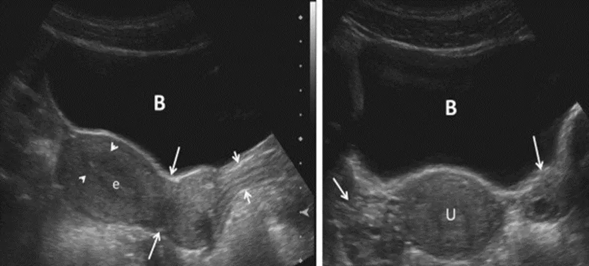
Which anatomical structures are labeled as (B), (U), and (e) in the context of pelvic ultrasound examination?
(B) bladder; (U) uterus; (e) endometrium.
(B) bowel; (U) urethra; (e) epithelium.
(B) bladder; (U) urethra; (e) epithelium.
(B) bowel; (U) uterus; (e) endometrium.
Why is it important for the bladder to be distended but not cover the fundus of the uterus during a pelvic ultrasound examination?
To ensure the bladder is visible in the sonographic evaluation.
To prevent compression and distortion of the anatomy.
To enhance the visibility of the uterus.
To avoid interference with the imaging of the endometrium.
How is the length of the uterus measured during a pelvic ultrasound examination?
From anterior to posterior walls
From transaxial or coronal view
From fundus to cervix
From lateral walls to midline
What is analyzed for thickness and echogenicity during a pelvic ultrasound examination?
Cervical diameters
Myometrium
Endometrium
Uterine walls
What is the myometrium evaluated for during a pelvic ultrasound examination?
Thickness and echogenicity
Contour changes, echogenicity, and masses
Cervical diameters
Uterine length
Where are ovaries identified during a pelvic ultrasound examination?
Posterior to the uterus
Anterior to internal iliac vessels
Lateral to the fallopian tubes
Inferior to the bladder
What should be documented regarding ovarian size during a pelvic ultrasound examination?
Length of the long axis and AP dimension measured parallel to length
Length of the long axis and AP dimension measured perpendicular to length
Width of the long axis and AP dimension measured perpendicular to width
Width of the long axis and AP dimension measured parallel to width
In which views is ovarian width measured during a pelvic ultrasound examination?
Sagittal and coronal views
Transaxial and coronal views
Transaxial and sagittal views
Coronal and transverse views
Which of the following is NOT a parameter documented during a pelvic ultrasound examination of the ovaries?
Size, shape, contour, echogenicity
Position relative to uterus
Ovarian size determined by measuring length of long axis with AP dimension measured perpendicular to length
Blood flow velocity in ovarian vessels
What additional calculation can be performed during a pelvic ultrasound examination of the ovaries?
Ovarian density
Ovarian volume
Ovarian echogenicity
Ovarian contour
What is the primary purpose of evaluating the cul-de-sac during a pelvic ultrasound examination?
To assess the presence of free fluid or mass
To measure the size of the uterus
To identify normal bowel loops
To detect abnormalities in the ovaries only
When a mass is detected during a pelvic ultrasound, what should be documented?
Size, position, shape, echographic pattern, and relationship to ovaries and uterus
Only the size and position of the mass
The echographic pattern and relationship to the bladder
The shape and relationship to the kidneys
Why is it important to differentiate normal loops of bowel during a pelvic ultrasound examination?
Identifying them as free fluid
Distinguishing them from masses
Measuring their echographic pattern
Comparing them to the ovaries
Which anatomical structure is located posterior to the bladder in the sonographic image?
Uterus.
Endometrium.
Myometrium.
Vagina.
In the sagittal image orientation, what does the term "inferior" refer to?
The area closer to the head.
The area closer to the feet.
The area closer to the front of the body.
The area closer to the back of the body.
What anatomic location is labeled as "superior" in the sonographic image orientation?
The area closer to the head.
The area closer to the feet.
The area closer to the back of the body.
The area closer to the front of the body.
What is one of the features to characterize a mass found on sonography?
Blood type
Location (uterine or extrauterine)
Patient's age
Heart rate
Which of the following describes the external contour of a mass found on sonography?
Well-defined, ill-defined, irregular borders
Cystic, complex, predominantly solid
Uterine or extrauterine
Large or small size
What does internal consistency of a mass found on sonography include?
Size of the mass
Cystic, complex, predominantly cystic, predominantly solid, solid
Location of the mass
External contour of the mass
Which of the following is NOT a feature used to characterize a mass found on sonography?
Size
Location
External contour
Blood pressure
What does "TA" stand for in the context of ultrasonography?
Transarterial
Transabdominal
Transalveolar
Transauricular
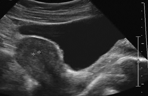
What is the primary organ being imaged in the provided transabdominal ultrasonography?
Liver
Uterus
Kidney
Heart
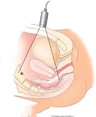
Which anatomical directions are labeled in a diagram of Transabdominal Sonography?
Anterior, Posterior, Cranial, Caudal
Superior, Inferior, Lateral, Medial
Dorsal, Ventral, Proximal, Distal
Left, Right, Superior, Inferior
What is the primary purpose of Transabdominal Sonography?
To visualize internal organs through the abdominal wall
To measure blood pressure in arteries
To examine the outer surface of the skin
To perform surgical procedures
Using a transvaginal image, in which direction is typically labeled as "Cranial"?
Towards the head
Towards the feet
Towards the back
Towards the abdomen
What anatomical location is typically known as 'Fundus'?
The upper part of the uterus
The lower part of the cervix
The bladder
The rectosigmoid colon
What is the primary purpose of Transabdominal Sonography?
To visualize internal organs through the abdominal wall
To measure blood pressure
To perform a biopsy
To treat infections
Which structure is located between the fundus and the cervix in the Transabdominal Sonography image?
Isthmus
Bladder
Myometrium
Rectosigmoid colon
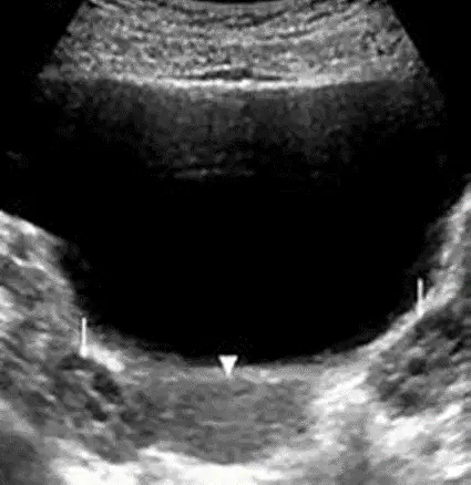
On the Transabdominal Sonography, what is primarily demonstrate in the transverse views of the adnexa?
uterus
ovary
The urinary bladder
The abdominal wall
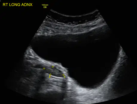
Which structure is labeled as "Urinary bladder" in the Transabdominal Sonography image?
A muscular organ that stores urine
A reproductive organ that produces eggs
A ligament supporting the uterus
A tube connecting the uterus to the ovaries
What is the purpose of Transabdominal Sonography in medical imaging?
To visualize the heart chambers
To examine the adnexa and pelvic organs
To assess the brain structure
To measure bone density
Which structure is located adjacent to the ovary in the Transabdominal Sonography image?
Abdominal wall
Fallopian tube and/or broad ligament
Urinary bladder
Uterus
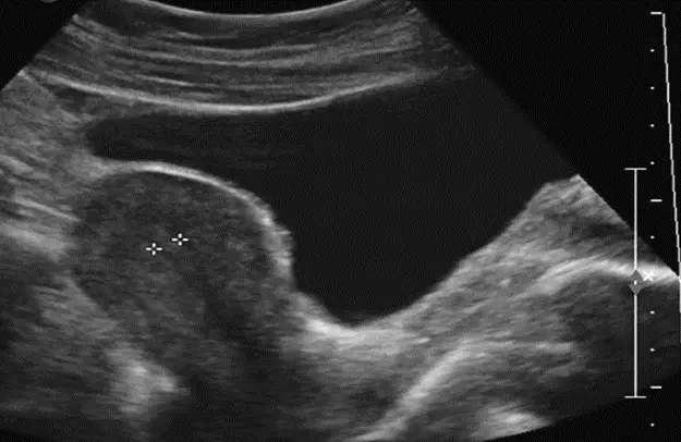
What is the primary imaging technique depicted in the diagram?
Transabdominal (TA) Sonography
Magnetic Resonance Imaging (MRI)
Computed Tomography (CT) Scan
X-ray Imaging
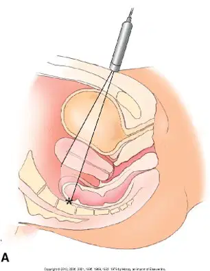
Which anatomical directions are labeled in the diagram of transabdominal sonography?
Anterior, Posterior, Left, Right
Superior, Inferior, Medial, Lateral
Proximal, Distal, Ventral, Dorsal
Cranial, Caudal, Medial, Lateral
What is the purpose of transabdominal sonography?
To visualize internal organs through the urinary bladder
To measure bone density
To detect uterus and ovary via the abdominal wall
To perform a biopsy
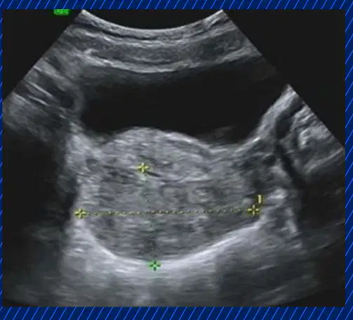
What is the primary purpose of transabdominal sonography using the image given
To measure the width and length of the uterus.
To examine the kidneys.
To assess the urinary bladder
To analyze the ovaries

Which anatomical structures are highlighted in a transverse image during transabdominal sonography?
Ovaries and uterus.
Kidneys and bladder.
Liver and pancreas.
Heart and lungs.
What is required for a transabdominal sonography to obtain clear transverse images?
A distended urinary bladder.
An empty stomach.
A full colon.
A relaxed diaphragm.
What is the purpose of measuring the length of the uterus in the transabdominal pelvic ultrasound protocol?
To determine the position of the bladder
To assess the uterus from fundus to cervix
To locate the iliacus muscle
To evaluate the area of the ovaries
Which anatomical structures are examined at the midline during the transabdominal pelvic ultrasound protocol?
Bladder, uterus, endometrium, cervix, vagina
Iliacus muscle, bladder, ovaries
Fundus, cervix, iliacus muscle
Right and left adnexal areas
What is the primary focus of the image titled 'Uterine Measurements'?
Measurement of uterine dimensions using ultrasound imaging
Diagnosis of uterine abnormalities
Monitoring fetal development
Identifying uterine blood flow

Which measurement is indicated as 'Ut-L' in the left ultrasound image?
7.90 cm
3.51 cm
4.60 cm
66.63 ml
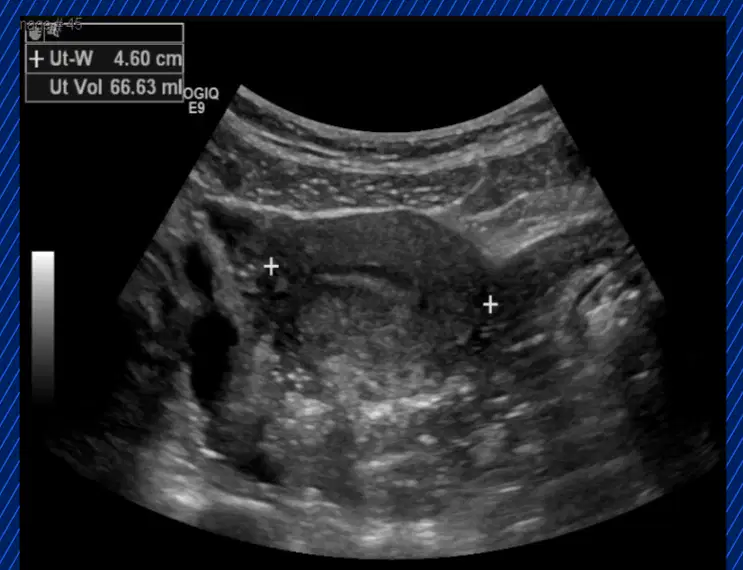
What is being measured in the ultrasound image given?
None of the given
Uterine length
Uterine width
Uterine height
Which imaging technique is commonly used to visualize longitudinal ovaries?
MRI
CT scan
Ultrasound
X-ray
What is the primary purpose of imaging longitudinal ovaries?
To assess ovarian function.
To measure ovarian size and structure.
To detect abnormalities in the ovaries.
All of the above.

What is 2 pointing to?
Fallopian tube
Cervix
Uterus
Vagina
Ovary

Using the image given, how is the transvaginal probed angled?
The transvaginal probe is angled horizontally.
The transvaginal probe is angled downwards.
The transvaginal probe is angled upwards.
The transvaginal probe is angled sideways.
Which of the following is best visualized using the transvaginal sonography approach?
Endometrial lining
Kidneys
Abdominal wall
Liver
During a pelvic ultrasound, which measurement is important for assessing ovarian volume?
Length, width, and anteroposterior diameter
Length and circumference only
Width and surface area only
Length and depth only
What preparation is typically required before a transabdominal pelvic ultrasound?
Patient should lie on their side
Patient should fast for 8 hours
Patient should avoid drinking water
Patient should have a full bladder

