
 Worksheets
Functions and Organization of the Nervous System
Total questions: 64
Worksheet time: 32mins
Which sequence best describes the three main functions of the nervous system from stimulus to action?
Sensory input, motor output, integration
Motor output, integration, sensory input
Sensory input, integration, motor output
Integration, sensory input, motor output
A hot surface is touched with the fingertip. Which step specifically involves the brain interpreting the meaning of the stimulus before deciding a response?
Motor output via efferent pathways
Integration within processing centers
Sensory input by receptors
Peripheral transmission to muscles
Select all statements that accurately characterize sensory input, integration, and motor output.
Motor output activates effectors like muscles
Integration interprets information centrally
Integration directly detects environmental stimuli
Sensory input collects data from receptors
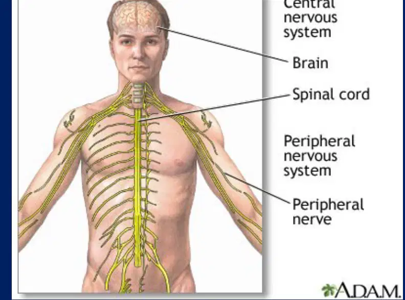
Which component pair forms the Central Nervous System?
Brain and spinal cord
Spinal cord and cranial nerves
Brain and peripheral nerves
Autonomic and somatic divisions
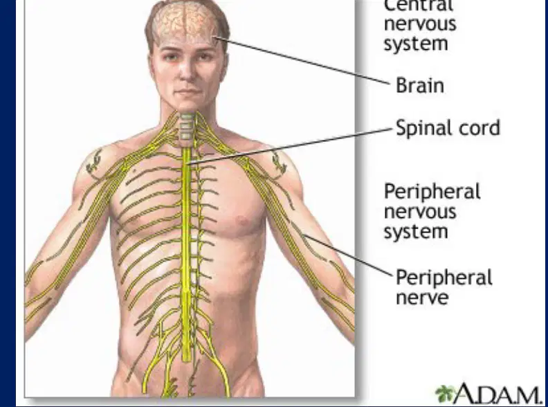
Which description best defines the Peripheral Nervous System at the gross anatomical level?
Processing centers in the brain
Neural tissue outside brain and spinal cord
Spinal cord gray and white matter
Cortical networks for integration
In a reflex arc, which elements primarily belong to the PNS?
Cerebral cortex association areas
Motor neurons to skeletal muscle
Spinal cord interneurons
Sensory receptors in skin

Which statement correctly links function to division of the nervous system?
CNS gathers sensory signals from receptors
PNS stores memories and forms decisions
PNS interprets and integrates incoming signals
CNS processes information and coordinates responses
Which statement best defines the sensory (afferent) division of the peripheral nervous system?
Balances sympathetic and parasympathetic tone
Carries impulses to the CNS from receptors
Controls voluntary skeletal muscle actions
Carries impulses from CNS to effectors
Which fibers are components of the sensory (afferent) division?
Somatic efferent fibers
Visceral afferent fibers
Somatic afferent fibers
Autonomic efferent fibers
The motor (efferent) division primarily does which of the following?
Monitors internal organ chemistries
Conveys impulses from CNS to effectors
Transduces stimuli to sensory nerves
Integrates signals within the spinal cord
Which systems comprise the motor (efferent) division?
Somatic nervous system
Special sensory pathways
Enteric endocrine system
Autonomic nervous system
Which description best matches the somatic nervous system?
Modulation of smooth muscle tone
Voluntary control of skeletal muscles
Involuntary control of cardiac rhythm
Regulation of glandular secretions
Which description best matches the autonomic nervous system?
Conscious control of skeletal muscle contraction
Transmission of sensory input to the cortex
Reflex arcs restricted to the spinal cord
Involuntary control of smooth and cardiac muscle
Which divisions make up the autonomic nervous system?
Somatic division
Enteric sensory division
Parasympathetic division
Sympathetic division

Which scenario best illustrates a sympathetic fight-or-flight response?
Quiet reading before bed
Daydreaming during class
Dozing after a large meal
Running from a threat rapidly
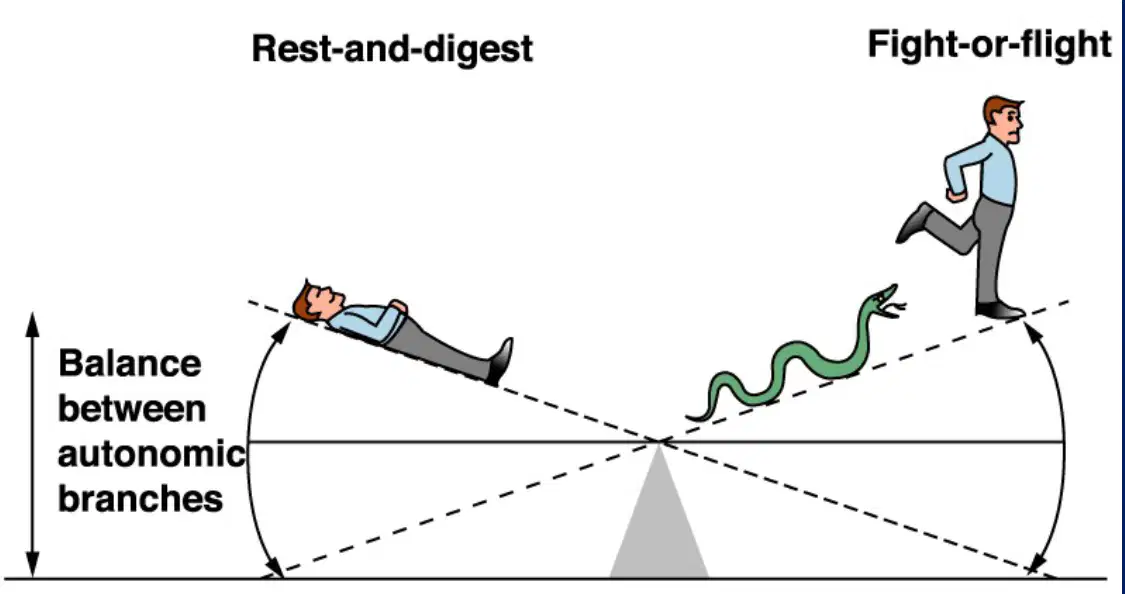
Which scenario best illustrates a parasympathetic rest-and-digest response?
Hyperventilating during fear
Digesting a meal calmly
Escaping a sudden hazard
Preparing for competition
Sympathetic stimulation of the heart typically causes which effects?
Suppressed pacemaker activity
Decreased conduction velocity
Increased force of contraction
Increased heart rate
Parasympathetic stimulation of the heart primarily causes which effect?
Accelerated conduction velocity
Enhanced ventricular contractility
Increased systolic ejection force
Decreased heart rate overall
What is the predominant sympathetic effect on most systemic arteries?
Constriction via alpha1 receptors
Dilation via M3 receptors
No change in basal tone
Vasodilation via histamine
Which autonomic effect occurs in bronchial smooth muscle under sympathetic activation?
Marked mucus hypersecretion
Bronchial muscle contraction
Ciliary beat suppression
Bronchial muscle relaxation
In the gastrointestinal tract, sympathetic stimulation generally causes which changes?
Increased peristaltic activity
Decreased motility overall
Contraction of sphincters
Relaxation of sphincters
Which hepatic processes are promoted by sympathetic activity?
Ketone clearance increases
Glycogenolysis increases
Gluconeogenesis increases
Glycogen synthesis increases
What is a primary parasympathetic effect on the liver?
Gluconeogenesis accelerates
Glycogen synthesis increases
Glycogenolysis accelerates
Lipolysis is stimulated
Which renal effect is associated with sympathetic stimulation?
Glomerular filtration ceases
Collecting duct contraction
Renin secretion increases
Aldosterone directly decreases
Which bladder changes reflect sympathetic activity?
Internal sphincter relaxation
Internal sphincter contraction
Detrusor relaxation
Detrusor contraction
Which uterine effect is mediated by sympathetic beta2 activation?
Cervical sphincter tightening
Endometrial secretion increase
Powerful uterine contractions
Relaxation of uterine smooth muscle
Which ocular effect is typical of sympathetic activation?
Pupillary dilation (mydriasis)
Lens accommodation spasm
Pupillary constriction (miosis)
Lacrimal secretion surges
Which statement best distinguishes supporting cells from neurons in nervous tissue?
Supporting cells transmit rapid electrical impulses
Neurons serve mainly as structural scaffolds
Neurons insulate and protect neighboring cells
Supporting cells wrap and surround the neurons
Which feature defines neurons as excitable cells?
They primarily wrap other cell types
They generate and transmit electrical signals
They store large reserves of glycogen
They divide to replace damaged tissue
Which combination correctly lists special characteristics of neurons?
Low metabolic rate with minimal oxygen need
Exceptional metabolic rate requiring continuous glucose
Extreme longevity across the lifespan
Amitotic nature with rare cell division
A neuron deprived of oxygen and glucose will fail quickly. Which property explains this vulnerability?
Extreme longevity under stress
Amitotic and nondividing behavior
Exceptionally high metabolic rate
Abundant glycogen storage capacity
Which statement about neuron longevity is most accurate?
Neurons divide regularly to renew themselves
Neurons survive only with myelin present
Neurons typically persist for a lifetime
Neurons live briefly and are frequently replaced
Why does nervous tissue heal poorly after injury?
Axons lack any protective sheathing
Supporting cells cannot proliferate locally
Neurons have low oxygen requirements
Neurons are amitotic and seldom regenerate
Which term refers to the neuronal cell body?
Nissl body
Perikaryon or soma
Myelin sheath layer
Neurofibril network
What are Nissl bodies primarily associated with in neurons?
Protein synthesis machinery
Membrane excitability control
Axonal transport pathways
Impulse conduction speed
Which structural element provides internal support within the neuron?
Dendritic spines
Synaptic vesicles
Myelin lamellae
Neurofibrils
Which pair correctly matches a neuron process with its typical function?
Dendrite—receives incoming signals
Myelin—surrounds axons to increase speed
Axon—propagates action potentials
Soma—wraps and insulates axons
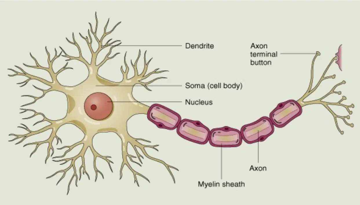
In the diagram, which labeled structure primarily receives synaptic inputs from other neurons?
Myelin sheath segments around axon
Nucleus located within the soma
Axon terminal button at the end
Dendrite branches near the soma

Which combination of structures together form the conductive pathway that carries action potentials away from the soma in the diagram?
Dendrite tree near cell body
Axon terminal button distal end
Axon along its length
Myelin sheath surrounding axon
Nucleus inside the soma

What is the primary role of the myelin sheath segments shown along the axon?
Increase conduction velocity via insulation
Store genetic material for the neuron
Anchor the soma to surrounding tissue
Receive chemical signals from presynaptic cells

Identify the structure labeled 'soma (cell body)' in the diagram.
Distal branching end of the axon
Central region containing the nucleus
Thin projection carrying action potentials
Insulating layers surrounding axon

Which labeled part in the diagram is most directly associated with neurotransmitter release onto a target cell?
Myelin sheath insulating segments
Nucleus within the cell body
Axon terminal button at the tip
Dendrite branches near the soma
Which structure is the specialized plasma membrane of an axon responsible for maintaining ionic gradients during signal conduction?
Collaterals
Axolemma
Telodendria
Axon hillock
At which region of the neuron are action potentials most commonly initiated due to a high density of voltage-gated channels?
Synaptic knob
Axolemma
Nodes of Ranvier
Axon hillock
What are collateral branches of an axon primarily responsible for?
Recycling synaptic vesicles
Generating action potentials
Synthesizing neurotransmitters
Diversifying target reach
Which term refers to the fine distal arborization of an axon ending in synaptic terminals?
Telodendria
Axon hillock
Axolemma
Nodes of Ranvier
Which structure contains synaptic vesicles that release neurotransmitters at chemical synapses?
Synaptic knob
Collaterals
Axon hillock
Nodes of Ranvier
Which statements about the myelin sheath are correct? Select all that apply.
It is formed by Schwann cells in the PNS
It decreases the rate of AP transmission
It electrically isolates the axon
It is a protein–lipid wrapping
Which comparison between myelinated and unmyelinated fibers is accurate?
Unmyelinated fibers have thicker axolemma
Myelinated fibers lack nodes of Ranvier
Myelinated fibers conduct impulses faster
Unmyelinated fibers are insulated by oligodendria
In the peripheral nervous system, which cell type forms the myelin sheath around axons?
Satellite cell
Schwann cell
Microglia
Astrocyte
What are nodes of Ranvier?
Branch points of collaterals
Protein channels in axolemma
Synaptic bulbs at terminals
Gaps without myelin along axons
Which feature enables saltatory conduction along myelinated axons?
Telodendria
Nodes of Ranvier
Synaptic vesicles
Axon hillock
In the illustrated sequence showing Schwann cell wrapping, which labeled outcome best represents a completed myelin sheath surrounding an axon?
Single membrane contact with axon
Concentric lamellae around axon
Terminal arborization of telodendria
Open groove with exposed axon
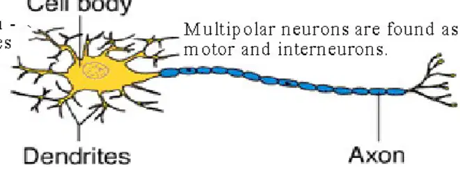
Which structural class of neuron typically has many dendrites and a single axon, as shown in the diagram with a star‑shaped cell body?
Bipolar neuron
Multipolar neuron
Pseudounipolar neuron
Unipolar neuron
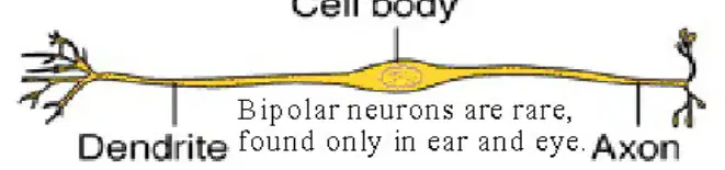
In structural terms, which neuron has one dendrite and one axon emerging from the cell body, commonly found in the retina and olfactory epithelium?
Multipolar neuron
Interneuron
Unipolar neuron
Bipolar neuron
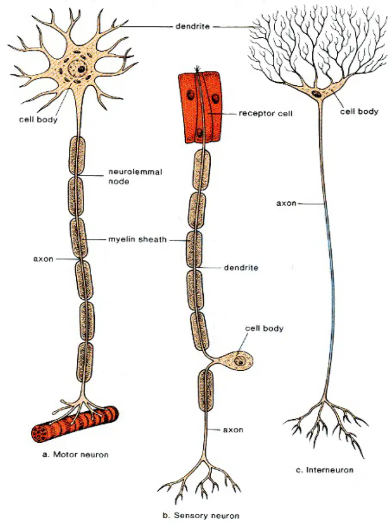
Which functional class carries information from receptors toward the CNS, as depicted with a receptor cell connected to a neuron?
Sensory (afferent) neuron
Motor (efferent) neuron
Interneuron
Autonomic neuron

Which functional class transmits commands from the CNS to effectors, illustrated by an axon ending at a muscle?
Motor (efferent) neuron
Bipolar neuron
Sensory (afferent) neuron
Interneuron

Which functional class primarily connects neurons within the CNS, as shown by a neuron with extensive branching but no receptor or effector depicted?
Interneuron (association)
Sensory (afferent)
Unipolar
Motor (efferent)
Match each structural class to a typical location or role.
Multipolar: common as motor neurons
Bipolar: rare, in ear and eye
Unipolar: most peripheral sensory neurons
Multipolar: mainly peripheral sensory receptors
Which statement best distinguishes bipolar from unipolar neuron architecture?
Unipolar has two somas connected
Bipolar has two processes from soma
Bipolar has many dendrites on soma
Unipolar has multiple axons from soma
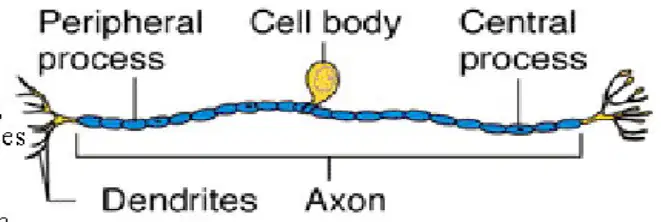
In the unipolar neuron diagram, where are dendrites typically located relative to sensory input?
Near the central process
At the axon hillock
On the cell body
At the receptor ending

Which combination correctly pairs structural and functional classifications commonly associated together?
Multipolar with motor/interneuron
Bipolar with motor neurons
Multipolar with peripheral receptors
Unipolar with interneurons

Which feature in the motor neuron diagram indicates rapid signal conduction to muscle?
Peripheral process split
Cell body off to side
Dendrites at receptor
Myelin sheath on axon

Select all statements that are accurate about sensory (afferent) neurons shown.
Synapse directly onto muscle
Associate with receptor cells
Carry impulses toward CNS
Often structurally unipolar

