
 Worksheets
RT AnP Final — Worksheet Questions (Extraction)
Total questions: 85
Worksheet time: 43mins
Where does gas exchange occur?
Alveoli
Trachea
Bronchi
Pleural cavity
What are the functions of the upper airway? Select all that apply.
Heat inhaled air
Humidify inhaled air
Protect the airway
Filter particulates
Taste
What are the functions of the upper airway? Select all that apply.
Smell
Talking
Gas exchange
Digestion via peristalsis
Blood pressure regulation
What are conducting airways?
Airways where gas exchange does not occur, such as the trachea and bronchi
The alveoli where gas exchange occurs
Pulmonary capillaries responsible for oxygen transport
The pleural space surrounding the lungs
What are the functions of the nose? Select all that apply.
Filter inhaled air
Warm inhaled air
Cool inhaled air
Enable gas exchange
Produce speech
If a patient is having nasal flaring and/or use of intercostal muscle contractions when breathing, what would this patient be showing signs of?
Respiratory distress
Normal breathing effort
Gastrointestinal distress
Cardiac tamponade
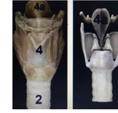
What structure in the upper airways is spoon shaped or leaf shaped and functions in protecting from aspiration?
Epiglottis
Thyroid cartilage
Cricoid cartilage
Trachealis muscle
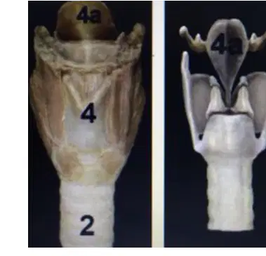
Epiglottis anatomy (picture of it): Identify the leaf-shaped structure at the top of the image.
Epiglottis
Thyroid cartilage
Cricoid cartilage
Arytenoid cartilages
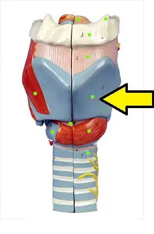
Thyroid cartilage anatomy : Identify the highlighted shield-shaped cartilage at the anterior larynx.
Thyroid cartilage
Cricoid cartilage
Epiglottis
Arytenoid cartilage

Cricoid cartilage anatomy (picture of it): Identify the ring-shaped cartilage inferior to the thyroid cartilage.
Cricoid cartilage
Thyroid cartilage
Epiglottis
Hyoid bone
If a patient needs an emergent airway to be placed, where would the incision for the emergent airway be?
Cricoid ligament between the thyroid and cricoid cartilage
Thyroid cartilage
Tracheal rings below the cricoid cartilage
Epiglottis
When orally intubating a patient, what anatomical landmark confirms you are in the right spot?
The breathing tube passes between the true vocal cords
The tube rests in the esophagus
The tube sits above the epiglottis
The tube is between the false vocal cords
If you are intubating a patient with an endotracheal tube and you place it too far, where would the breathing tube end up being located?
Right mainstem bronchus
Left mainstem bronchus
Trachea above the carina
Nasopharynx
If a patient comes in with croup, what would be the most common finding you would expect to see?
Nothing, they are fine
Slow or fast heart rate
Swollen epiglottis
Abdomen problems
When taking an x-ray, what problem is considered the thumb sign on the image?
Swollen epiglottis
Pneumothorax
Pulmonary edema
Atelectasis
When taking an x-ray, what would you expect when you see a church steeple sign?
Swollen epiglottis
Collapsed lungs
Fluid in the lungs
Thinned airway
When ventilating a patient, what position should they be placed in?
Sniffing
Semi‑Fowler’s
Tripod
Supine
A patient presents with a neck injury and you need to open the airway to intubate without worsening the injury. Which maneuver should you use?
Jaw thrust
Head tilt–chin lift
Cricoid pressure only
Bag‑valve‑mask ventilation without airway maneuver
At what part of the airways does the trachea bifurcate and split into two?
Carina
Epiglottis
Alveolar duct
Laryngeal inlet
Through which anatomical zone does air pass with no gas exchange?
Conducting zone
Respiratory zone
Alveolar sacs
Pulmonary capillary bed
In which anatomical zone does gas exchange occur?
Respiratory zone
Conducting zone
Trachea
Terminal bronchioles
Where do oxygen and carbon dioxide cross to move in and out of the alveoli?
Alveolar–capillary membrane
Pleural space
Bronchial smooth muscle
Nasal mucosa
If the lungs have no surfactant, what will happen?
Alveoli collapse leading to respiratory distress
Increased alveolar expansion and hyperinflation
Enhanced mucociliary clearance
Immediate bronchodilation
What carries blood away from the heart?
Arteries
Veins
Capillaries
Venules
What carries blood towards the heart?
Veins
Arteries
Arterioles
Capillaries
A patient has an injury at the C4 spinal region. What would you most expect?
Difficulty swallowing
Problem blinking due to impaired eye‑movement nerve
Heart pumping problem requiring a pacemaker
Patient needs to be placed on a ventilator
A heavy smoker is short of breath. Which noninvasive method could you instruct to help relieve the shortness of breath?
Tripod position
Supine position
Trendelenburg position
Prone position
What does it indicate if a patient is using accessory muscles to breathe? Select all that apply.
Increased work of breathing
Possible respiratory distress
Effective diaphragmatic relaxation
Normal resting ventilation
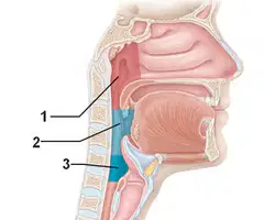
On the sagittal upper‑airway diagram, which numbered label corresponds to the Nasopharynx?
1
2
3
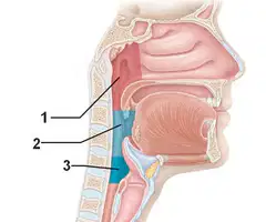
On the sagittal upper‑airway diagram, which numbered label corresponds to the Oropharynx?
1
2
3

On the sagittal upper‑airway diagram, which numbered label corresponds to the Larynx?
1
2
3
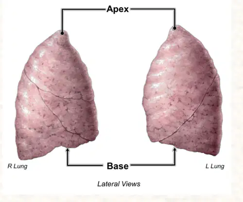
What is the top of the lungs and the bottom of the lungs referred to as?
Top = apex; bottom = base
Top = base; bottom = apex
Top = hilum; bottom = diaphragm
Top = lobe; bottom = fissure
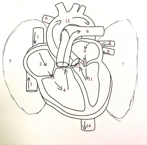
What is the flow of blood through the heart (picture)?
Vena cava → right atrium → tricuspid valve → right ventricle → pulmonic valve → pulmonary arteries to lungs → pulmonary veins from lungs → left atrium → mitral valve → left ventricle → aortic valve → aorta to body tissues → back to vena cava
Vena cava → right ventricle → tricuspid valve → right atrium → pulmonary veins → lungs → left atrium → aortic valve → left ventricle → mitral valve → aorta → back to vena cava
Pulmonary veins → right atrium → mitral valve → right ventricle → aortic valve → aorta → lungs → pulmonary arteries → left atrium → tricuspid valve → left ventricle → vena cava
Aorta → left ventricle → mitral valve → left atrium → pulmonary arteries → lungs → pulmonary veins → right atrium → tricuspid valve → right ventricle → vena cava
What is the sac that encloses the heart known as?
Pericardium
Endocardium
Epicardium
Myocardium
What is an inflammation of the heart sac know as?
Pericarditis
Myocarditis
Endocarditis
Pleuritis
What formula is used to calculate Systemic Vascular Resistance?
SVR=COMAP−RAP
SVR=COMAP+RAP
SVR=SVMAP−CVP
SVR=COHR×MAP
What term is used when referencing the amount of air going in and out of the lungs per breath?
Tidal volume
Minute ventilation
Inspiratory reserve volume
Residual volume
What is considered the normal adult tidal volume?
500 mL
350 mL
800 mL
1,000 mL
What is the normal Barometric pressure when at sea level?
760 mmHg
700 mmHg
800 mmHg
600 mmHg
What is know as no breathing for more than 20 seconds?
Apnea
Dyspnea
Bradypnea
Hyperpnea
What is the term used to describe a respiratory rate of more than 20 breaths per minute?
Tachypnic
Eupneic
Bradypnic
Apneic
If your patient has a respiratory rate of 9 what kind of breathing is this called?
Bradypnic
Tachypnic
Apneic
Eupneic
What is the normal ventilation to perfusion ratio?
0.8
1.2
0.5
1.0
If a baby has underdeveloped lung what would you expect to be needed?
Surfactant (artificial surfactant)
Bronchodilator
Diuretic
Anticoagulant
What is known as the volume of air in and out of the lungs per minute?
Minute ventilation
Tidal volume
Residual volume
Vital capacity
How do you calculate Minute Ventilation?
VE=fB×VT
VE=HR×SV
VE=VT−IRV
VE=MAP−RAP
If you have a patient with a Respiratory Rate of 12 and a tidal volume of 500. What is this patients minute ventilation?
6 L per minute
3 L per minute
9 L per minute
12 L per minute
What is anatomical dead space and what is an example of anatomical dead space?
Volume in the conducting zone that is part of the body; example: a giraffe has more anatomical dead space because of a long neck
Alveolar volume that participates in gas exchange; example: increased diffusion surface
Artificial tubing outside the body; example: snorkel or breathing tube
Collapsed alveoli; example: atelectasis
What is mechanical dead space and what is an example of mechanical dead space?
Volume in the conducting zone that is artificial; example: snorkel or breathing tube
Natural conducting zone in the body; example: trachea and bronchi
Non-ventilated perfused alveoli; example: shunt
Ventilated alveoli participating in exchange; example: healthy lung
What are signs of respiratory distress?
Nasal flaring
Use of intercostal muscles when breathing
Pink frothy sputum
Pursed-lip breathing
If breathing tube too deep where does it go?
Right mainstem bronchi (right lung)
Left mainstem bronchi (left lung)
Esophagus
Nasopharynx
What should the breathing tube pass through?
Vocal cords (glottis)
Esophagus
Right mainstem bronchi
Nasal conchae
If the breathing tube is in the wrong spot where might it be instead of the trachea?
Esophagus
Right main bronchus
Nasopharynx
Laryngeal vestibule
What is the Carina?
Where the trachea bifurcates and splits into two bronchi
The cartilage ring that closes the glottis
The narrowest point of the trachea
A term for the upper airway above the vocal cords
What happens in the alveoli?
Gas exchange occurs
Mucus is produced
Air is humidified
Cough reflex is initiated
What carries oxygen in the blood?
Hemoglobin in red blood cells; a small amount dissolves in plasma
Platelets transport most oxygen
Albumin binds the majority of oxygen
Carbon dioxide carries oxygen to tissues
If three hemoglobins have oxygen attached, what percent is oxygenated? (use the four boxes: 1 box has O2 and Hb = 25%, 2 boxes = 50%, 3 boxes = 75%, 4 boxes = 100%)
25%
50%
75%
100%
If a patient is on oxygen but the O2 sat still will not increase, what problem might need correction to get the O2 up?
Low red blood cell count requiring blood transfusion
Upper airway obstruction requiring bronchodilator
Hyperventilation causing respiratory alkalosis
Excess hemoglobin causing polycythemia
A patient from a suspected house fire is using accessory muscles to breathe and has blue finger tips. A finger pulse oximeter reads 100%. What BEST action should the RT take?
Try another O2 sat location (ear, forehead, toe)
Nothing is wrong because the O2 is good
Get an ABG to clarify oxygen
Check breath sounds
What are factors that may shift the oxygen demand on the curve chart?
Temperature
pH
BPG (2,3-bisphosphoglycerate)
Carbon monoxide
Which statement correctly describes the 40-50-60 / 70-80-90 rule relating PaO2 to SaO2?
If PaO2 is 40, SaO2 is 70; if PaO2 is 50, SaO2 is 80; if PaO2 is 60, SaO2 is 90
If PaO2 is 40, SaO2 is 90; if PaO2 is 50, SaO2 is 80; if PaO2 is 60, SaO2 is 70
If PaO2 is 60, SaO2 is 70; if PaO2 is 50, SaO2 is 80; if PaO2 is 40, SaO2 is 90
SaO2 is always equal to PaO2 plus 10
What is pulmonary shunting?
Blood passes alveoli without becoming oxygenated, leading to poor oxygenation
Air bypasses the trachea into the esophagus
Excessive ventilation of well-perfused alveoli
Blood flow stops completely to a lung segment
What is anatomic shunting?
Blood moves from the unoxygenated to the oxygenated side through vascular pathways without oxygenating the blood
Alveoli overinflate, increasing dead space
Bronchoconstriction decreases airflow to alveoli
Mucus plugs block ventilation causing atelectasis
What kind of blood do arteries carry?
Oxygenated (bright red) blood
Deoxygenated (dark red) blood
Mixed venous blood
Carboxyhemoglobin-rich blood
What kind of blood do veins carry?
Deoxygenated (dark red or "blue") blood
Oxygenated (bright red) blood
Mixed arterial blood
Highly oxygen-saturated blood
What term defines low O2 in arterial blood?
Hypoxemia
Hypoxia
Hypercapnia
Hypocapnia
What is the term used to define low O2 for tissue metabolism?
Hypoxia
Hypercapnia
Hyperoxia
Apnea
PaO2 vs PAO2: select the correct descriptions as printed on the worksheet.
PaO2 is the partial pressure of oxygen in the arteriol
PAO2 is the partial pressure of oxygen in the Alveoli.
PaO2 is the partial pressure of carbon dioxide in the arteries.
PAO2 is the partial pressure of oxygen in the venous system.
What is CO2 primarily carried by in the blood?
Bicarbonate (HCO3−)
Dissolved plasma only
Hemoglobin-bound oxygen
Platelets
What is a noninvasive way to measure how a patient is ventilating?
Capnometry (Capnometer) to measure CO2
Arterial blood gas sampling
Bronchoscopy
Pulse oximetry alone
Which rhythm is characterized by sawtooth wave patterns on the ECG?
Atrial flutter
Atrial fibrillation
Sinus rhythm
Ventricular fibrillation
Identify the rhythm shown on the ECG strip.
Atrial fibrillation
Atrial flutter
Sinus tachycardia
Ventricular fibrillation
Identify the rhythm shown on the ECG strip.
Sinus rhythm
Atrial fibrillation
Ventricular tachycardia
Sinus bradycardia
Identify the rhythm shown on the ECG strip.
Sinus tachycardia
Atrial flutter
Sinus bradycardia
Ventricular fibrillation
Identify the rhythm shown on the ECG strip.
Sinus bradycardia
Sinus tachycardia
Atrial fibrillation
Ventricular tachycardia
Which colloquial descriptor is associated on the worksheet with ventricular fibrillation?
"Tombstones"
"Sawtooth"
"Shark-fin"
"Delta waves"
Select all life‑threatening rhythms indicated by the ECG strip as labeled on the worksheet.
Asystole
Pulseless electrical activity (PEA)
Ventricular tachycardia
Atrial flutter
If your patient is in atrial flutter, how would you fix this rhythm?
Synchronized cardioversion
Defibrillation
Adenosine bolus
Carotid massage
If your patient has a heart rate of 48, what would the RT’s best action be?
Tell them to exercise
Tell them to bear down and activate vasovagal response
Administer atropine
Administer morphine
In emergent situations, what medicine is almost always given?
Epinephrine
Atropine
Morphine
Nitroglycerin
How do you calculate heart rate on an EKG that has a regular rhythm?
Count the big boxes between peak R waves and divide 300 by the number of boxes.
Count the R‑wave peaks in 6 seconds and multiply by 10.
Divide the number of small boxes between R waves by 150.
Use the PR interval to estimate beats per minute.
How do you calculate heart rate on an EKG that has an irregular rhythm?
Count the R‑wave peaks in 6 seconds and multiply by 10.
Divide 300 by the number of big boxes between R waves.
Use the QT interval to estimate beats per minute.
Count small boxes between R waves and divide by 150.
What is a noninvasive way to temporarily lower heart rate?
Vasovagal response by telling the patient to bear down
Administer atropine
Give epinephrine
Synchronized cardioversion
What is a noninvasive way to lower a baby’s heart rate?
Apply ice to the forehead
Administer atropine
Start chest compressions
Give racemic epinephrine
What is the first thing the RT should do when asystole is seen on the EKG?
Check another lead
Begin defibrillation immediately
Administer morphine
Perform carotid massage

