
 Worksheets
Congenital Hip Dislocation (Luxația Congenitală de Șold)
Total questions: 81
Worksheet time: 41mins
What is the definition of congenital hip dislocation (luxația congenitală de șold)? Fill in the blank: Congenital hip dislocation is the loss of the right of residence of the femoral head, gradually, due to _______ of the acetabulum and femoral head.
dysplasia
fracture
infection
inflammation
Which of the following is a consequence of dysplasia and can occur earlier or later after birth due to a dysplastic acetabulum?
Fracture
Congenital hip dislocation
Arthritis
Sprain
Fill in the blank: One of the causes of congenital hip dislocation is the maintenance of the _______ in adduction.
thighs
arms
feet
hands
Placing the body's weight on the _______ can lead to congenital hip dislocation.
femoral head
acetabulum
ischial tuberosity
pubic symphysis
From birth to 7-8 months, what is a characteristic of congenital hip dislocation?
The femoral neck is anteverted and the femoral heads are not contained in the acetabular cavities, being placed in front of them.
The femoral neck is retroverted and the femoral heads are deeply contained in the acetabular cavities.
The femoral neck is fractured.
The femoral heads are fused.
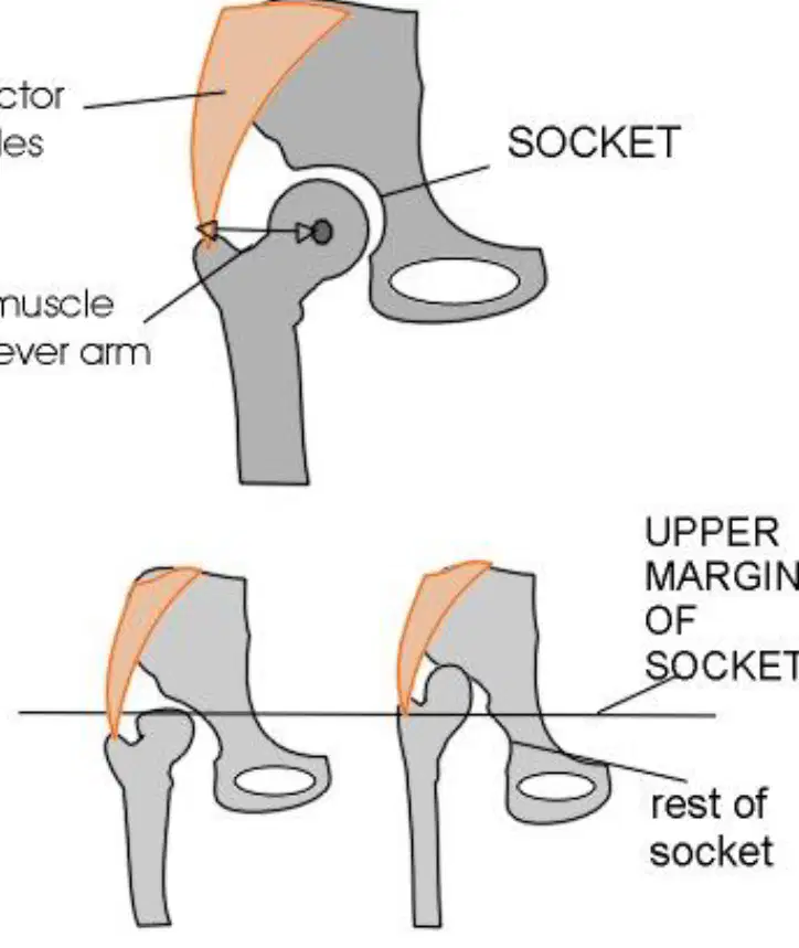
Refer to the diagram of the hip anatomy. Which of the following is labeled as the muscle lever arm in the normal hip?
Abductor muscles
Socket
Muscle lever arm
Upper margin of socket
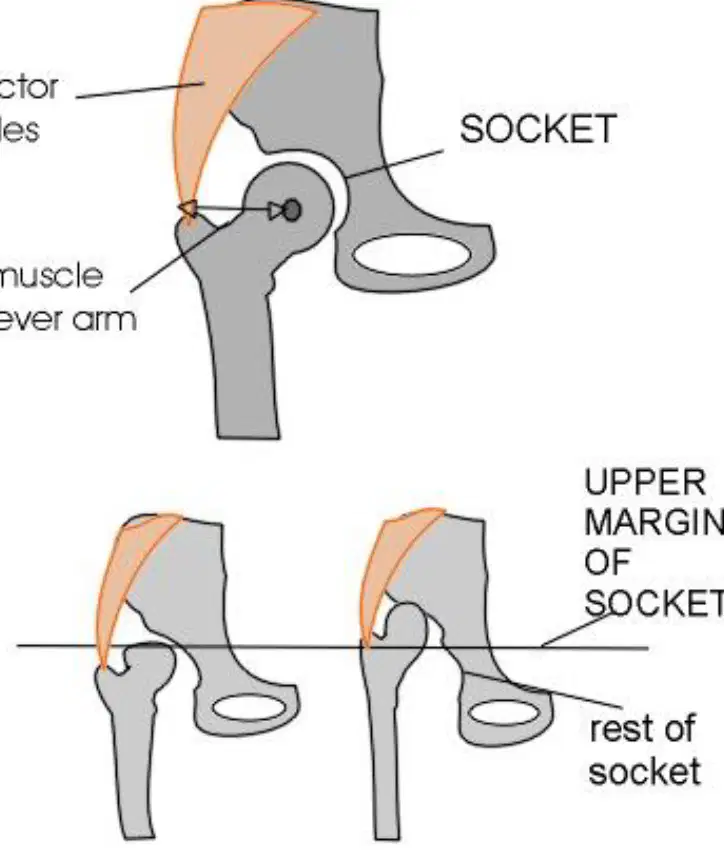
Based on the diagram, which type of hip shows the upper margin of the socket above the rest of the socket?
Normal Hip
Dysplastic Hip
Dislocated Hip
All of the above
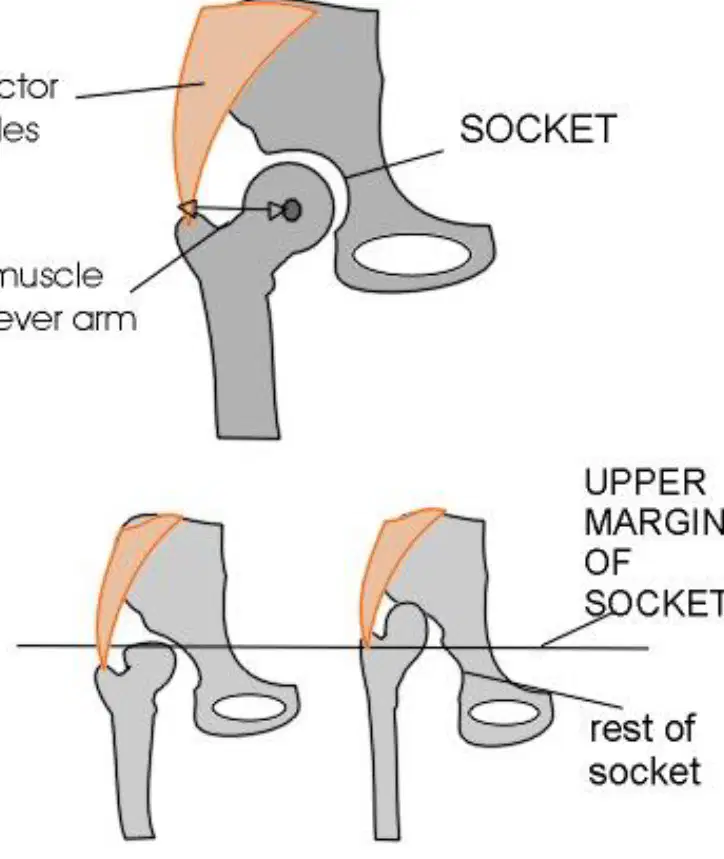
Fill in the blank: In the normal hip diagram, the ________ are shown above the socket and muscle lever arm.
abductor muscles
adductor muscles
ligaments
tendons
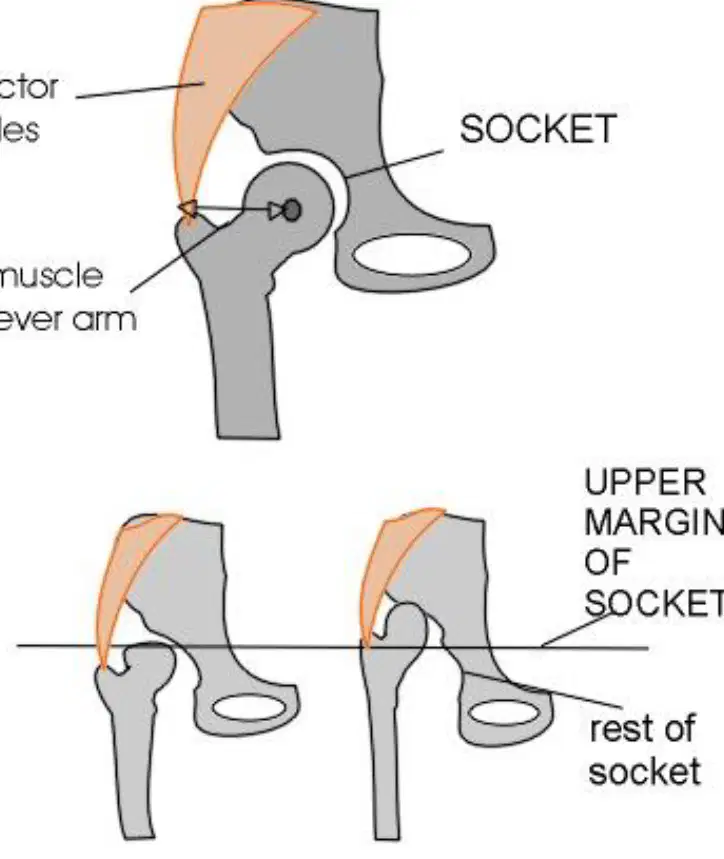
Which hip condition is depicted when the socket is not aligned with the femoral head, as shown in the diagram?
Normal Hip
Dysplastic Hip
Dislocated Hip
None of the above
Observe the X-ray image provided. Which side of the hip joint is labeled as 'normal'?
Left side
Right side
In the X-ray image, what does the annotation 'out' on the right side of the hip joint likely indicate?
It likely indicates that the hip joint is dislocated or not in its normal position.
It likely indicates that the hip joint is fractured.
It likely indicates that the hip joint is inflamed.
It likely indicates that the hip joint is perfectly normal.
Based on the X-ray image, which side of the hip joint appears abnormal?
Left side
Right side
Fill in the blank: Congenital hip dislocation has a clear ______ character.
familial
infectious
traumatic
metabolic
Fill in the blank: The hereditary transmission of congenital hip dislocation does not exceed ______%.
10
25
50
70
Fill in the blank: Congenital hip dislocation appears ______.
sporadically
frequently
rarely
universally
Fill in the blank: Congenital hip dislocation predominates in the ______ sex (7/1).
female
male
neutral
intersex
Which of the following is NOT a theory for the cause of congenital hip dislocation?
Traumatic
Inflammatory
Muscular
Genetic
Anthropological
Fill in the blank: One theory for the cause of congenital hip dislocation is the ______ theory, which suggests injury as a cause.
traumatic
genetic
hormonal
mechanical
Fill in the blank: The ______ theory suggests that inflammation may cause congenital hip dislocation.
inflammatory
genetic
mechanical
vascular
Fill in the blank: The ______ theory suggests that muscle issues may cause congenital hip dislocation.
muscular
genetic
vascular
neural
Fill in the blank: The theory of ______ in the development of the hip joint is one explanation for congenital hip dislocation.
arrest in development
overgrowth
calcification
ossification
What is the condition of the acetabulum in congenital hip dislocation? Fill in the blank: The acetabulum is ________, does not have depth, its opening plane appears normal, but its antero-superior and postero-superior edges are rounded and ________.
aplastic, aplastic
deep, sclerotic
normal, hypertrophic
shallow, thickened
What is the orientation of the femoral neck in congenital hip dislocation? Fill in the blank: The femoral neck is ________ and faces forward at an angle of ________ degrees.
anteverted, 60
retroverted, 30
neutral, 45
anteverted, 30
Which clinical sign is described as 'ORTOLANI sign' and is present up to 3 months in congenital hip dislocation?
ORTOLANI sign (semnul resortului)
Barlow sign
Trendelenburg sign
Galeazzi sign
What clinical sign in congenital hip dislocation refers to the limitation of abduction of the thighs?
Limitation of abduction of the thighs (semnul limitării abducției coapselor)
Ortolani sign (semnul Ortolani)
Galeazzi sign (semnul Galeazzi)
Trendelenburg sign (semnul Trendelenburg)
In congenital hip dislocation, exaggeration of rotational movements (Gourdon's sign) can reach up to 180 degrees, while normally it is 90 degrees.
True
False
What is the name of the clinical sign involving the ischiopubic branch in congenital hip dislocation?
Ischiopubic branch sign (semnul ramurii ischiopubiene)
Trendelenburg sign
Galeazzi sign
Ortolani sign
Which clinical sign in congenital hip dislocation involves asymmetry of skin folds on the inner thigh and subgluteal region?
Asymmetry of skin folds (asimetria pliurilor tegumentare de pe fața internă a coapselor și din regiunea subfesieră)
Galeazzi sign (semnul Galeazzi)
Ortolani sign (semnul Ortolani)
Limited abduction of the hip (limitarea abducției șoldului)
Congenital hip dislocation is frequently associated with congenital valgus foot, especially in girls.
True
False
What clinical sign is observed after the child has started walking and the hip dislocation has occurred, involving shortening of the lower limb?
Shortening of the luxated lower limb (scurtarea membrului inferior luxat)
Swelling of the knee joint
Redness of the ankle
Pain in the upper limb
After hip dislocation in a walking child, which direction does the vulvar slit deviate?
Towards the luxated side (spre partea luxată)
Away from the luxated side (spre partea opusă luxației)
No deviation
Upwards
What clinical sign is described as 'ascension of the trochanter' in congenital hip dislocation after the child has started walking?
Ascension of the trochanter (semne de ascensiune trohanteriene)
Galeazzi sign
Ortolani sign
Barlow sign
Look at the diagram showing the hip joint. Which bone is dislocated in the image?
A) Femur (thighbone)
B) Tibia
C) Fibula
D) Humerus
Fill in the blank: The socket part of the hip joint is called the _________.
Acetabulum (hip socket)
Femoral head
Glenoid cavity
Patella
Based on the photograph of the child's legs, what condition is likely being shown?
Hip dislocation
Fracture
Sprain
Bruising
In an infant, which of the following is a sign of Developmental Dysplasia of the Hip (DDH)?
Limitation of abduction
Increased abduction
Swelling of the knee
Redness of the skin
In an infant, Galeazzi's sign is used to detect which condition?
Developmental Dysplasia of the Hip (DDH)
Clubfoot
Scoliosis
Fracture
In an infant, the signs of DDH depend on whether the hip is reducible or non reducible.
True
False
In an infant, what should be looked for in the number of thigh folds when assessing for DDH?
Asymmetry or increased number of thigh folds may indicate DDH.
Symmetrical and decreased number of thigh folds always indicate DDH.
No change in thigh folds is relevant to DDH assessment.
Increased number of thigh folds always rules out DDH.
What is one paraclinical investigation for congenital hip dislocation?
ECHO articular
ECG
Chest X-ray
Blood glucose test
At what age is Rx-grafie usually performed to establish a certain diagnosis for congenital hip dislocation?
3-4 months
6-8 months
1-2 months
12 months
Which anatomical feature is present in a normal hip but broken in a dislocated hip?
Acetabular Index
Hilgenreiner Line
Shenton Line
Perkin Line
Fill in the blank: The ________ is intact in a normal hip and broken in a dislocated hip.
Shenton Line
Hilgenreiner Line
Pectineal Line
Intertrochanteric Line
Which line is used to assess the position of the femoral head in the hip joint according to the diagram?
Hilgenreiner Line
Perkin Line
Shenton Line
Acetabular Index
The Acetabular Index is labeled on the side of the normal hip in the diagram.
True
False
Refer to the X-ray image comparing a Normal Hip and a Dislocated Hip. Which side of the image shows the dislocated hip?
Left
Right
Both
Neither
In the X-ray image, what does the label 'P' most likely represent in the context of hip anatomy?
Pelvis
Pubis
Patella
Phalanx
Fill in the blank: In the X-ray image, the hip on the left side is labeled as _________.
Normal Hip
Fractured Hip
Dislocated Hip
Arthritic Hip
Refer to the diagram showing three hip joint conditions. Which part is labeled as the 'Acetabulum (socket)'?
The ball-shaped end of the femur
The socket in the pelvis
The shaft of the femur
The cartilage around the joint
Based on the diagram, what is the difference between 'Normal', 'Subluxation', and 'Dislocation' of the hip joint? Fill in the blank: In 'Dislocation', the head of femur is ________ the acetabulum.
completely out of
partially inside
touching the edge of
within
Observe the two X-ray images of the pelvic region. What abnormality is highlighted by the red box in the right image?
Fracture
Dislocation
Normal bone structure
Infection
Compare the left and right X-ray images. Which image shows a normal hip joint alignment?
The left image shows a normal hip joint alignment.
The right image shows a normal hip joint alignment.
Both images show a normal hip joint alignment.
Neither image shows a normal hip joint alignment.
What is the purpose of the red box in the right X-ray image?
To highlight the area of abnormality or concern, specifically the dislocated hip joint.
To indicate the location of a fracture in the spine.
To show the region of normal bone structure.
To mark the area for a future surgical incision.
Fill in the blank: The alpha angle measured in the ultrasound image is approximately ____ degrees.
45
30
60
75
Fill in the blank: In the coronal view, the ultrasound transducer is placed parallel to the ______ aspect of the infant's hip.
lateral
medial
anterior
posterior
Identify the anatomical structure labeled as 'Labrum' in the diagram.
Labrum
Mandible
Maxilla
Palp
Which of the following is NOT labeled in the hip joint diagram?
Labrum
Femoral Head
Acetabulum
Greater Trochanter
What is required for a positive diagnosis of congenital hip dislocation? Fill in the blank: Corroboration of several clinical signs with at least one ______ sign.
radiologic
clinical
neurological
vascular
Which of the following is a differential diagnosis for congenital hip dislocation?
Coxa vara congenitală
Diabetes mellitus
Asthma
Hypertension
Which of the following is a differential diagnosis for congenital hip dislocation?
Luxația traumatică de șold
Pneumonia
Epilepsy
Migraine
Which of the following is a differential diagnosis for congenital hip dislocation?
Luxațiile patologice ale șoldului (TBC, osteomielită, poliomielită)
Fracture of the humerus
Appendicitis
Otitis media
Which of the following is a differential diagnosis for congenital hip dislocation?
Fracturi de col femural (sechele)
Fracture of the tibia
Bronchitis
Gastritis
Which of the following is a differential diagnosis for congenital hip dislocation?
Epifizioliza capului femural (sechele)
Fracture of the radius
Sinusitis
Dermatitis
What is the correct position for a child with luxating hip dysplasia during treatment?
With the thigh in abduction and internal rotation
With the thigh in adduction and external rotation
With the thigh in flexion and extension
With the thigh in neutral position
What procedure is performed on the adductors during the treatment of luxating hip dysplasia?
Tenotomy
Suturing
Injection
Massage
How long does the treatment for luxating hip dysplasia last? Fill in the blank: The duration of treatment is ________, with X-ray every 30 days.
3-4 months
1-2 weeks
6-8 months
2-3 days
When does the treatment for luxating hip dysplasia stop? Treatment stops ONLY when ________ disappear.
Rx-graphic signs
Clinical symptoms
Pain
Swelling
Which of the following is a treatment method for congenital hip dislocation?
Orthopedic
Surgical
Physiokinetotherapy
All of the above
Is orthopedic treatment a method for congenital hip dislocation?
True
False
Is physiokinetotherapy a method for congenital hip dislocation?
True
False
Look at the image of the child wearing an orthopedic device for congenital hip dislocation. What is the name of this device?
Pavlik harness
Spica cast
Frejka pillow
Denis Browne splint
Look at the image of the orthopedic device used for congenital hip dislocation. What is the name of this device?
Pavlik harness
Spica cast
Frejka pillow
Denis Browne splint
Look at the image of the orthopedic device used for congenital hip dislocation. What is the name of this device?
Pavlik harness
Spica cast
Frejka pillow
Denis Browne splint
What is the name of the medical device shown in the image that is used for treating hip dysplasia in infants?
Pavlik harness
Spica cast
Hip abduction brace
Clubfoot brace
What is the name of the strap labeled at the top of the Pavlik harness diagram?
Shoulder Strap
Waist Strap
Leg Strap
Chest Strap
Which strap is located across the chest in the Pavlik harness diagram?
Chest Strap
Shoulder Strap
Leg Strap
Waist Strap
What is the name of the strap that is attached to the legs in the Pavlik harness diagram?
Leg Strap
Shoulder Strap
Waist Strap
Chest Strap
Which strap is responsible for abduction in the Pavlik harness diagram?
Abduction Strap
Flexion Strap
Extension Strap
Rotation Strap
Treatments for hip dysplasia include:
Pavlik harness
Short leg hip spica cast
Both A and B
Which treatment for hip dysplasia is shown on the left side of the treatments diagram?
Pavlik harness
Hip spica cast
Physical therapy
Surgical intervention
Which treatment for hip dysplasia is shown on the right side of the treatments diagram?
Short leg hip spica cast
Pavlik harness
Open reduction surgery
Physical therapy

