
 Worksheets
Spleen and GI tract HW
Total questions: 82
Worksheet time: 1hrs 13mins
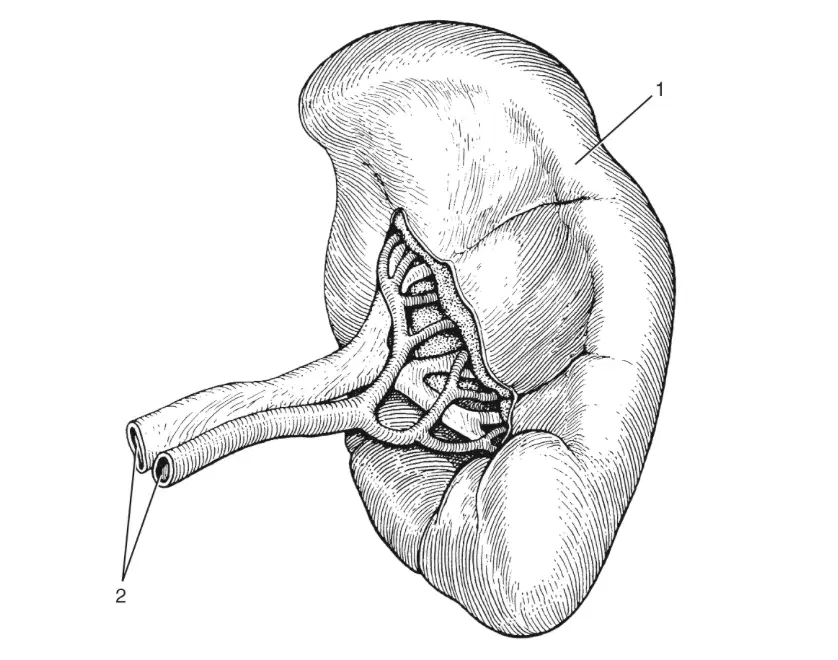
In this image, what is labeled 1?
(a)
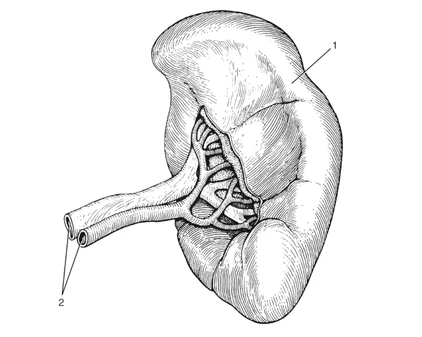
In this image, what is labeled 2?
(a)
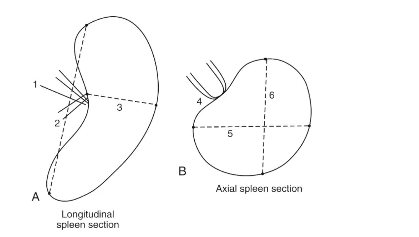
In this image, what is labeled 1?
(a)
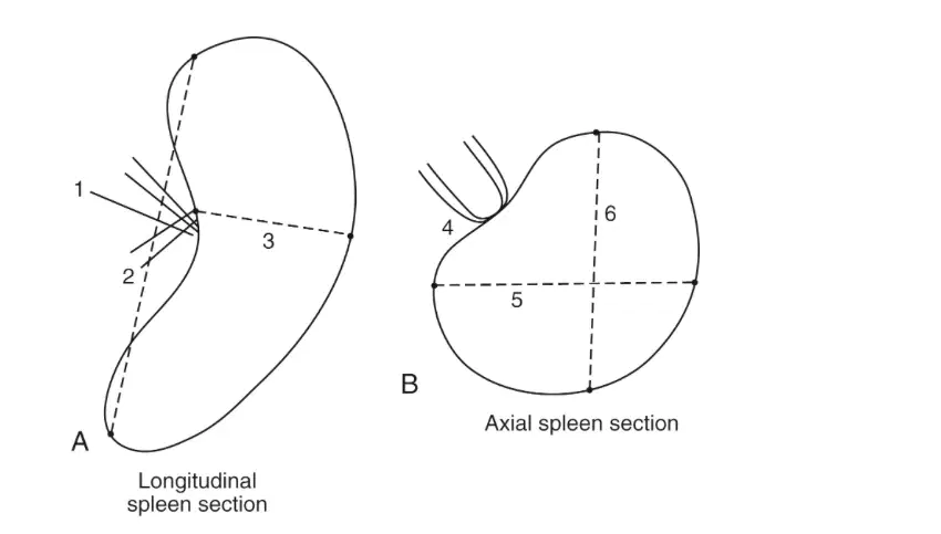
In this image, what is labeled 2?
(a)
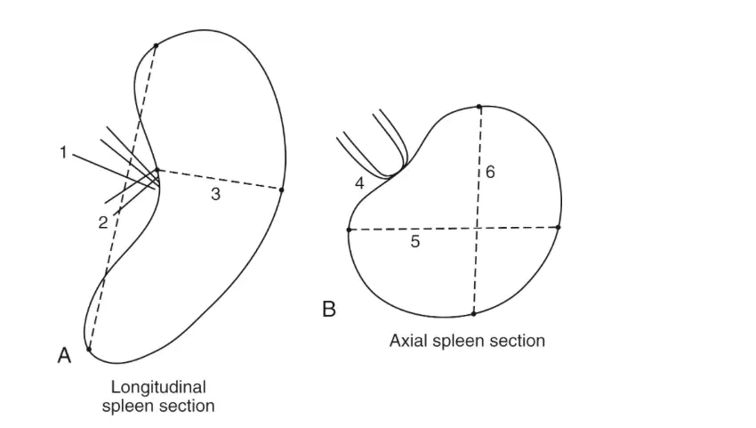
In this image, what is labeled 3?
(a)
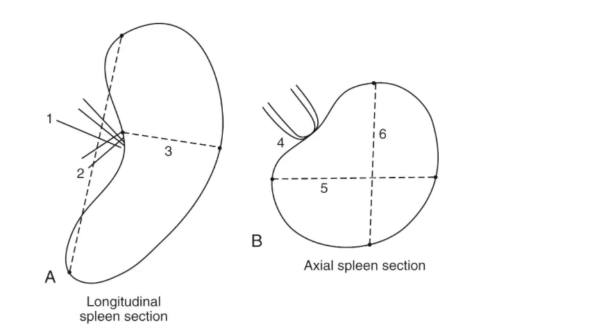
In this image, what is labeled 4?
(a)
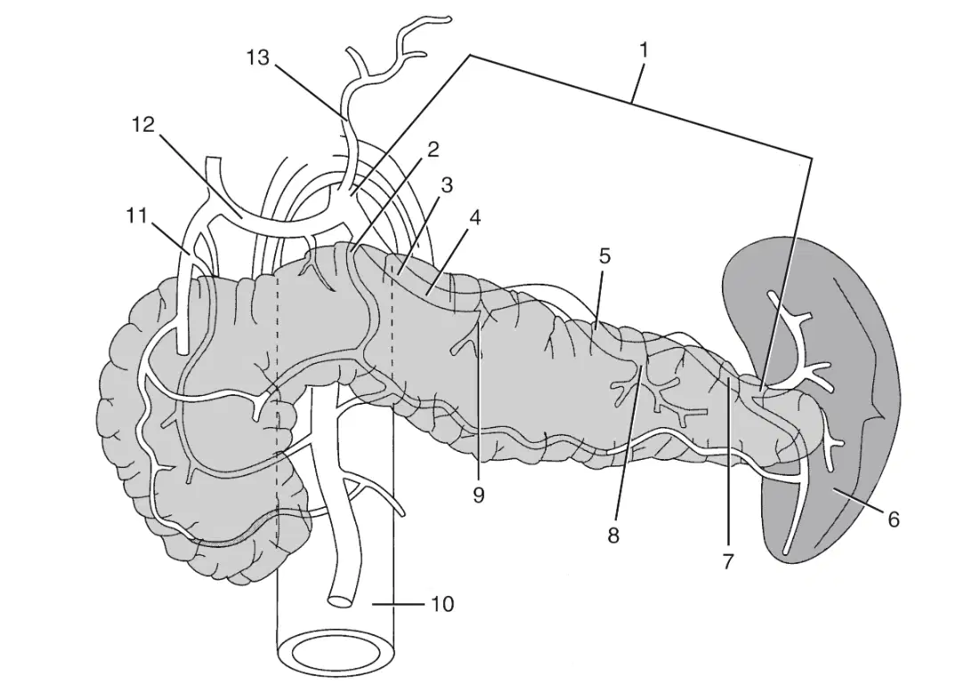
In this image what is labeled 1?
(a)

In this image what is labeled 2?
(a)
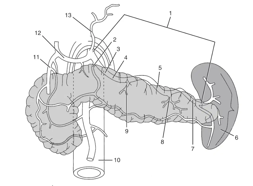
In this image what is labeled 3?
(a)
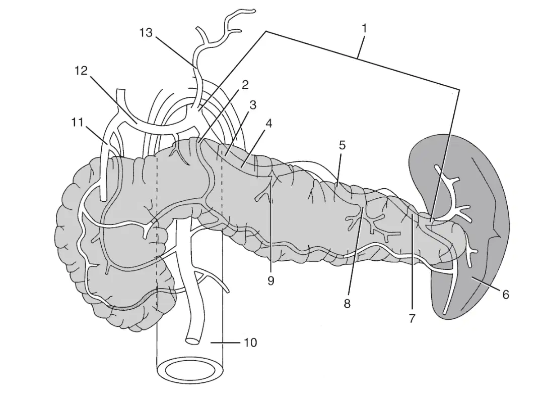
In this image what is labeled 4?
(a)
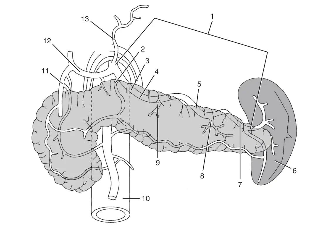
In this image what is labeled 5?
(a)
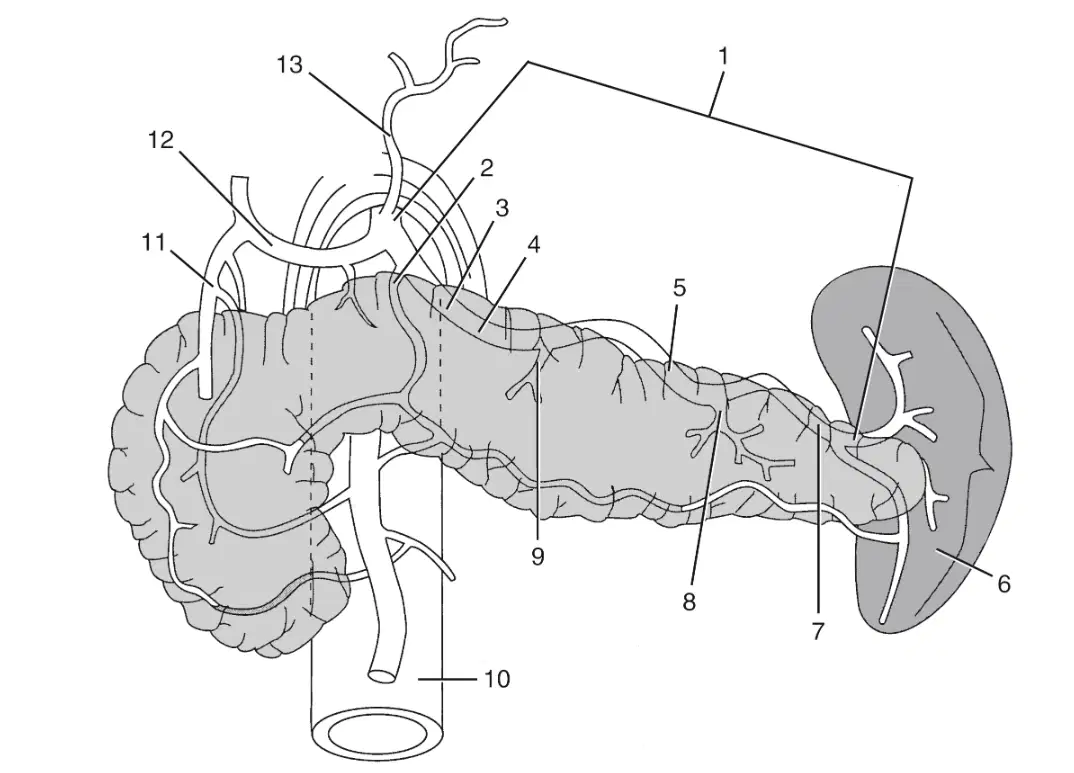
In this image what is labeled 6?
(a)
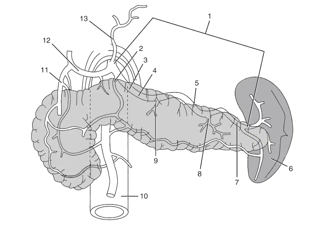
In this image what is labeled 7?
(a)
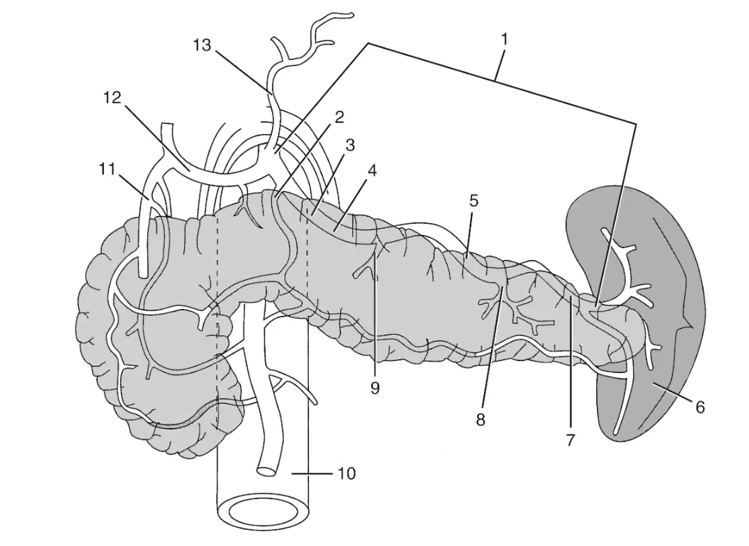
In this image what is labeled 8?
(a)
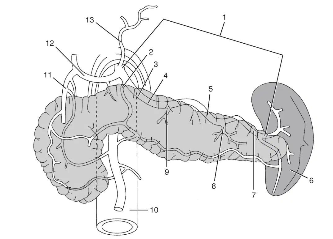
In this image what is labeled 9?
(a)
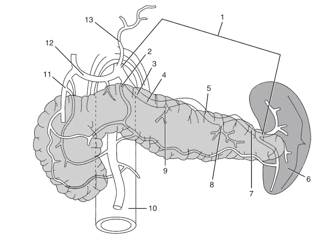
In this image what is labeled 10?
(a)
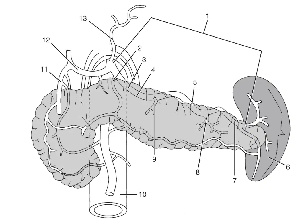
In this image what is labeled 11?
(a)
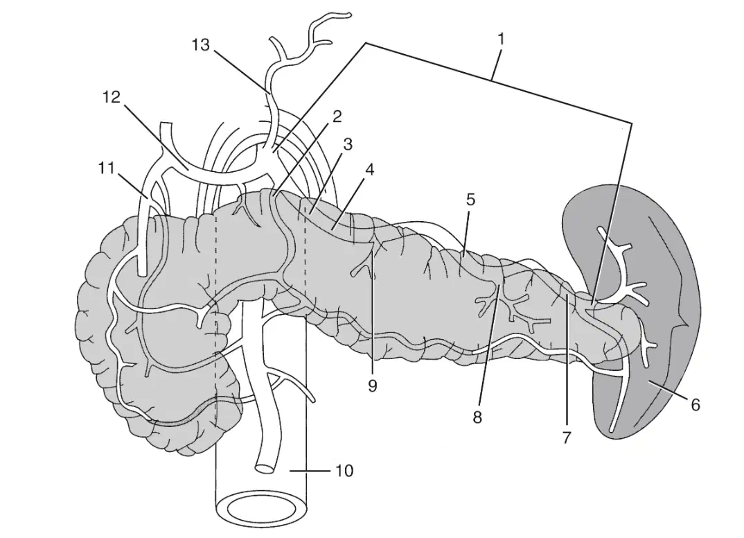
In this image what is labeled 12?
(a)
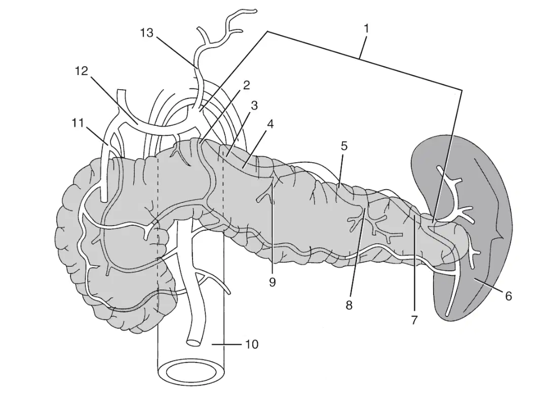
In this image what is labeled 13?
(a)
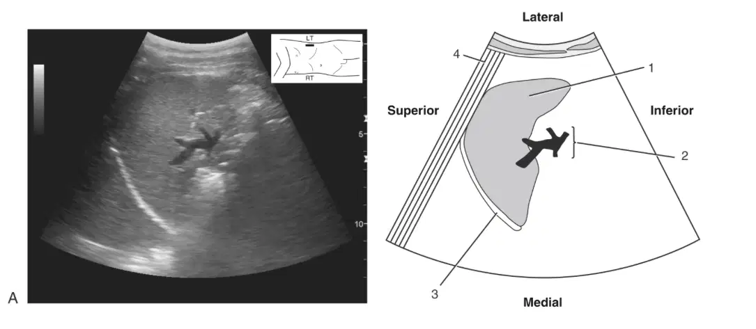
In this image what is labeled 1?
(a)

In this image what is labeled 2?
(a)
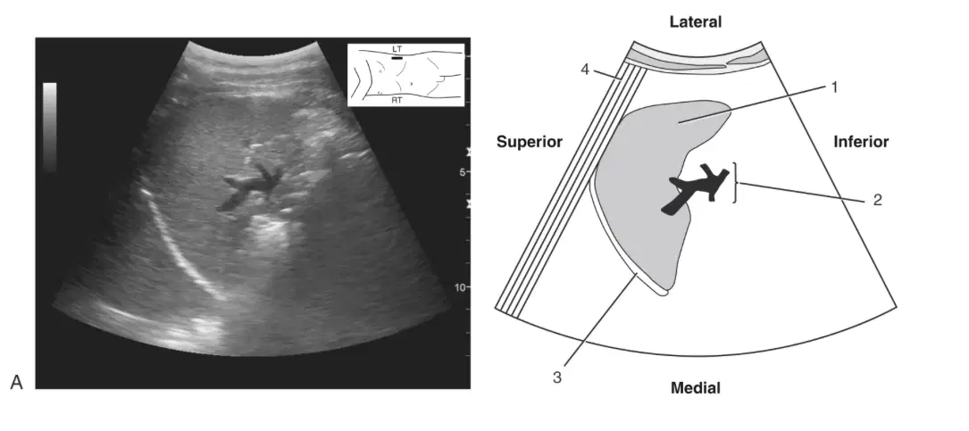
In this image what is labeled 3?
(a)

In this image what is labeled 1?
(a)
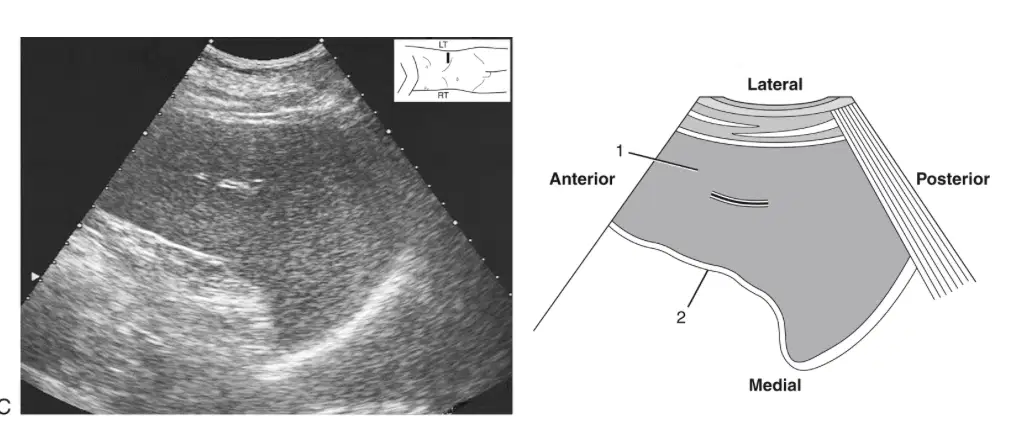
In this image what is labeled 2?
(a)
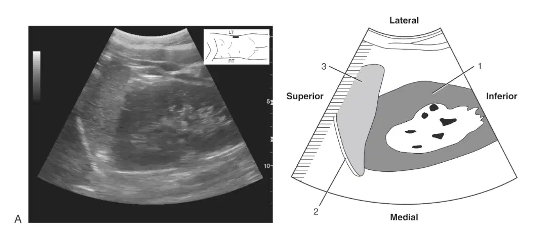
In this image what is labeled 1?
(a)
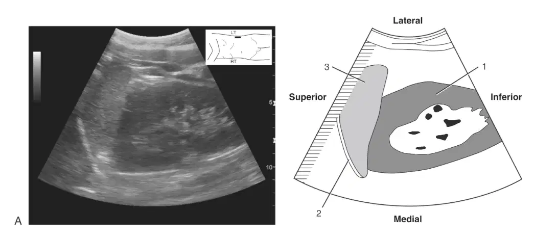
In this image what is labeled 3?
(a)
The splenic vein conveys venous blood from the spleen and courses along the gastrolineal ligament to its confluence with the ___
SMA
Splenic Artery
SMV
MPV
All of the following are functions of the spleen except:
Defense
Hematopoiesis
Serves as blood reservoir
Stores bile
The process of removing abnormal RBC's by the spleen
Erthyrocyte
Phagocytosis
Hematopoiesis
Culling
Oxygen carrying and iron containing pigment of RBC's
Hemoglobin
Pitting
White pulp
Culling
The process that produces erythrocytes and white blood cells in developing fetus
Phagocytosis
Pitting
Hematopoiesis
Culling
The term for red blood cell
pitting
Erythrocyte
White pulp
Hemoglobin
The process of removing nuclei from old RBC's
Pitting
Phagocytosis
White pulp
Hematopoiesis
Responsible for phagocytosis of damaged or old cells
Reticuloendothelial System
Phagocytosis
Hematopoiesis
Culling
Found in the spleen, consists of lymphatic tissue, and is where immune functions take place
Culling
White pulp
Pitting
Reticuloendothelial System
The process of removing old worn out and abnormal red blood cells and platelets from the bloodstream by phagocyte cells in the spleen
Cutting
Hematopoiesis
Pitting
Phagocytosis
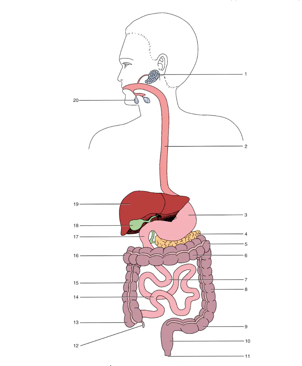
In this image what is labeled 1?
(a)

In this image what is labeled 2?
(a)
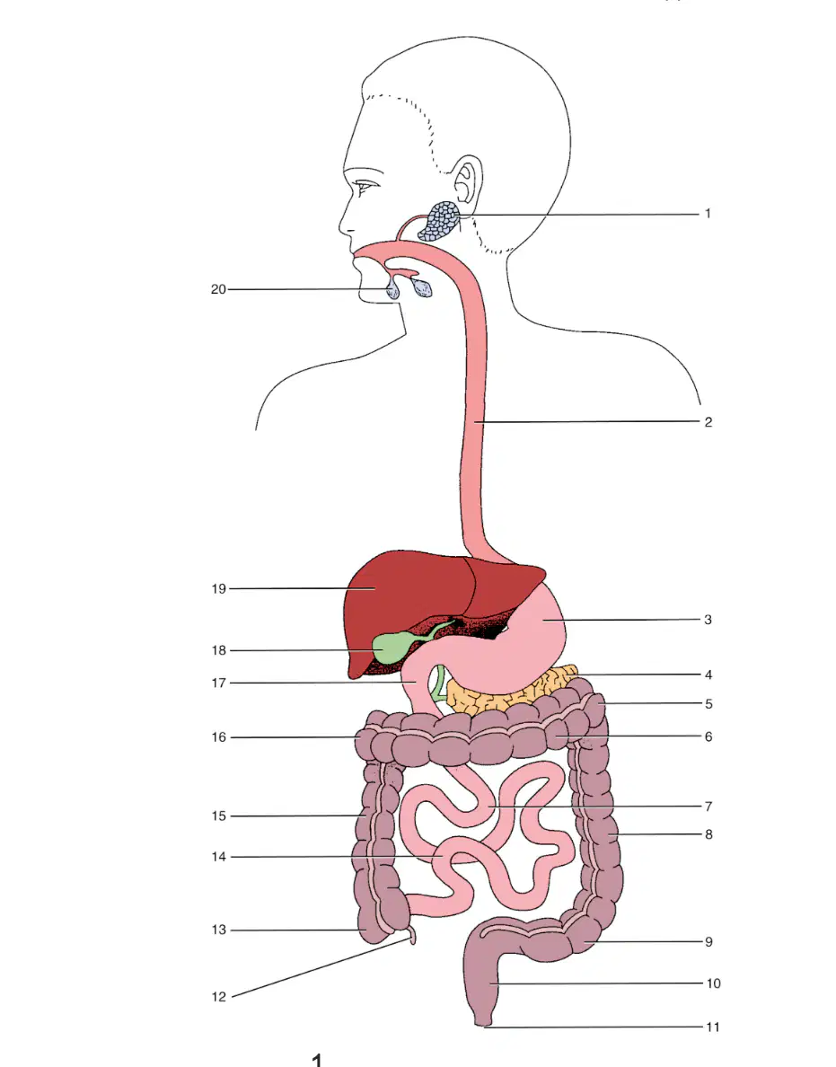
In this image what is labeled 3?
(a)

In this image what is labeled 4
(a)

In this image what is labeled 5
(a)

In this image what is labeled 6
(a)
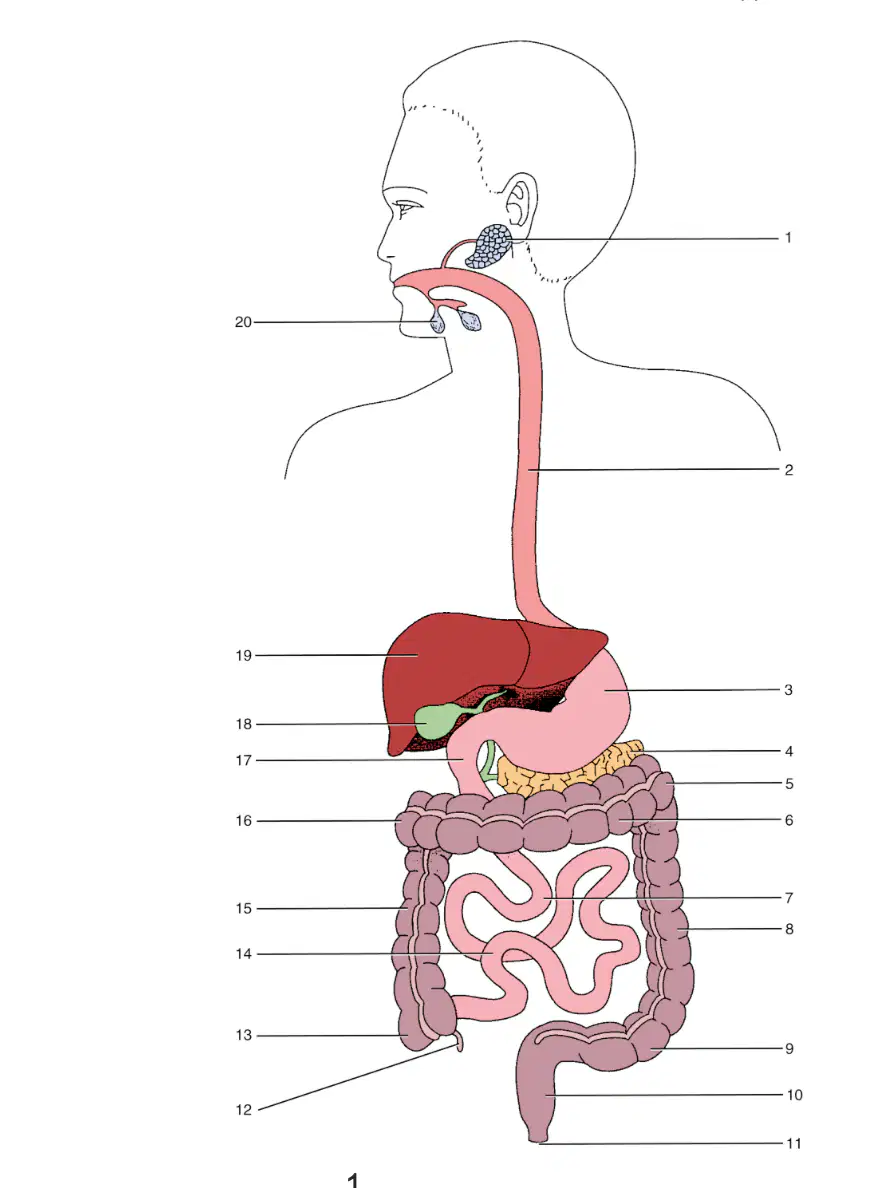
In this image what is labeled 7
(a)
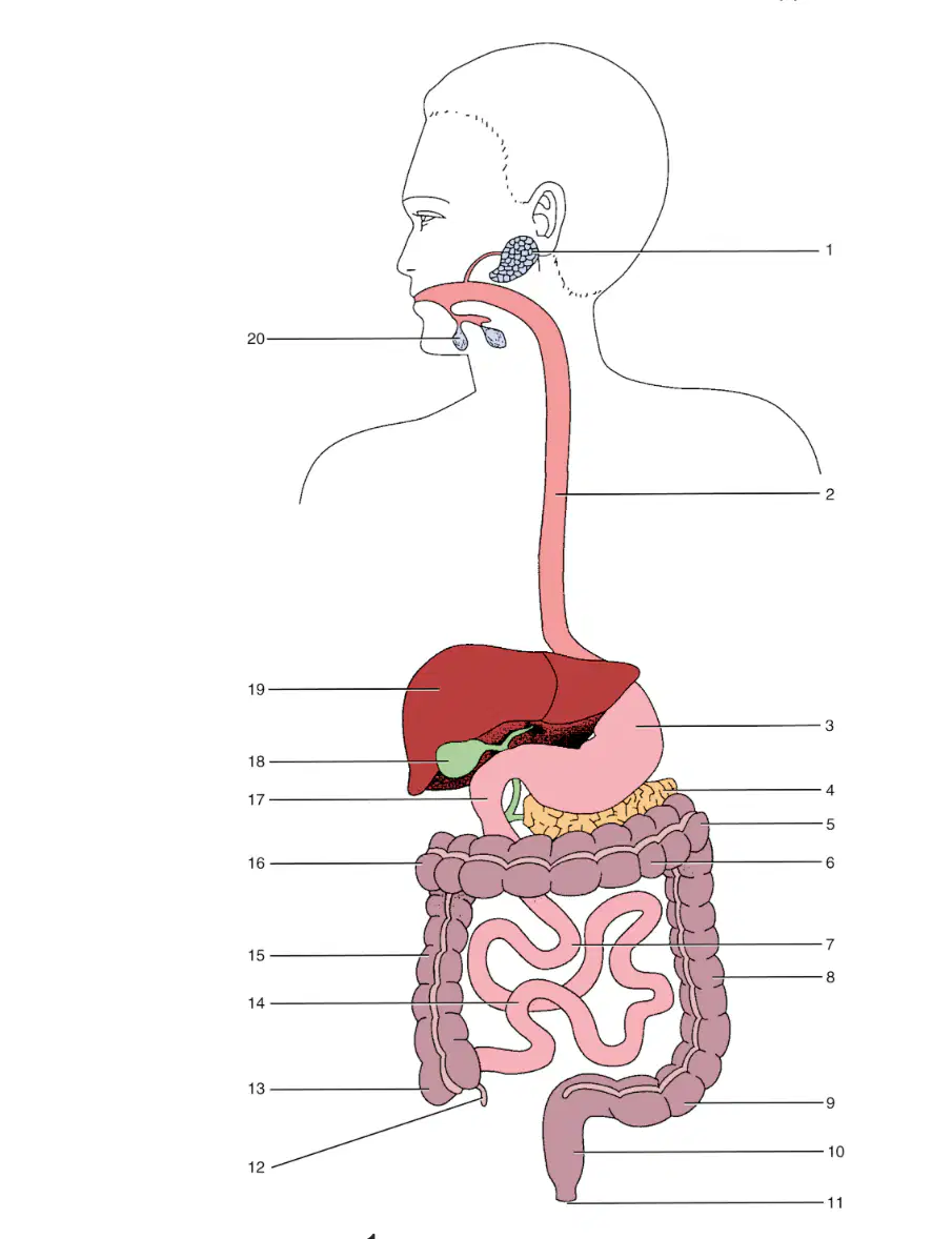
In this image what is labeled 8
(a)
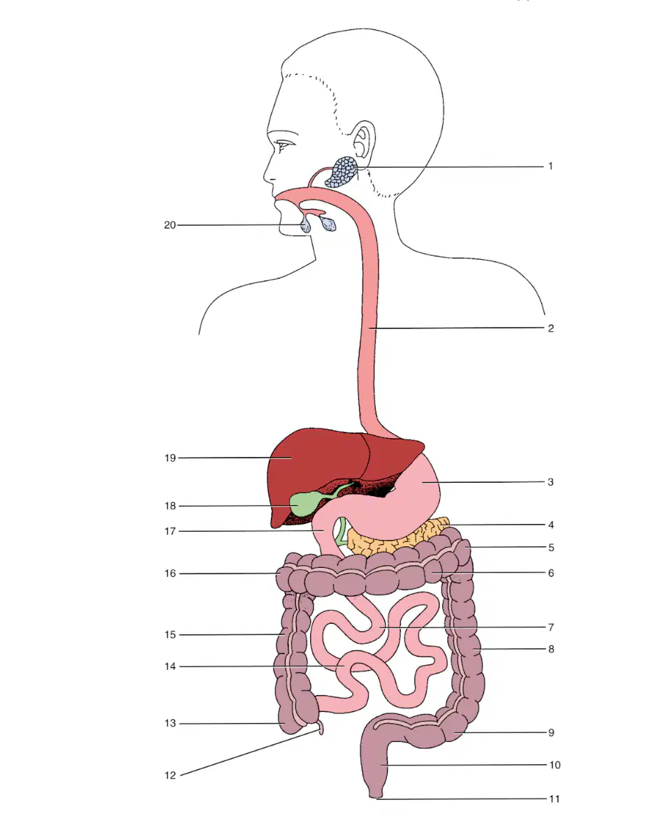
In this image what is labeled 9
(a)

In this image what is labeled 10
(a)
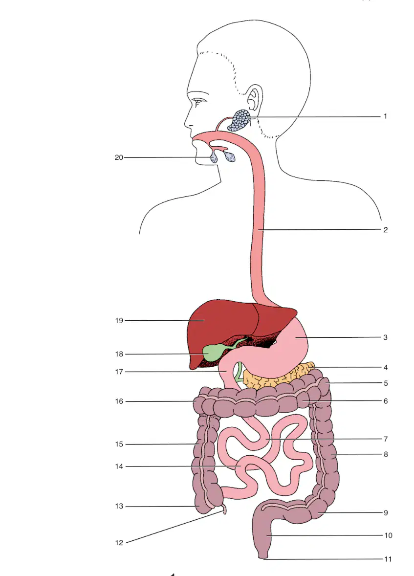
In this image what is labeled 11
(a)
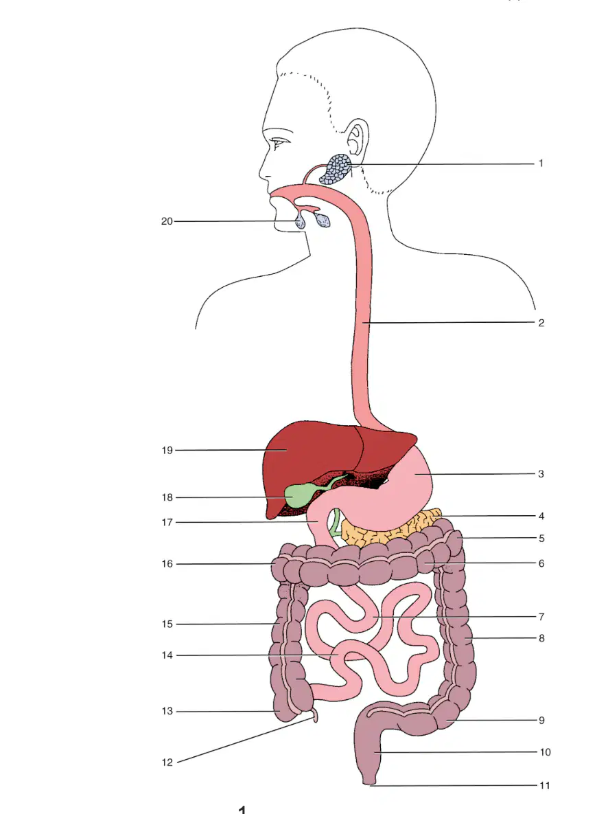
In this image what is labeled 12
(a)

In this image what is labeled 13
(a)

In this image what is labeled 14
(a)

In this image what is labeled 15
(a)

In this image what is labeled 16
(a)
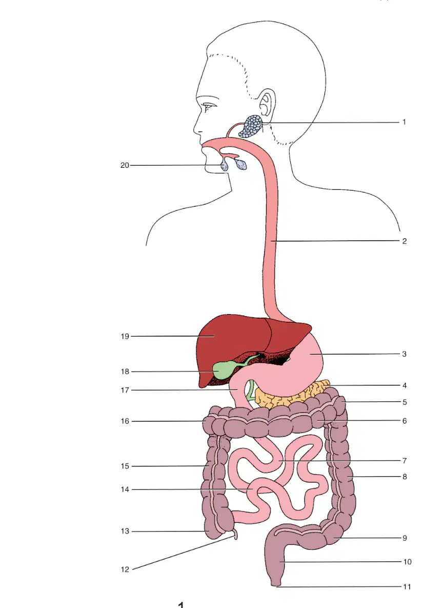
In this image what is labeled 17
(a)
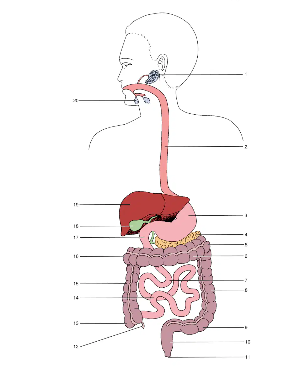
In this image what is labeled 18
(a)

In this image what is labeled 19
(a)
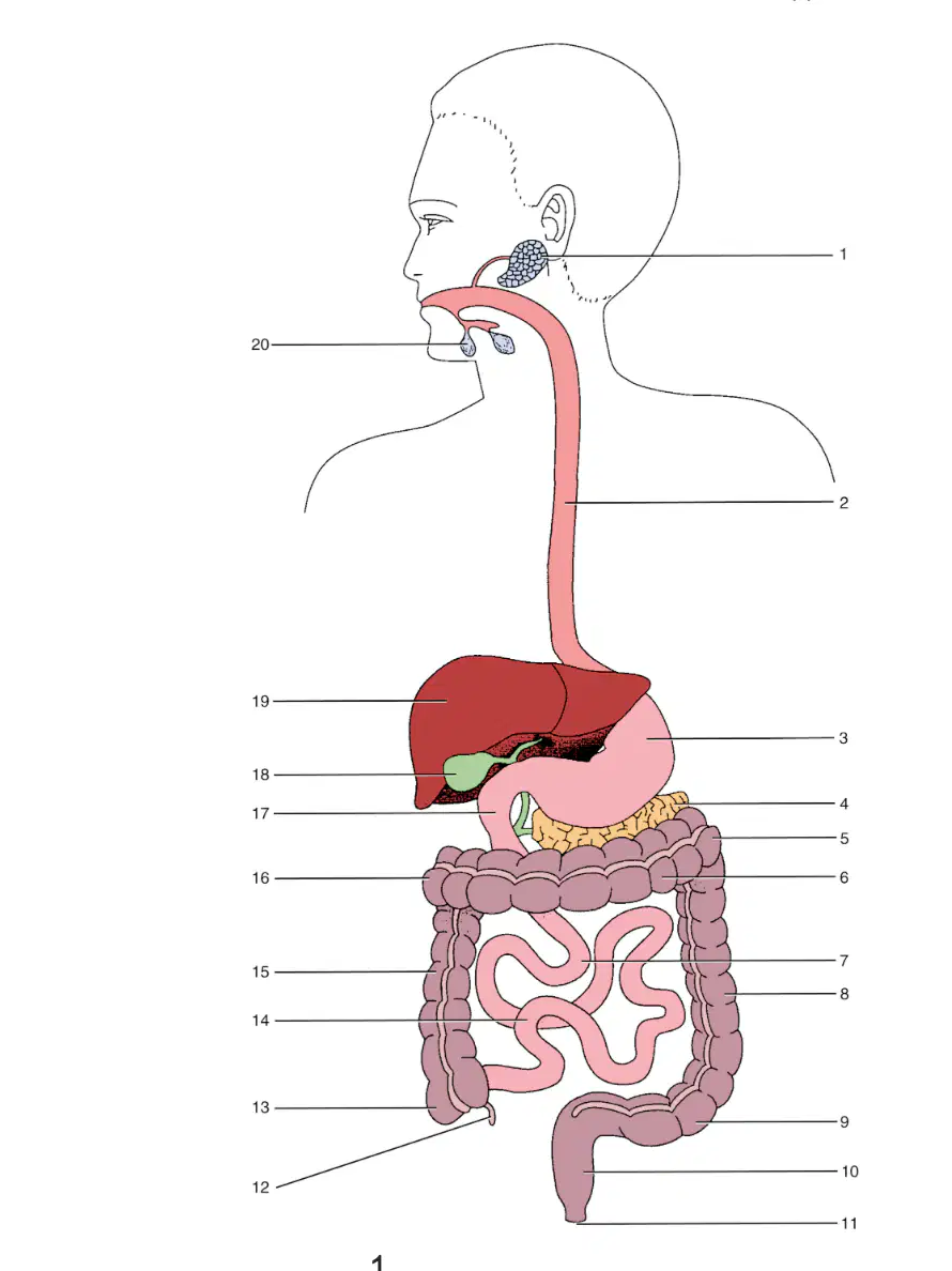
In this image what is labeled 20
(a)
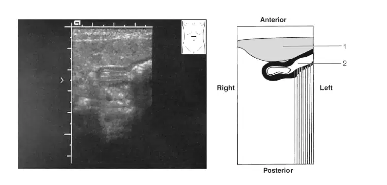
In this image what is labeled 1
(a)
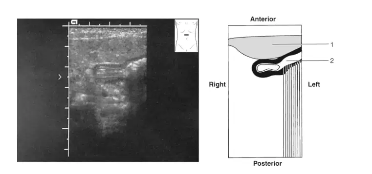
In this image what is labeled 2
(a)
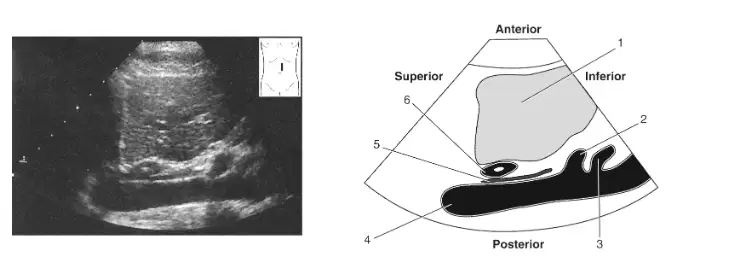
In this image what is labeled 1
(a)
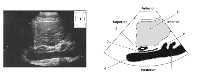
In this image what is labeled 2
(a)
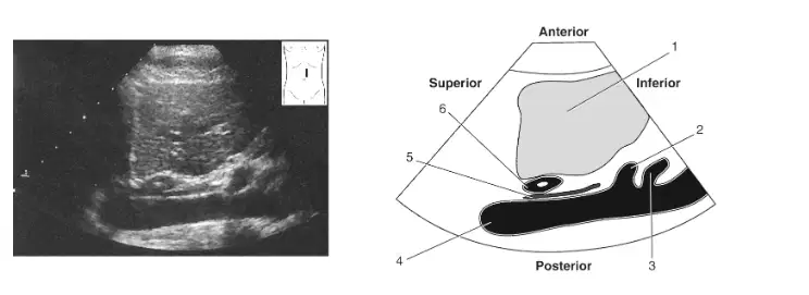
In this image what is labeled 3
(a)
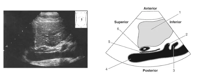
In this image what is labeled 4
(a)
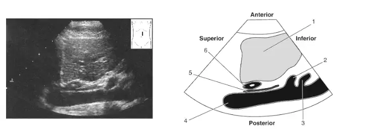
In this image what is labeled 5
(a)
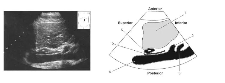
In this image what is labeled 6
(a)
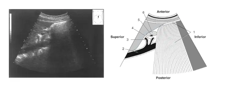
In this image what is labeled 2
(a)
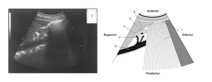
In this image what is labeled 3
(a)

In this image what is labeled 4
(a)
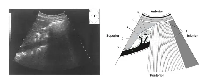
In this image what is labeled 5
(a)
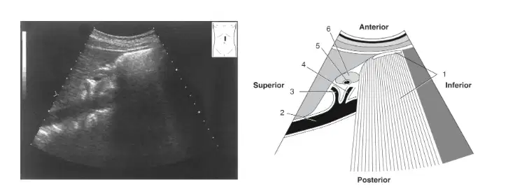
In this image what is labeled 6
(a)
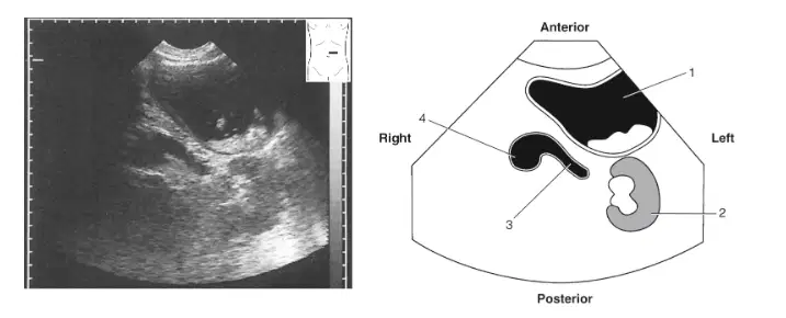
In this image what is labeled 1
(a)
In this image what is labeled 2
(a)
In this image what is labeled 3
(a)
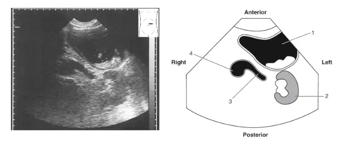
In this image what is labeled 4
(a)
Another name for the gastrointestinal tract is the
Parenteral Canal
Alimentary Canal
Mucosal canal
Medistinum
The characteristics appearance of the bowel wall can be described by a term known as the
omentum
hastura
gut signature
mesentery
The parietal peritoneum covers the anterior, lateral, and posterior walls of the abdominopelvic cavity and forms a closed sac
true
false
The deepest fold of the peritoneal cavity in females is called the
Rectouterine pouch
Posterior cul de sac
Pouch of Douglas
All are correct
The esophagus is a part of the GI tract that can be seen on a longitudinal image of the left lobe of the liver, just inferior to the heart and anterior to the aorta. This segment is commonly referred to as the
Cardiac antrum
Lesser omentum
Esophageal crus
Esophagastric junction
The visceral layer of the peritoneum lines the walls of the abdominopelvic and the parietal peritioneum covers the organs
True
False
The space between the two layers of peritoneum is known as the peritoneal cavity. It is divided into two parts. Which of the following is one of those parts
Greater omentum
Mediastinum
Epiploic foramen
Lesser sac
An open window opening between the two parts of the peritoneal cavity is called:
Greater omentum
Mediastinum
Epiploic foramen
Lesser sac
Which of the structures shown below will NOT be found in the peritoneum?
Stomach
Pancreas
cecum
uterine fundus

