
 Worksheets
Respiratory System
Total questions: 98
Worksheet time: 49mins
What is ventilation in the context of the respiratory system?
The movement of air into and out of the lungs
The exchange of O2 and CO2 between the air and the blood
The sensation of smell
The production of sound
What is the function of external respiration?
The movement of air into and out of the lungs
The exchange of O2 and CO2 between the air in the lungs and the blood
The regulation of blood pH
The production of chemical mediators
Gas transport in the respiratory system refers to the transport of ______ and ______ in the blood to and from cells.
O2 and CO2
O2 and H2O
CO2 and N2
N2 and H2O
Internal respiration is the exchange of O2 and CO2 between the ______ and the ______.
blood; tissues
lungs; blood
alveoli; capillaries
trachea; bronchi
Which function of the respiratory system allows the respiratory system to alter blood pH by changing blood CO2 levels?
Regulation of blood pH
Voice production
Olfaction
Protection
The lungs produce an enzyme called angiotensin-converting enzyme (ACE), which regulates blood pressure. This is an example of which function of the respiratory system?
Regulation of blood pH
Production of chemical mediators
Voice production
Olfaction
Air moving past the vocal folds makes sound and speech possible. This describes which function of the respiratory system?
Regulation of blood pH
Voice production
Olfaction
Protection
The sensation of smell occurs when airborne molecules are drawn into the nasal cavity. This function is called ______.
Olfaction
Gustation
Respiration
Perception
Which function of the respiratory system provides protection against some microorganisms by preventing them from entering the body and removing them from respiratory surfaces?
Regulation of blood pH
Voice production
Olfaction
Protection
What is the main function of the external nose in the respiratory system?
Encloses the chamber for air inspiration
Cleans, warms, and humidifies inspired air
Serves as a shared passageway for food and air
Keeps the airway open or patent
The nasal cavity is responsible for ______, ______, and ______ inspired air.
cleaning, warming, humidifying
filtering, cooling, drying
moistening, cooling, purifying
warming, drying, filtering
The pharynx is commonly called the ______ and serves as a shared passageway for food and air.
throat
nose
esophagus
larynx
The larynx is also known as the ______ and keeps the airway open or patent.
voice box
windpipe
throat cartilage
nasal passage
What is the function of the trachea in the respiratory system?
The trachea is commonly known as the windpipe. It is an air-cleaning tube to funnel inspired air to each lung.
The trachea is responsible for pumping blood throughout the body.
The trachea helps in the digestion of food.
The trachea produces hormones to regulate metabolism.
Which tubes direct air into the lungs?
Bronchi
Alveoli
Esophagus
Tracheoles
What is the site of gas exchange between the air and the blood in the lungs?
Alveoli
Bronchi
Trachea
Pleura
Which of the following is NOT a division of the respiratory system?
Upper respiratory tract
Lower respiratory tract
Digestive tract
Conducting zone
The upper respiratory tract includes structures from the nose to the _______.
larynx
trachea
bronchi
alveoli
The lower respiratory tract includes structures from the trachea through the _______ in the lungs.
alveoli
bronchioles
pleura
diaphragm
The conducting zone consists of structures from the nose to the smallest air tubes within the lungs used strictly for _______.
ventilation
digestion
filtration
absorption
The respiratory zone consists of small air tubes in the lungs and the alveoli where _______ occurs.
gas exchange
digestion
blood clotting
nerve transmission
What is the visible structure that forms a prominent feature of the face and is composed mainly of hyaline cartilage?
External nose
Mandible
Zygomatic arch
Maxilla
Which part of the nasal cavity divides it into left and right parts?
Nasal septum
Nasal conchae
Olfactory bulb
Hard palate
What forms the floor of the nasal cavity, separating it from the oral cavity?
Hard palate
Soft palate
Nasal septum
Vomer bone
What is the function of the conchae in the nasal cavity?
Increase surface area
Decrease surface area
Produce mucus
None of the above
Paranasal sinuses are air-filled spaces within bone that open into the nasal cavity and are lined with _______.
mucous membrane
serous membrane
cartilage
epithelial tissue
Nasolacrimal ducts carry tears from the eyes and open into the _______.
nasal cavity
oral cavity
ear canal
pharynx
What is the pharynx a common passageway for?
The pharynx is a common passageway for the respiratory and digestive systems.
The pharynx is a common passageway for the circulatory and nervous systems.
The pharynx is a common passageway for the excretory and reproductive systems.
The pharynx is a common passageway for the muscular and skeletal systems.
Which region of the pharynx is located posterior to the choanae and superior to the soft palate?
Nasopharynx
Oropharynx
Laryngopharynx
Hypopharynx
Which tissue lines the nasopharynx?
Pseudostratified Ciliated Columnar Epithelium
Stratified Squamous Epithelium
Simple Cuboidal Epithelium
Transitional Epithelium
Which region of the pharynx extends from the uvula to the epiglottis and houses the palatine and lingual tonsils?
Oropharynx
Nasopharynx
Laryngopharynx
Hypopharynx
Which region of the pharynx extends from the epiglottis to the esophagus and allows food and drink to pass through?
Laryngopharynx
Nasopharynx
Oropharynx
Trachea
Which tissue lines the laryngopharynx?
Stratified Squamous Epithelium and Ciliated Columnar Epithelium
Simple Cuboidal Epithelium
Transitional Epithelium
Pseudostratified Columnar Epithelium
What is the largest cartilage of the larynx, also called the Adam's Apple?
Thyroid Cartilage
Cricoid Cartilage
Arytenoid Cartilage
Epiglottis
Which cartilage is the most inferior cartilage of the larynx?
Cricoid Cartilage
Thyroid Cartilage
Arytenoid Cartilage
Epiglottis
The larynx consists of an outer casing of nine cartilages connected to one another by muscles and ligaments. How many of these cartilages are unpaired?
Three
Four
Two
Five
What is the function of the epiglottis in the human respiratory system?
The epiglottis is a flap that covers the larynx during swallowing to prevent food and liquids from entering the respiratory system.
The epiglottis produces mucus to trap dust particles in the air.
The epiglottis helps in the exchange of gases between the lungs and blood.
The epiglottis regulates the rate of breathing by controlling airflow.
What are vestibular folds also known as?
False vocal cords
True vocal cords
Epiglottis
Arytenoid cartilage
What are vocal folds also known as?
True vocal cords
False vocal cords
Laryngeal muscles
Pharyngeal bands
Which structure is known as the 'Guardian of the Airway'?
Epiglottis
Trachea
Larynx
Pharynx
The windpipe is also known as the ________.
Trachea
Bronchi
Esophagus
Larynx
What is the main function of the tracheal rings?
To support the trachea and keep it from collapsing.
To aid in the exchange of gases.
To produce mucus for trapping dust.
To regulate the temperature of inhaled air.
The trachea divides into right and left main (primary) ________ in the lungs at the carina.
Bronchi
Alveoli
Pleura
Capillaries
The esophagus lies along the back, cartilage-free part of the trachea.
True
False
True or False: Vestibular folds are the source of voice production.
True
False
What type of tissue lines the bronchioles and helps move mucus and trapped particles upward to keep the airways clear?
Ciliated simple columnar epithelium
Stratified squamous epithelium
Transitional epithelium
Simple cuboidal epithelium
Which main bronchus is more horizontal and why?
The left main bronchus is more horizontal because it is displaced by the heart.
The right main bronchus is more horizontal because it is shorter.
The left main bronchus is more horizontal because it is longer.
The right main bronchus is more horizontal because it is displaced by the liver.
Foreign objects that enter the trachea usually lodge in which main bronchus and why?
Right main bronchus, because it is wider, shorter, and more vertical than the left main bronchus and is more in direct line with the trachea.
Left main bronchus, because it is wider, shorter, and more vertical than the right main bronchus and is more in direct line with the trachea.
Right main bronchus, because it is narrower, longer, and more horizontal than the left main bronchus and is less in direct line with the trachea.
Left main bronchus, because it is narrower, longer, and more horizontal than the right main bronchus and is less in direct line with the trachea.
What is the function of the carina in the trachea?
It is a ridge of cartilage at the point of division, serving as a landmark for reading x-rays.
It produces mucus to trap dust particles.
It controls the opening and closing of the trachea.
It is responsible for the exchange of gases between blood and air.
Which of the following is NOT a part of the conducting zone?
Primary bronchi
Secondary bronchi
Respiratory bronchioles
Terminal bronchioles
How many secondary bronchi are on the right and left sides?
Three on the right and two on the left.
Two on the right and three on the left.
Four on the right and one on the left.
One on the right and four on the left.
What is the main function of the terminal bronchioles?
They are the last part of the conducting zone before the air reaches the respiratory zone, lined with ciliated simple cuboidal epithelium to maintain clean air passages.
They are responsible for gas exchange between the alveoli and blood vessels.
They produce surfactant to reduce surface tension in the lungs.
They filter blood before it enters the pulmonary capillaries.
Which muscle action causes bronchodilation?
Smooth muscle contracts
Smooth muscle relaxes
What happens during an asthma attack in terms of bronchiole diameter?
Contraction of terminal bronchioles leads to reduced air flow.
Dilation of terminal bronchioles leads to increased air flow.
Bronchiole diameter remains unchanged, but mucus production increases.
Bronchioles expand, allowing more oxygen to enter the lungs.
Which tissue type lines the alveolar ducts?
Simple squamous epithelium with no cilia
Stratified squamous epithelium
Pseudostratified ciliated columnar epithelium
Transitional epithelium
What is the function of alveolar sacs?
They are chambers connected to two or more alveoli at the end of an alveolar duct.
They filter oxygen from the blood.
They produce mucus to trap dust particles.
They control the rate of breathing.
Label the following parts - Trachea, Primary bronchi, Secondary bronchi, Tertiary bronchi.
Trachea, Primary bronchi, Secondary bronchi, Tertiary bronchi (as shown in the diagram)
Trachea, Secondary bronchi, Primary bronchi, Tertiary bronchi (as shown in the diagram)
Primary bronchi, Trachea, Secondary bronchi, Tertiary bronchi (as shown in the diagram)
Tertiary bronchi, Secondary bronchi, Primary bronchi, Trachea (as shown in the diagram)
What type of tissue lines the alveoli?
Simple squamous epithelium with no cilia
Stratified columnar epithelium with cilia
Transitional epithelium
Cuboidal epithelium with microvilli
Which of the following are sites of external respiration?
Bronchioles
Alveoli
Capillaries
Trachea
The alveolar wall consists of two types of cells. Name them.
Squamous epithelial cells (type I pneumocytes) and Surfactant-secreting cells (type II pneumocytes)
Cuboidal epithelial cells and Goblet cells
Columnar epithelial cells and Ciliated cells
Macrophages and Fibroblasts
The respiratory membrane of the lungs is where gas exchange between air and blood takes place.
True
False
List the six layers of the respiratory membrane in order.
1. A thin layer of fluid lining the alveolus 2. The alveolar epithelium, composed of simple squamous epithelium 3. The basement membrane of the alveolar epithelium 4. A thin interstitial space 5. The basement membrane of the capillary endothelium 6. The capillary endothelium, composed of simple squamous epithelium
1. The capillary endothelium, composed of simple squamous epithelium 2. The basement membrane of the capillary endothelium 3. A thick interstitial space 4. The alveolar epithelium, composed of cuboidal epithelium 5. The basement membrane of the alveolar epithelium 6. A thick layer of fluid lining the alveolus
1. The alveolar epithelium, composed of cuboidal epithelium 2. The basement membrane of the alveolar epithelium 3. A thick interstitial space 4. The basement membrane of the capillary endothelium 5. The capillary endothelium, composed of simple squamous epithelium 6. A thick layer of fluid lining the alveolus
1. A thin layer of fluid lining the alveolus 2. The alveolar epithelium, composed of cuboidal epithelium 3. The basement membrane of the alveolar epithelium 4. A thick interstitial space 5. The basement membrane of the capillary endothelium 6. The capillary endothelium, composed of cuboidal epithelium
Which of the following is NOT a component of the thoracic wall?
Thoracic Vertebrae
Ribs
Liver
Sternum
The thoracic wall consists of the following: Thoracic Vertebrae, Ribs, Costal Cartilages, Sternum, and Associated muscles.
True
False
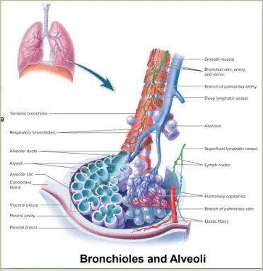
Identify the structure where gas exchange occurs.
Alveoli
Bronchi
Trachea
Capillaries
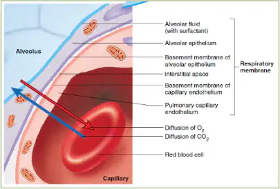
Refer to the diagram labeled 'Alveolus'. Which layer is responsible for the diffusion of O2 and CO2?
Respiratory membrane
Smooth muscle layer
Cartilaginous layer
Connective tissue layer
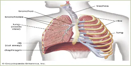
What is the space enclosed by the thoracic wall and the diaphragm called?
Thoracic cavity
Abdominal cavity
Pelvic cavity
Cranial cavity
What is the primary organ of respiration?
Lungs
Heart
Kidney
Liver
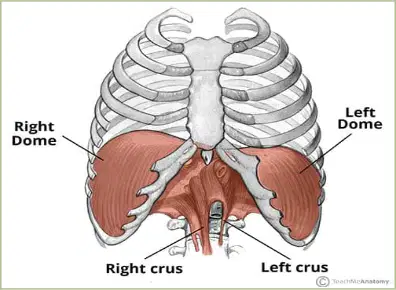
Which muscle separates the thoracic cavity from the abdominal cavity?
Diaphragm
Rectus abdominis
Intercostal muscle
Pectoralis major
The diaphragm and skeletal muscles of the thoracic wall change thoracic volume during ventilation.
True
False
Which lung has three lobes?
Right lung
Left lung
Both lungs
Neither lung
The left lung has how many lobes?
One
Two
Three
Four
What is the indentation on the medial surface of each lung where bronchi, blood vessels, nerves, and lymphatic vessels enter and exit called?
Hilum
Alveolus
Pleura
Carina
The right lung is larger than the left lung.
True
False

What is the name of the indentation in the left lung to accommodate the heart?
Cardiac notch
Oblique fissure
Lingula
Costal groove
What is the main pathway for gas exchange in the lungs?
Bronchial Circulation
Pulmonary Circulation
Systemic Circulation
Coronary Circulation
Fill in the blank: The pulmonary arteries carry _______ blood from the right ventricle of the heart to the lungs.
deoxygenated
oxygenated
nutrient-rich
carbon dioxide-rich
Which vessels return oxygenated blood back to the left atrium of the heart?
Pulmonary veins
Bronchial veins
Aorta
Superior vena cava
Bronchial circulation provides oxygenated blood to nourish the tissues of the lungs themselves.
True
False
What are the two lymphatic supplies to the lungs?
Superficial and Deep lymphatic vessels
Pulmonary and Bronchial arteries
Parietal and Visceral pleura
Alveolar and Capillary vessels
The lungs are contained within the thoracic cavity. In addition, each lung is surrounded by a separate _______.
pleural cavity
pericardial cavity
abdominal cavity
cranial cavity
Which part of the pleura lines the thoracic wall?
Visceral Pleura
Parietal Pleura
Pleural Cavity
Alveolar Membrane
The pleural fluid acts as a lubricant and helps hold the pleural membranes together.
True
False
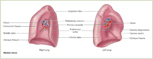
Which structure is labeled as the space around each lung within the thoracic cavity?
Pleural Cavity
Alveoli
Bronchi
Capillaries
What are the two aspects of ventilation?
Actions of the muscles of respiration and air pressure gradients
Inspiration and expiration
Diaphragm and intercostals
Thoracic and abdominal muscles
Name the two phases of ventilation.
Inspiration (Inhalation) and Expiration (Exhalation)
Contraction and relaxation
Elevation and depression
Passive and active
Fill in the blank: The process of moving air in and out of the lungs is called _________.
ventilation
respiration
diffusion
circulation
Which muscle is the primary muscle of inspiration?
A. Diaphragm
B. External intercostals
C. Pectoralis minor
D. Abdominal muscles
Match the following muscles with their function:
Contracts and moves downward
1-A
Elevates ribs
2-B
Depresses ribs
3-C
Assists in expiration by forcing diaphragm upward
4-D
The relaxation of the abdominal muscles allows the abdominal organs to move downward, giving space for the lungs to expand.
True
False
During inspiration, the thoracic cavity ________ in volume, allowing air to flow into the lungs.
increases
decreases
remains the same
contracts
Which muscles are involved in expiration?
Diaphragm (relaxes)
Internal Intercostals
Transverse Thoracic
Abdominal muscles
In quiet breathing, expiration is a ________ process.
passive
active
voluntary
forced
What happens to the thoracic cavity during expiration?
Increases in volume
Decreases in volume
Stays the same
Expands vertically
Which muscles contract to pull the ribs downward and inward during forced expiration?
Internal intercostal muscles
External intercostal muscles
Diaphragm
Pectoralis minor
What is the difference between quiet breathing (eupnea) and labored breathing (forced breathing)?
Quiet breathing is normal, relaxed breathing at rest, while labored breathing occurs during physical exertion, stress, or respiratory problems when breathing becomes deeper and faster.
Quiet breathing is only possible during sleep, while labored breathing happens only when awake.
Quiet breathing involves holding your breath, while labored breathing means rapid shallow breaths.
Quiet breathing is always faster than labored breathing.
What is the function of a spirometer?
A spirometer is a device that measures pulmonary volumes.
A spirometer is used to measure blood pressure.
A spirometer is used to monitor heart rate.
A spirometer is used to test vision.
What is the approximate value of tidal volume?
About 500mL
About 100mL
About 2000mL
About 50mL

