
 Worksheets
Tissues
Total questions: 93
Worksheet time: 47mins
What is the study of cells known as?
Histology
Cytology
Anatomy
Physiology
What is the study of tissues known as?
Cytology
Histology
Pathology
Anatomy
A tissue is a collection of cells having similar origin, structure, and _______.
function
size
color
weight
Which of the following is NOT a function of epithelial tissues?
Protection
Absorption
Excretion
Conduction
Epithelial tissues are classified into two types, ______ and ______, based on arrangement of cells in number of layers.
simple, stratified
cuboidal, columnar
squamous, columnar
glandular, transitional
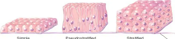
Which type of epithelium has a single layer of cells?
Simple
Pseudostratified
Stratified
None of the above
Which type of simple epithelium is usually found on absorptive surfaces?
Squamous epithelium
Cuboidal epithelium
Columnar epithelium
Ciliated epithelium
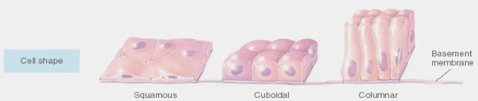
Refer to the diagram showing cell shapes and basement membrane. Which type of epithelium consists of a single layer of flat cells and is also called pavement epithelium?
Squamous epithelium
Cuboidal epithelium
Columnar epithelium
Ciliated epithelium
Which function is associated with squamous epithelium?
Protection
Diffusion and filtration
Secretion
Absorption
Fill in the blank: Squamous epithelium is found in the lungs, bowman’s capsule, heart, blood vessels, and lymph vessels as an __________.
endothelium
mesothelium
epidermis
pericardium
Which type of epithelium consists of cells with large, spherical central nuclei fitting closely together on a basement membrane?
Squamous epithelium
Cuboidal epithelium
Columnar epithelium
Ciliated epithelium
Cuboidal epithelium is actively involved in which of the following functions?
Diffusion and filtration
Secretion, absorption, and excretion
Protection
Ciliary action
Columnar epithelium is mainly found in which part of the body?
Lungs
Digestive tract (stomach to anal canal)
Kidney tubule
Heart
Fill in the blank: The main functions of columnar epithelium are __________, absorption, and secretion.
protection
movement
insulation
contraction
Ciliated epithelium cells have about 15-25 hair-like structures on each cell. These structures are known as __________.
cilia
flagella
microvilli
pili
Which function is performed by ciliated epithelium?
Diffusion
Filtration
Secretion of mucus, enzymes, and other substances; propulsion of mucus by ciliary action
Absorption
Stratified epithelium is mainly divided into two types. What are they?
A) Squamous and Cuboidal
B) Stratified squamous epithelium and Transitional epithelium
C) Columnar and Ciliated
D) Pavement and Glandular
What is the description of non-keratinized stratified squamous epithelium?
Cells are arranged in multilayers and are not filled with keratin.
Cells are arranged in a single layer and are filled with keratin.
Cells are arranged in multilayers and are filled with keratin.
Cells are arranged in a single layer and are not filled with keratin.
What is the function of non-keratinized stratified squamous epithelium?
This is found on wet surfaces subjected to wear and tear, provide protection from drying.
It secretes hormones for metabolic regulation.
It absorbs nutrients from digested food.
It produces mucus to trap pathogens.
Where is non-keratinized stratified squamous epithelium located?
Esophagus, mouth, vagina, conjunctiva of eyes, pharynx.
Skin, nails, hair, cornea.
Trachea, bronchi, alveoli, larynx.
Stomach, small intestine, colon, rectum.
What is the description of keratinized stratified squamous epithelium?
Cells are arranged in multilayers and cells in superficial layers filled with keratin. These cells are generally dead cells.
Cells are arranged in a single layer and are all living cells.
Cells are arranged in multilayers but do not contain keratin in any layer.
Cells are arranged in a single layer and contain keratin in all cells.
What is the function of keratinized stratified squamous epithelium?
These form a tough, relatively waterproof protective layer that prevents drying of the live cells underneath. The surface layer of skin is rubbed off and is replaced from below layers.
It produces hormones that regulate metabolism.
It is responsible for the absorption of nutrients in the small intestine.
It helps in the transmission of nerve impulses across the skin.
Where is keratinized stratified squamous epithelium located?
Skin, hair and nails.
Liver and pancreas.
Lungs and trachea.
Intestines and stomach.
What is the description of transitional epithelium?
It consists of 3-4 layers of cells. It is known as transitional because the height of cells varies with situation. The upper layers of cells are large and flat. Lower layer of cells are polyhedral cells.
It consists of a single layer of squamous cells. It is known for rapid diffusion and filtration.
It consists of columnar cells with microvilli on the surface. It is specialized for absorption.
It consists of cuboidal cells arranged in rings. It is mainly found in glandular tissue.
Where is transitional epithelium located?
Pelvis of kidney, uterus, urinary bladder and urethra.
Lining of alveoli, blood vessels, and heart.
Inner surface of small intestine and stomach.
Outer layer of skin and lining of mouth.
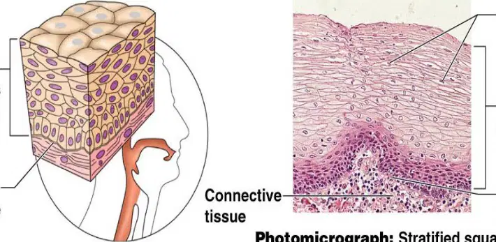
What type of tissue is shown and what is its main function?
Stratified squamous epithelium; its main function is protection from wear and tear and drying.
Simple cuboidal epithelium; its main function is absorption and secretion.
Adipose tissue; its main function is energy storage and insulation.
Cardiac muscle tissue; its main function is contraction to pump blood.
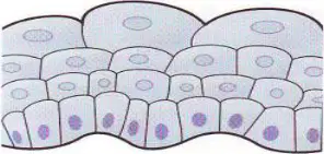
What does the diagram illustrate about transitional epithelium?
The diagram illustrates that transitional epithelium changes shape depending on whether it is relaxed or stretched.
The diagram illustrates that transitional epithelium is always a single layer of flat cells.
The diagram illustrates that transitional epithelium is found only in the lungs.
The diagram illustrates that transitional epithelium does not change its shape under any condition.
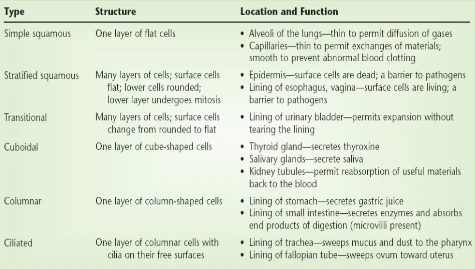
Which type of epithelial tissue lines the alveoli of the lungs to permit diffusion of gases?
Simple squamous
Stratified squamous
Transitional
Cuboidal

Fill in the blank: The lining of the stomach, which secretes gastric juice, is made up of ________ epithelial cells.
Columnar
Squamous
Cuboidal
Transitional
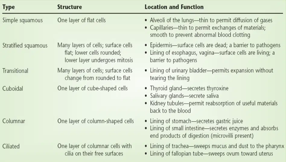
Which type of epithelial tissue sweeps mucus and dust to the pharynx in the trachea?
Simple squamous
Stratified squamous
Ciliated
Cuboidal
What are the four main functions of connective tissue?
A. Binding and support, Protection, Transport, Insulation
B. Digestion, Absorption, Secretion, Excretion
C. Movement, Sensation, Growth, Reproduction
D. Filtration, Respiration, Circulation, Excretion
Fill in the blank: The origin of all connective tissues is ________, an embryonic tissue.
mesenchyme
ectoderm
endoderm
epithelium
Which of the following is NOT a function of connective tissue?
A. Binding and support
B. Protection
C. Transport
D. Photosynthesis
Fill in the blank: Ground substance and fibers make up the ________ matrix in connective tissue.
extracellular
intracellular
cytoplasmic
nuclear
Which protein controls the fiber organization in the extracellular matrix?
Collagen
Fibrillin
Elastin
Keratin
Which of the following is common in many types of connective tissue?
Chondrocytes
Osteocytes
Fibrocytes
Adipocytes
Which cell type is found in cartilage?
Chondrocytes
Osteocytes
Adipocytes
Blood cells
Fill in the blank: ________ are fat storing and metabolizing cells found in connective tissue.
Adipocytes
Fibroblasts
Chondrocytes
Osteocytes
Which type of collagen fiber provides tensile strength in dense connective tissues of the skin dermis, tendons, and ligaments?
Type I
Type II
Type III
Fill in the blank: Type II collagen fibers are predominant in ________ and elastic cartilage.
hyaline
fibrocartilage
compact bone
spongy bone
Which protein is secreted by fibroblasts and chondroblasts to give elasticity to supporting tissues?
Collagen
Elastin
Fibrillin
Histamine
Fat cells (adipocytes) act as storage sites of ________.
energy
oxygen
protein
calcium
Which cells produce histamine and heparin to mediate the inflammatory response?
Plasma cells
Mast cells
Fat cells
Fibrocytes
Plasma cells secrete ________ and migrate to connective tissues from the circulation.
antibodies
hormones
enzymes
neurotransmitters

Refer to the diagram of connective tissue classification. Which cell type gives rise to connective tissue proper?
Chondroblast
Fibroblast
Osteoblast
Pleuriopotent stem cell
Areolar connective tissue is found in almost every part of the body.
True
False
List two functions of areolar connective tissue.
Elasticity, Tensile strength
Support, Protection
Transport, Storage
Contraction, Movement
Areolar connective tissue connects and supports other tissues. Name two locations where it is found.
Under the skin, Between muscles
Inside bones, In the brain
In the heart, In the liver
On the surface of teeth, In the cornea
Adipose tissue is characterized by abundant ______ which store and metabolize fat.
adipocytes
erythrocytes
chondrocytes
osteocytes
Which type of fibers support adipose tissue?
Collagen type I fibers
Elastic fibers
Reticular fibers
Cartilage fibers
There are two main types of adipose tissue: White adipose tissue and Brown adipose tissue. Which type of adipose tissue acts as insulation (under the skin) and as a shock absorber (e.g., around kidney)?
White adipose tissue
Brown adipose tissue
There are two main types of adipose tissue: White adipose tissue and Brown adipose tissue. Which type of adipose tissue contributes to thermoregulation of newborns by producing heat?
White adipose tissue
Brown adipose tissue
Reticular tissue forms a delicate supporting framework of reticular fibres (type III collagen). Reticular tissue is classified as a type of ______ connective tissue due to the sparsity of its fibres.
loose
dense
elastic
adipose
Dense connective tissue contains more fibers and fewer cells than loose connective tissue.
True
False
Fibrous tissue is characterized by a dense arrangement of collagen fibers and reduced ground substance compared to areolar connective tissue.
True
False
Dense regular connective tissues such as tendons and ligaments have collagen bundles in ordered, parallel orientations that provide maximum tensile strength.
True
False
Dense irregular connective tissues have interwoven collagen bundles to resist tension from many directions and provide tough physical support and protection.
True
False
Elastic tissue is capable of considerable extension and recoil. It is found in organs where stretching or alteration of shape is required. Which of the following is NOT an example of an organ where elastic tissue is found?
Large blood vessels
Trachea
Kidney
Bronchi
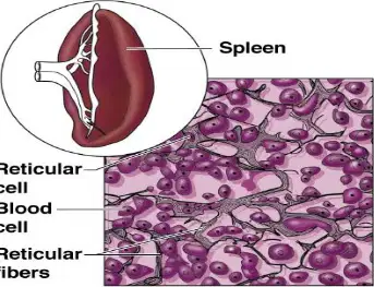
What type of cell is shown along with reticular fibers and blood cells?
Reticular cell
Plasma cell
Macrophage
Fibroblast

Refer to the photomicrograph labeled 'Dark-staining network of reticular connective tissue (400x)'. Which type of cell is indicated along with reticular fibers?
White blood cell (lymphocyte)
Red blood cell (erythrocyte)
Fibroblast
Adipocyte
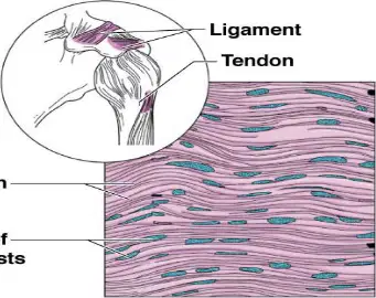
Refer to the diagram labeled 'Dense fibrous'. Which fibers are present in this tissue?
Collagen fibers
Elastic fibers
Reticular fibers
Muscle fibers
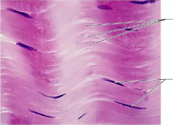
Refer to the photomicrograph labeled 'Dense fibrous connective tissue from a tendon (500x)'. What structures are labeled in this image?
Collagen fibers, Nuclei of fibroblasts
Elastic fibers, Nuclei of chondrocytes
Reticular fibers, Nuclei of adipocytes
Muscle fibers, Nuclei of neurons
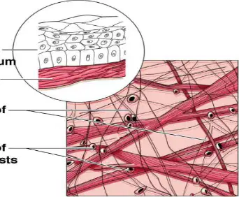
Refer to the diagram labeled 'Areolar'. Which type of cell's nuclei are shown in the matrix?
Nuclei of fibroblasts
Nuclei of mast cells
Nuclei of adipocytes
Nuclei of macrophages
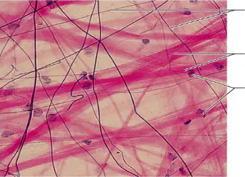
Refer to the photomicrograph labeled 'Areolar connective tissue, a soft packaging tissue of the body (330x)'. Which fibers are present in this tissue?
Elastic fibers, Collagen fibers
Reticular fibers, Muscle fibers
Nerve fibers, Collagen fibers
Adipose fibers, Elastic fibers
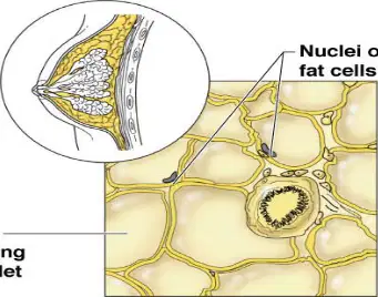
Refer to the diagram labeled 'Adipose'. What does the vacuole contain?
Fat droplet
Water
Protein
Carbohydrate
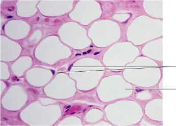
Refer to the photomicrograph labeled 'Adipose tissue from the subcutaneous layer beneath the skin (330x)'. What is labeled inside the vacuole?
Fat droplet
Nucleus
Mitochondria
Cytoplasm
What are the cells of cartilage called?
Chondrocytes
Osteocytes
Erythrocytes
Leukocytes
Cartilage is characterized by proteoglycan-containing ground substance complexed to a fine collagen (mainly Type II) fiber latticework.
True
False
Which of the following is NOT a main type of cartilage?
Hyaline cartilage
Fibro cartilage
Elastic cartilage
Spongy cartilage
Hyaline cartilage provides which of the following functions?
Flexibility
Support
Smooth surfaces for movement at joints
All of the above
Fill in the blank: Hyaline cartilage is a smooth ______-white tissue.
bluish
greenish
yellowish
reddish
Where is hyaline cartilage commonly found? Select all that apply.
Ends of long bones that form joints
Coastal cartilages attaching ribs to the sternum
Forming part of pharynx, larynx, and trachea
Between articulating surfaces of the bones of the knee joint
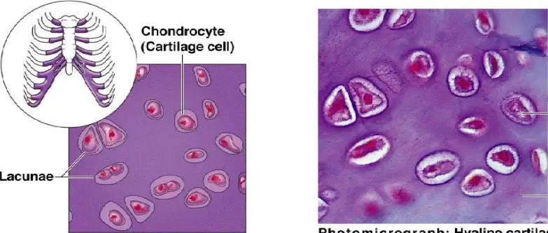
Identify the cell type labeled in the hyaline cartilage diagram.
Chondrocyte
Osteocyte
Fibroblast
Erythrocyte
Fibro cartilage consists of dense masses of white ______ fibers in a matrix similar to that of hyaline cartilage.
collagen
elastic
reticular
actin
Which of the following is a location where fibro cartilage is found?
As intervertebral discs
As ligaments joining bones
Between articulating surfaces of the bones of the knee joint
All of the above
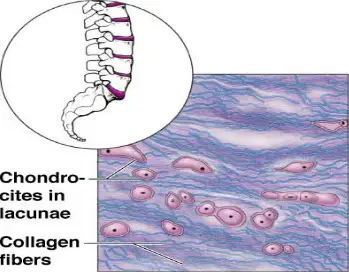
What is the function of elastic cartilage?
Provides support and maintains shape
Stores fat
Produces blood cells
Protects organs
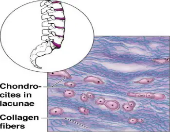
Where is elastic cartilage located?
Lobe of the ear, epiglottis, and tunica media of blood vessel walls
Nose, trachea, and bronchi
Ribs and sternum
Vertebral column
Fill in the blank: Among all the connective tissues, ______ is the hardest connective tissue.
bone
cartilage
adipose tissue
blood
The extracellular matrix of bone is composed of 25% water, 50% mineral salts, and 25% ______ fibers.
collagen
elastin
reticular
keratin
What are the open spaces between trabeculae in living bones filled with?
Red or yellow bone marrow
Blood vessels
Nerve fibers
Cartilage
The tubular shaft of a long bone is called the ______.
diaphysis
epiphysis
periosteum
metaphysis
The bone ends of a long bone are called ______.
epiphysis
diaphysis
periosteum
metaphysis
The structural unit of compact bone is called ______ or ______ system.
osteon or haversian
canaliculi or Volkmann's
trabeculae or lamellar
periosteum or endosteum
Which of the following is NOT a function of the haversian canal?
Entry of blood vessels
Entry of nerves
Storage of fat
Formation of concentric circles
What is the function of blood as a connective tissue?
Blood provides communication between different parts of the body and with the external environment. It is involved in transportation, regulation, and protection functions.
Blood acts as a structural support for bones and muscles.
Blood stores energy for long-term use in the body.
Blood is responsible for transmitting nerve impulses throughout the body.
Which pigment gives blood its red color?
Hemoglobin (Hb)
Chlorophyll
Melanin
Carotene
Name the three types of muscle tissue described in the passage.
Skeletal muscle, Smooth muscle, Cardiac muscle
Skeletal muscle, Connective tissue, Cardiac muscle
Smooth muscle, Nervous tissue, Cardiac muscle
Skeletal muscle, Epithelial tissue, Cardiac muscle
Which type of muscle is voluntary in nature?
Skeletal muscle
Cardiac muscle
Smooth muscle
Involuntary muscle
Which types of muscle are involuntary in nature?
Smooth muscle and Cardiac muscle
Skeletal muscle and Cardiac muscle
Skeletal muscle and Smooth muscle
Skeletal muscle only
What is the description of skeletal muscle according to the passage?
Long, cylindrical, unbranched, striated, multinucleated cells.
Short, spindle-shaped, non-striated, single nucleus cells.
Branched, striated, single nucleus cells.
Flat, non-striated, multinucleated cells.
Why are skeletal muscles called 'striated'?
Striations can be seen on microscopic examination.
They are attached to bones.
They help in involuntary movements.
They are smooth in appearance.
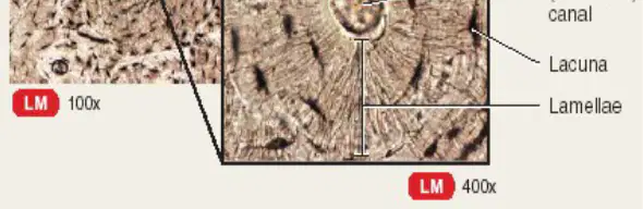
What is the function of canaliculi in bone tissue?
Canaliculi provide routes for nutrients to reach osteocytes and for wastes to leave them.
Canaliculi store calcium and phosphate ions for bone mineralization.
Canaliculi are responsible for bone growth and elongation.
Canaliculi produce collagen fibers for bone strength.
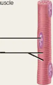
Refer to the diagram of skeletal muscle. Which of the following features is NOT present in smooth muscle cells? Choose the correct option.
A) Striations
B) Central nuclei
C) Spindle shape
D) Loose connective tissue

