
Font size
 Worksheets
Unit 5 Bone Basics
Total questions: 90
Worksheet time: 45mins
What two types of tissue make up your skeleton?
Cartilage and bone
Muscle and tendon
Ligament and cartilage
Bone and muscle
What type of tissue is your skeleton made of before it is bone?
Cartilage
Ligament
Tendon
Adipose tissue
Bones are living.
True
False
Bones are made of cells.
True
False
How many bones are in the adult human skeleton?
196
206
216
250
Which of the major four types of tissue includes bone?
Epithelial tissue
Muscular tissue
Nervous tissue
Connective tissue
Does bone have a matrix? If it does, what is this matrix called?
Osteoid
Sarcolemma
Perimysium
Chondrolemma
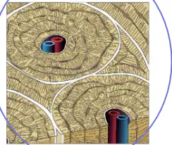
Which of the following is a picture of bone tissue under a microscope? Choose the correct letter from the four micrographs labeled A–D.
A
B
C
D
Which of the following are functions of bone tissue? Select all that apply.
Support
Protection
Anchorage
Digestion
Respiration
Which of the following are functions of bone tissue? Select all that apply.
Mineral storage
Blood cell formation
Fat storage
Hormone production
Vision
What organs in your body are best protected by bone? Select all correct organ–bone pairings.
Brain — skull
Spinal cord — vertebrae
Heart and lungs — ribcage
Internal reproductive organs — pelvis
Intestines — clavicle
Which other type of tissue do bones work together with to cause movements in the body?
Adipose tissue
Skeletal muscle tissue
Nervous tissue
Epithelial tissue
What do we call bones of the skeleton that fall on the midline (sagittal plane)?
Appendicular skeleton
Axial skeleton
Cranial skeleton
Peripheral skeleton
What do we call bones of the skeleton that are not on the midline?
Axial skeleton
Appendicular skeleton
Cranial skeleton
Central skeleton
Classify each item as Axial or Appendicular skeleton.
Skull
Axial
Limbs
Appendicular
Vertebral column
Axial
Scapula
Appendicular
Ribs
Axial
Bone Types: Which type of bone does each item fit?
Carpals (wrist bones)
Short
Femur (thigh bone)
Long
Vertebrae (backbones)
Irregular
Patella (kneecap)
Sesamoid
Sternum (breastbone)
Flat
Circle the 10 examples of long bones from the list. Select all long bones.
Clavicle
Scapula
Femur
Ribs
Ulna
Circle the 10 examples of long bones from the list. Select all long bones.
Tibia
Fibula
Radius
Carpals
Phalanges
Circle the 10 examples of long bones from the list. Select all long bones.
Metacarpals
Metatarsals
Sternum
Mandible
Nasal bone
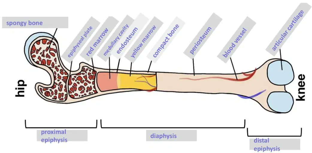
Label the following long bone. According to the diagram, which tissue lines the medullary cavity?
Endosteum
Periosteum
Articular cartilage
Spongy bone
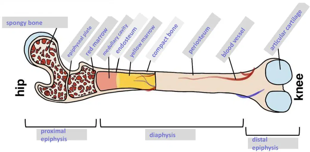
Label the following long bone. On the diagram, which structure is found at the ends of the bone and composed of trabecular tissue?
Spongy bone
Compact bone
Periosteum
Epiphyseal plate
Fill-in-the-Blank – Anatomy of a Long Bone: Match each description to the correct structure of a long bone.
A connective tissue membrane that covers the outside of the bones
Periosteum
A connective tissue membrane that lines the medullary cavity
Endosteum
A tiny hole that the blood vessel threads through
Nutrient foramen
Adipose tissue found within the medullary cavity
Yellow marrow
Allows newly formed blood to exit the bone and enter the bloodstream; allows the bone tissue to receive nutrients, oxygen, and hormones
Nutrient foramen
Fill-in-the-Blank – Anatomy of a Long Bone: Match each description to the correct structure of a long bone.
Filled with yellow marrow
Medullary cavity
Found inside the spongy tissue at the ends of our bones
Red marrow
Full of bone-growing cells (osteoblasts) that allow our bones to grow in width
Periosteum
Full of stem cells that produce red blood cells, most white blood cells, and platelets
Red marrow
Growth plates
Epiphyseal plates
Fill-in-the-Blank – Anatomy of a Long Bone: Match each description to the correct structure of a long bone.
Hollow space inside the diaphysis
Medullary cavity
Knobby ends of the bone
Epiphyses
Made of hyaline cartilage that ossifies and regrows repeatedly as you grow in height; ossify permanently between ages 18 and 25
Epiphyseal plates
Name literally means “inside-bone”
Endosteum
Fill-in-the-Blank – Anatomy of a Long Bone: Match each description to the correct structure of a long bone.
Name literally means “outside-bone”
Periosteum
Protects the ends of our bones from friction and shock as joints move and we put weight on them
Articular cartilage
Shaft of the bone
Diaphysis
The epiphysis that is closer to the trunk
Proximal epiphysis
Fill-in-the-Blank – Anatomy of a Long Bone: Match each description to the correct structure of a long bone.
The epiphysis that is further from the trunk
Distal epiphysis
Type of bone found in the epiphyses
Spongy bone
Type of bone that makes up the diaphysis
Compact bone
When it is lost, arthritis develops
Articular cartilage
If someone loses a significant amount of blood, how does their marrow in their bones change?
Yellow marrow changes to red marrow to increase blood production
Red marrow changes to yellow marrow to conserve energy
Compact bone converts to spongy bone to hold more blood
Periosteum thickens to produce blood cells
Is bone vascular or avascular? Select all statements that explain why it must be this way.
Bone is vascular so nutrients and oxygen can reach bone tissue
Bone is vascular so blood created in the marrow can leave the bone
Bone is avascular because cartilage covers all bones
Bone is avascular so bones remain lightweight
Fill-in-the-Blank – Types of Bone Cells: Match each description to the correct type of bone cell.
This type of bone cell allows your bones to perform their daily functions
Osteocyte
This type of bone cell becomes an osteocyte
Osteoblast
This type of bone cell can do mitosis
Osteogenic cells
This type of bone cell creates osteoblasts
Osteogenic cells
This type of bone cell creates the matrix of bones
Osteoblast
Fill-in-the-Blank – Types of Bone Cells: Match each description to the correct type of bone cell.
This type of bone cell gets buried and becomes another type of bone cell
Osteoblast
This type of bone cell is a stem cell
Osteogenic cells
This type of bone cell is considered a “mature” bone cell
Osteocyte
This type of bone cell is in the epiphyseal plates and on the periosteum
Osteoblast
This type of bone cell is in the periosteum and endosteum (don’t pick osteogenic cells)
Osteoclast
Fill-in-the-Blank – Types of Bone Cells: Match each description to the correct type of bone cell.
This type of bone cell is located inside the spongy bone and compact bone
Osteocyte
This type of bone cell removes dead bone after a fracture
Osteoclast
This type of bone cell shaves away old bone when your bones are growing wider
Osteoclast
This type of bone cell translates into “bone break”
Osteoclast
Fill-in-the-Blank – Types of Bone Cells: Match each description to the correct type of bone cell.
This type of bone cell translates into “bone cell”
Osteocyte
This type of bone cell translates into “bone produce”
Osteoblast
This type of bone cell creates osteoid
Osteoblast
Why are osteoblasts located on the periosteum?
To build new bone that widens bones and aids in fracture repair and remodeling
To dissolve old bone so the medullary cavity grows wider
To transport blood from marrow into the bloodstream
To cushion joints and reduce friction
Why are osteoblasts located on epiphyseal plates?
To build new bone that allows bones to grow in length
To clean away old bone after a fracture
To create adipose tissue within the medullary cavity
To form compact bone around the diaphysis
Why are osteoclasts located on the periosteum?
To clean away old bone during bone remodeling or fracture repair
To lay down osteoid to form new bone
To produce red blood cells
To create articular cartilage
Why are osteoclasts located within the endosteum?
To shave away bone and widen the medullary cavity as bones grow in width, preventing bones from becoming too heavy
To build bone that increases the length of the bone
To produce osteoid that becomes compact bone
To store yellow marrow
How are osteogenic cells, osteocytes, and osteoblasts related in terms of how each cell is created?
Osteogenic cells undergo mitosis and become osteoblasts; osteoblasts create osteoid and become buried as osteocytes
Osteoblasts divide to form osteogenic cells, which mature into osteoclasts
Osteocytes divide to become osteoblasts, which then create osteoclasts
Osteoclasts form osteoid that turns into osteogenic cells and later osteocytes
What is bone remodeling?
The removal of old, damaged, worn-out bone and its replacement with new healthy bone
The initial formation of bone during fetal development
The conversion of spongy bone to compact bone with age
The deposition of calcium without changing old bone
Why is bone remodeling necessary?
To prevent bones from becoming brittle and breaking
To increase the number of joints in the skeleton
To make bones heavier for better support
To convert cartilage directly into compact bone
How often is the human skeleton fully replaced by bone remodeling?
Every 10 years
Every 2 years
Every 25 years
Every 5 months
How do osteoclasts and osteoblasts work together to remodel bones?
Osteoclasts resorb old bone; osteoblasts lay down new osteoid and become osteocytes
Osteoblasts remove old bone; osteoclasts deposit new bone
Osteoclasts create articular cartilage; osteoblasts widen the medullary cavity
Osteoblasts produce red marrow; osteoclasts form periosteum
What is endochondral ossification?
Formation of bone within a fibrous membrane
Formation of bone from hyaline cartilage
Replacement of bone with elastic cartilage
Calcification of existing compact bone
At what ages does endochondral ossification begin?
2 to 4 weeks in the womb
6 to 8 weeks in the womb
At birth
During adolescence
What specific material are our bones made of at the beginning of endochondral ossification? Be sure to mention which type.
Hyaline cartilage
Elastic cartilage
Fibrocartilage
Spongy bone
What material are our bones made of at the end of endochondral ossification?
Bone
Hyaline cartilage
Elastic cartilage
Fibrocartilage
List the 6 steps of endochondral ossification in order. Summarize what happens in each step.
Cartilage model forms → Perichondrium becomes periosteum → Primary ossification center develops → Medullary cavity formed → Secondary ossification centers develop → Ossification is complete
Perichondrium becomes periosteum → Cartilage model forms → Secondary ossification centers develop → Primary ossification center develops → Medullary cavity formed → Ossification is complete
Cartilage model forms → Primary ossification center develops → Perichondrium becomes periosteum → Secondary ossification centers develop → Medullary cavity formed → Ossification is complete
Primary ossification center develops → Cartilage model forms → Perichondrium becomes periosteum → Medullary cavity formed → Ossification is complete → Secondary ossification centers develop
Which type of cell starts the process of endochondral ossification—osteoblasts, osteocytes, chondrocytes, or osteoclasts—and what is their role?
Osteoblasts; they dissolve cartilage to form a model
Osteocytes; they divide to create cartilage rings
Chondrocytes; they form a model of where the bone will eventually be
Osteoclasts; they secrete osteoid to start bone building
What is the perichondrium?
A vascular membrane inside the medullary cavity
A layer of cartilage around the original cartilage model
The bony sleeve surrounding compact bone
The connective tissue lining the articular surfaces
When the perichondrium turns to bone, what is it now called?
Endosteum
Periosteum
Perimysium
Perineurium
The formation of the periosteum causes what to occur within the developing bone, and why?
It increases cartilage growth because nutrients flow more easily
It cuts off nutrients to the cartilage model, causing the cartilage within the model to die and be replaced by bone
It expands the marrow cavity by adding more cartilage
It converts elastic cartilage into fibrocartilage to begin ossification
What must form within the epiphyses and diaphysis for ossification to begin taking place, and why must these form?
Bone marrow; it provides stem cells for cartilage growth
Ligaments; they stabilize joints for cartilage deposition
Blood vessels; they deliver nutrients that osteoblasts need to create osteoid
Nerves; they stimulate chondrocyte division
Where is the primary ossification center found?
Epiphysis
Diaphysis
Metaphysis
Articular surface
Where is the secondary ossification center found?
Diaphysis
Epiphyses
Medullary cavity
Periosteum
When do the secondary ossification centers form?
Early embryonic period
Around the time of birth
Late childhood
During adulthood
Which ossifies first—the diaphysis or the epiphyses?
Diaphysis
Epiphyses
What is the role of osteoblasts in endochondral ossification?
They build new bone in the primary and secondary ossification centers, causing ossification
They digest bone to enlarge the marrow cavity
They maintain existing bone without forming new tissue
They convert cartilage directly into elastic fibers
What is the role of osteoclasts in endochondral ossification?
They lay down osteoid to begin ossification
They clean away newly formed bone in the diaphysis, forming a medullary cavity
They produce cartilage for the growth plates
They create the periosteum from the perichondrium
Does ossification occur from the outside-in or the inside-out?
Outside-in
Inside-out
What cartilages are left at the end of endochondral ossification?
Articular cartilage and epiphyseal plates
Elastic cartilage and fibrocartilage
Hyaline cartilage throughout the diaphysis
No cartilage remains anywhere
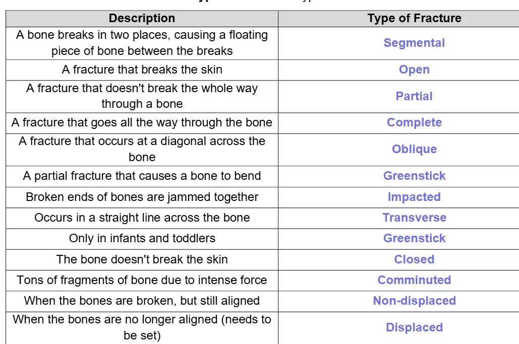
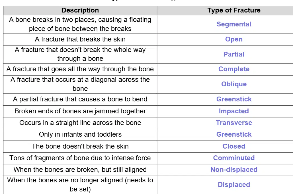
Match each description to the correct fracture type.
A bone breaks in two places, causing a floating piece of bone between the breaks
Segmental
A fracture that breaks the skin
Open
A fracture that doesn't break the whole way through a bone
Partial
A fracture that goes all the way through the bone
Complete
A fracture that occurs at a diagonal across the bone
Oblique
Match each description to the correct fracture type.
A partial fracture that causes a bone to bend
Greenstick
Broken ends of bones are jammed together
Impacted
Occurs in a straight line across the bone
Transverse
Only in infants and toddlers
Greenstick
The bone doesn't break the skin
Closed
Match each description to the correct fracture type.
Tons of fragments of bone due to intense force
Comminuted
When the bones are broken, but still aligned
Non-displaced
When the bones are no longer aligned (needs to be set)
Displaced
At the greatest risk of infection
Open
Which fracture types would describe the picture? Pick 3 terms that apply.
Partial
Closed
Non-displaced
Which fracture types would describe the picture? Pick 4 terms that apply.
Complete
Transverse
Closed
Displaced
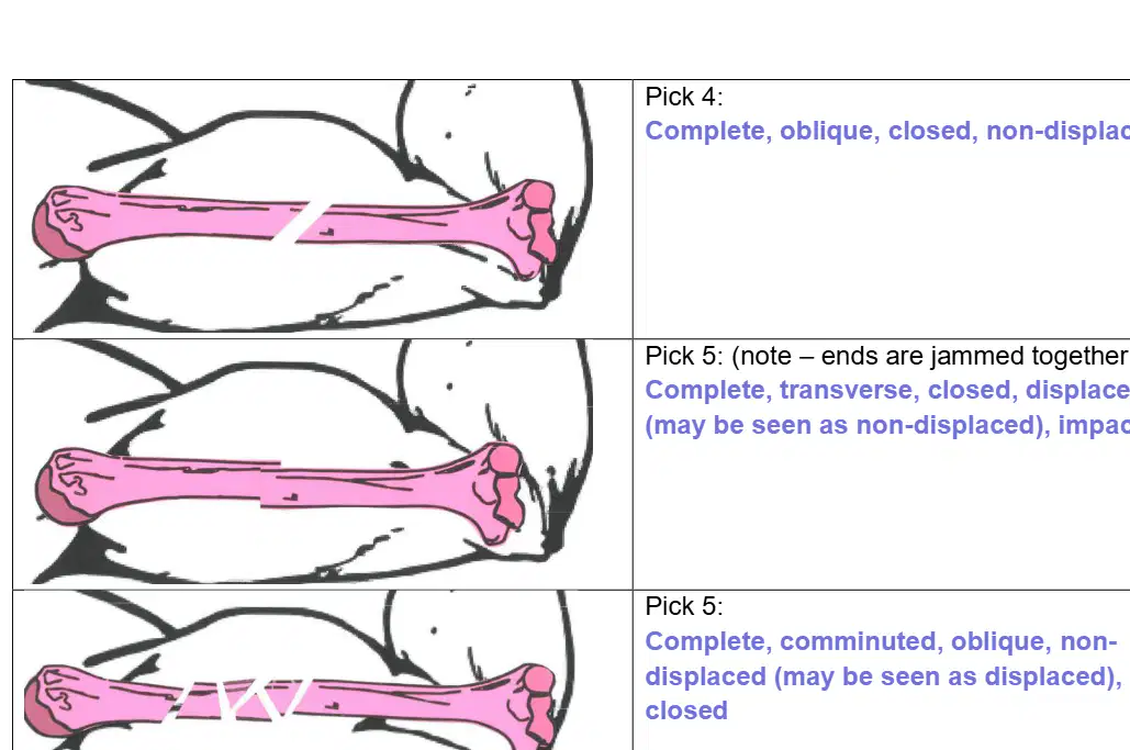
Which fracture types would describe the picture? Pick 4 terms that apply.
Complete
Oblique
Closed
Non-displaced
Which fracture types would describe the picture? Pick 5 terms that apply.
Complete
Transverse
Closed
Displaced
Impacted
Which fracture types would describe the picture? Pick 5 terms that apply.
Complete
Comminuted
Oblique
Non-displaced
Closed
Which fracture types would describe the picture? Pick 4 terms that apply.
Complete
Transverse
Open
Displaced
Which fracture types would describe the picture? Pick 5 terms that apply.
Complete
Transverse
Closed
Non-displaced
Segmental
Which fracture types would describe the picture? Pick 3 terms that apply.
Partial
Greenstick
Closed
Displaced
Most of the repair of a fracture occurs due to the action of the body itself. What is the doctor’s role in ensuring that a fracture is repaired properly?
Realigns the broken bone ends to restore proper alignment
Administers antibiotics to prevent bone infection
Applies heat therapy to increase blood flow
Prescribes calcium supplements to speed healing
What term means realignment of broken bone ends?
Reduction
Fixation
Subluxation
Traction
What is the difference between a closed and an open reduction?
Closed reduction uses manual manipulation; open reduction requires surgical intervention with hardware
Closed reduction is performed under general anesthesia; open reduction is performed without anesthesia
Closed reduction treats only soft tissue; open reduction treats only bone
Closed reduction is slower than open reduction but has a higher complication rate
What is another term for a closed reduction?
External reduction
Internal reduction
External fixation
Open fixation
What is another term for an open reduction?
Internal reduction
External reduction
Closed fixation
Manual alignment
How long does the average fracture take to heal?
6 to 8 weeks
2 to 3 weeks
10 to 12 weeks
1 week
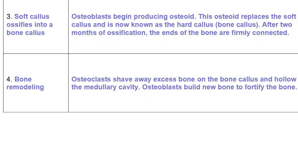
List the 4 steps of fracture repair in order. Match each step with its summary.
Hematoma forms
Blood vessels in bone and periosteum tear; leaked blood clots form a hematoma and some bone cells die
Soft callus forms
New blood vessels grow; chondroblasts create cartilage matrix forming a temporary connection between bone ends
Soft callus ossifies into a bone callus
Osteoblasts produce osteoid that replaces the soft callus, forming a hard callus; after two months the bone ends are firmly connected
Bone remodeling
Osteoclasts remove excess callus and hollow the medullary cavity; osteoblasts build new bone to fortify the bone
What is a hematoma?
A blood clot at the injury site
A pocket of pus formed by infection
A calcium-rich bone spur
A scar formed after healing
How long does it take a hematoma to form after a bone is fractured?
6 to 8 hours
1 to 2 days
Within a week
Immediately at the moment of fracture
Why do bone cells start to die after a fracture?
Blood vessels are broken, stopping oxygen and nutrients
The immune system attacks healthy bone cells
Calcium dissolves from the bone too quickly
Muscle contraction compresses and kills bone cells
Do blood vessels regrow after a fractured bone, and if so, how long does it take?
Yes; they regrow within a few days
Yes; they regrow after 6 to 8 weeks
No; blood vessels do not regrow
Yes; they regrow only after the hard callus forms
What type of cells produce the soft callus?
Chondroblasts
Osteoclasts
Fibrocytes
Erythrocytes
Is the soft callus temporary or permanent?
Temporary
Permanent
What replaces the soft callus during fracture repair?
Bone callus (hard callus)
Ligamentous scar tissue
Fibrous capsule
Cartilage pad
How long after a fracture do osteoblasts start repairing the broken bone?
Within a week
Within a few hours
After three weeks
After two months
What is the role of osteoblasts in formation of the hard callus?
They produce osteoid (bone matrix)
They resorb dead bone tissue
They form blood vessels
They compress the soft callus
How long does it take for the hard callus to firmly connect the broken ends of the bones?
Approximately 2 months
Approximately 2 weeks
6 to 8 months
About 5 days
What are reasons that bone remodeling is necessary at the end of fracture repair? Select all that apply.
Excess bone in the hard callus must be removed
The medullary cavity must be hollowed out
Cartilage must be converted back to muscle tissue
Blood vessels must be permanently sealed to prevent bleeding
What is the role of osteoclasts in remodeling a broken bone?
They absorb injured and dead bone tissue
They deposit new bone at the fracture site
They form the soft callus
They produce osteoid for the hard callus
What is the role of osteoblasts in remodeling a broken bone?
They lay down new bone tissue in the fracture site
They dissolve the bone callus to reopen the marrow cavity
They constrict blood vessels to reduce swelling
They convert cartilage directly into muscle

