
 Worksheets
CNS path, immuo, & NTDs
Total questions: 90
Worksheet time: 45mins
A 34-year-old woman with MS is started on a monoclonal antibody therapy that blocks leukocyte entry into the CNS. Six months later, she develops progressive cognitive decline, hemiparesis, and vision loss. MRI shows multifocal demyelinating lesions, and CSF PCR is positive for JC virus/PML. Blockade of which interaction most likely predisposed her to this infection?
CXCL12 & CXCR4
ICAM-1 and LFA-1
VCAM-1 and VLA-4
PECAM-1 and CD31
CCL19 and CCR7
A researcher injects a fluorescent tracer into the bloodstream of a mouse. All peripheral organs stain except the brain. Which endothelial cell feature most directly explains this finding?
Increased fenestrations
High rates of transcytosis
Tight junction-associated electrical resistance
Absence of pericytes
High expression of MHC II
A patient with bacterial meningitis develops severe cerebral edema and herniation. Which feature of CNS inflammation most directly explains the lethality of this condition?
Excessive antibody deposition
Limited lymphatic drainage capacity
Neutrophil-mediated synaptic pruning
Reduced microglial phagocytosis
Increased MHC expression on neurons
A neonate is born with an abnormally enlarged brain due to excessive synaptic connectivity. Dysregulated activity of which CNS immune cell type most likely contributed to this phenotype during development?
Microglia
Oligodendrocytes
Astrocytes
Endothelial cells
Pericytes
A viral CNS infection leads to increased BBB permeability and leukocyte infiltration. Which cytokine is most directly responsible for downregulating tight junction proteins in this setting?
IFN-β
IFN-α
IL-10
IFN-γ
TGF-β
A mouse model of viral meningitis shows massive neutrophil recruitment and fatal BBB breakdown. Which immune cell population initiated this cascade?
Astrocytes
Microglia
Mast cells
Plasma cells
Endothelial cells
A medical student averages 3–4 hours of sleep nightly for one year. Which BBB change is most likely present?
Decreased claudin-5 expression
Increased GLUT-1 expression
Reduced microglial number
Increased tight junction stability
Reduced CNS cytokine signaling
A 29-year-old man receives an experimental therapy that selectively blocks chemokine-driven immune surveillance at the choroid plexus while leaving inflammatory recruitment pathways intact. Several months later, he develops a low-grade CNS infection that progresses slowly without marked neutrophilic inflammation. Analysis of his CSF shows a reduction in the dominant immune cell population normally present under steady-state conditions.
Which cell population is most likely reduced in this patient’s CSF?
Naïve CD8⁺ T lymphocytes
Memory CD4⁺ T lymphocytes
Neutrophils
Eosinophils
Microglia
A neuroscientist labels circulating leukocytes and tracks their CNS entry in a healthy animal. She later induces encephalitis and repeats the experiment. She observes that leukocyte entry occurs through different anatomic routes under these two conditions.
Which pairing correctly matches the primary route of leukocyte entry during steady-state immune surveillance versus inflammatory conditions?
Steady state: cortical capillaries | Inflammation: choroid plexus
Steady state: choroid plexus | Inflammation: perivascular space
Steady state: arachnoid granulations | Inflammation: dural sinuses
Steady state: perivascular space | Inflammation: cortical capillaries
Steady state: meninges | Inflammation: ventricular ependyma
A monoclonal antibody selectively neutralizes two chemokines responsible for maintaining CNS immune surveillance without affecting chemokines involved in inflammatory recruitment. Following treatment, CSF immune monitoring shows a sharp decline in CNS-patrolling T cells, but patients do not develop acute inflammatory demyelination.
Which chemokine pair was most likely targeted?
CCL19 and CCL20
CXCL10 and CXCL12
CCL2 and CCL5
IL-8 and TNF-α
IFN-γ and IFN-β
Two patients with viral CNS infections show different patterns of BBB dysfunction:
Patient A has preserved tight junction integrity with minimal leukocyte infiltration. Patient B develops widespread BBB breakdown and secondary immune-mediated damage.
Differences in signaling by which cytokine best explain Patient B’s BBB pathology?
IFN-β
IFN-α
IFN-γ
IL-10
TGF-β
A researcher compares microglial gene expression profiles across brain regions in adult mice. She finds that one region exhibits disproportionately high expression of immune response genes, making it more susceptible to inflammatory damage during infection.
Which brain region most likely demonstrates this microglial phenotype?
Prefrontal cortex
Hippocampus
Basal ganglia
Cerebellum
Brainstem
A drug prevents leukocytes from completing diapedesis across CNS endothelial cells but does not impair rolling or firm adhesion. Histology shows leukocytes arrested at endothelial junctions.
Which molecule’s function is most directly inhibited?
ICAM-1
VCAM-1
PECAM-1
CXCR3
VLA-4
A tracer injected into the brain parenchyma is later detected in peripheral lymphoid tissue. This finding contradicts the traditional concept of absolute CNS immune privilege.
Where is this tracer most likely detected?
Deep cervical lymph nodes
Axillary lymph nodes
Thoracic duct
Spleen
Superior sagittal sinus
A patient with progressive multiple sclerosis shows extensive oxidative injury and worsening BBB disruption despite immunosuppressive therapy. Postmortem analysis reveals dominance of a microglial activation state associated with ROS production and pro-inflammatory cytokines.
Which microglial phenotype most likely predominated?
M2, IL-10-dominant
TGF-β–dependent regulatory
IGF-1–driven developmental
M1, classically activated
VEGF-mediated angiogenic
A 41-year-old man with traumatic brain injury is enrolled in a clinical trial evaluating immune modulation after CNS injury. Brain biopsy performed weeks later shows a predominance of microglia secreting IL-10 and TGF-β, with reduced oxidative stress markers and evidence of axonal preservation.
Which additional finding would most likely be present in this patient’s CNS environment?
Downregulation of tight junction proteins and increased BBB permeability
Increased reactive oxygen species and nitric oxide production
Enhanced phagocytosis of stressed but viable neurons
Upregulation of CXCL10 leading to recruitment of cytotoxic T cells
Suppression of pro-inflammatory cytokine cascades and promotion of tissue repair
A researcher compares antigen presentation capacity between peripheral macrophages and CNS-resident microglia under steady-state conditions. She finds that microglia exhibit markedly reduced ability to activate naïve T cells despite expressing antigen-processing machinery.
Which feature most directly explains this difference?
Complete absence of MHC class I molecules on microglia
Low baseline expression of MHC class I and class II molecules in the CNS
Inability of microglia to phagocytose antigen
Defective proteasomal antigen processing
Irreversible suppression of costimulatory molecules during inflammation
A 36-year-old woman with autoimmune encephalitis shows marked T-cell accumulation within the brain parenchyma rather than confinement to perivascular spaces. Histologic analysis demonstrates reactive astrocytes surrounding inflamed vessels. Which astrocyte-derived factor most directly enables leukocyte migration from the perivascular space into brain parenchyma?
CCL19
CCL21
CXCL10
ICAM-1
PECAM-1
A patient develops worsening neurologic deficits despite intact endothelial tight junctions on biopsy. Imaging reveals immune cells accumulating beyond the perivascular space. Which cellular dysfunction most likely explains continued immune cell penetration?
Loss of endothelial ICAM-1
Astrocytic chemokine dysregulation
Pericyte apoptosis
Decreased integrin expression on leukocytes
Reduced choroid plexus fenestration
A circulating bacterial toxin reaches specific hypothalamic nuclei despite an otherwise intact blood–brain barrier. The toxin enters through a region with fenestrated capillaries but epithelial tight junctions.
Which structure most likely allowed toxin entry?
Area postrema
Corpus callosum
Hippocampus
Thalamus
Internal capsule
A patient presents with intractable vomiting triggered by circulating toxins. MRI shows no parenchymal lesions. The responsible structure lacks a normal BBB and monitors blood-borne signals.
Which structure is implicated?
Pineal gland
Subfornical organ
Median eminence
Area postrema
Posterior pituitary
A genetic knockout mouse fails to develop normal CNS-resident macrophages despite intact peripheral monocyte populations.
Loss of which signaling molecule most directly explains this phenotype?
IFN-γ
GM-CSF
TGF-β
IL-10
VEGF
During early neurodevelopment, viable neurons are eliminated without undergoing apoptosis. Complement proteins are found coating intact synapses.
Which process best explains this phenomenon?
Classical apoptosis
Autophagy
Necroptosis
Phagoptosis
Excitotoxicity
A patient with parasitic CNS infection develops marked eosinophilic infiltration of the meninges without neutrophil predominance.
Which pathogen type most likely caused this pattern?
Pyogenic bacteria
Neurotropic virus
Migrating helminths
Prion
JC virus
Following traumatic brain injury, a patient shows improved neuronal survival despite evidence of T-cell infiltration. No secondary demyelination occurs.
Which explanation best accounts for this finding?
Breakdown of immune privilege always worsens outcomes
T-cells exclusively promote autoimmunity in CNS
Microglial activation is universally neurotoxic
BBB disruption prevents repair mechanisms
Controlled CNS immune responses can support neuronal survival
A patient with altered gut microbiota would experience which CNS immune response?
Decreased claudin-5 and occludin, resulting in increased number of dysfunctional microglia
M1-mediated neuroinflammation, resulting in accelerated motor neuron loss
M1-mediated neuroinflammation, resulting in worsening amyloid pathology.
Upregulated M2-mediated response for inflammation resolution and tissue repair
What is/are the type(s) of microglia responsible for regulating vessel growth, branching, and patterning of CNS vasculature?
VEGF-C
VEGF-3
VEGF-C and VEGF-D
VEGF-C, VEGF-D, and VEGF-3
What is the main difference between development of CNS microglia and peripheral microglia?
CNS microglia self-renew locally and are maintained by TGF-β
Peripheral microglia self-renew locally and are maintained by TGF-β
CNS and peripheral microglia develop and are maintained the same way
CNS microglia perform phagocytosis, while peripheral microglia perform phagoptosis
In the intracranial infection lymphocytic choriomeningitis, CD8 T-cells coordinate with what kind of cell and for what purpose?
Type I interferons to decrease BBB permeability/tighten the BBB
Mast cells to recruit PMNs to meninges for BBB breakdown
Type II interferon to increase permeability to recruit more CXCL10
Integrins for diapedesis to enable immune cell entry
A fetus is found to have complete absence of the cranial vault and disorganized neural tissue exposed to amniotic fluid. Maternal serum AFP is markedly elevated. Review of embryologic development suggests failure occurred during a period when neural tissue is particularly vulnerable to chemical injury.
Which mechanism most directly explains the lethality of this condition?
Failure of neural crest migration leading to autonomic dysfunction
Inability of mesoderm to form the vertebral arches
Progressive necrosis of exposed neuroepithelium due to amniotic fluid contact
Absence of CSF circulating leading to hydrocephalus
Impaired segmentation of the paraxial mesoderm
A newborn presents with flaccid paralysis of the lower extremities, loss of bowel and bladder control, and hindbrain herniation through the foramen magnum. Prenatal screening showed elevated AFP.
Which embryologic failure best explains the coexistence of these findings?
Abnormal secondary neurulation affecting the caudal cell mass
Failure of neural plate induction by the notochord
Incomplete closure of the cranial neuropore
Defective primary neurulation with exposed neural tissue
Abnormal neural crest cell differentiation and migration
A fetus is diagnosed with a lumbosacral spinal defect covered by skin, with a normal AFP level and preserved motor function. Later in childhood, the patient develops progressive lower-extremity weakness due to tethering of the spinal cord.
Which developmental process was most likely abnormal?
Neural fold elevation during week 3
Closure of the cranial neuropore
Canalization of the caudal cell mass
Differentiation of the alar plate
Migration of neural crest cells
A pregnant woman with poorly controlled diabetes gives birth to an infant with sacral agenesis and lower limb deformities. The defect occurred despite intact neural plate folding and normal cranial development.
Which feature distinguishes this condition from primary neurulation defects?
Neural tissue exposure
Elevated maternal AFP
Occurrence during weeks 3-4
Secondary neurulation involvement
Lethality in utero
A fetus is noted to have a posterior skull defect with herniation of meninges and neural tissue. Postnatal outcome depends heavily on the amount of neural tissue involved.
Which embryologic event most directly failed?
Fusion of cranial neural folds
Closure of the surface ectoderm over the neural tube
Differentiation of paraxial mesoderm into somites
Ventral patterning by SHH
Neural crest cell migration
A newborn has a midline facial cleft, hypotelorism, and a single ventricular cavity on imaging. Genetic testing reveals a mutation affecting a morphogen responsible for midline CNS specification during neural plate stages.
Which signaling pathway was disrupted?
BMP4/7 from non-neural ectoderm
FGF8 from the anterior neural ridge
SHH from the prechordal plate
WNT1 from the isthmic organizer
EN1/EN2 gradient signaling
An infant has a tuft of hair over the lumbar spine but normal neurologic function. MRI later reveals tethering of the spinal cord.
Which screening result would most likely have been normal prenatally?
Ultrasound detection of spinal abnormality
Maternal serum AFP
Fetal MRI of neural tissue
Cranial suture development
Vertebral segmentation
A fetus demonstrates failure of neural tube closure along the entire length of the neuraxis, resulting in massive necrosis of exposed neural tissue.
Which diagnosis best explains this finding?
Anencephaly
Myelomeningocele
Craniorachischisis
Encephalocele
Caudal regression syndrome
A pregnant patient taking valproic acid presents for counseling after abnormal prenatal screening suggests an open neural tube defect.
The teratogenic effect of this medication most directly interferes with which preventive mechanism?
Folate-dependent DNA synthesis
Neural crest cell survival
SHH-mediated ventralization
FGF8-driven neural plate expansion
CSF circulation
A newborn with a large myelomeningocele undergoes early surgical repair. Despite intervention, long-term care is required.
Which complication is most likely to require ongoing multidisciplinary management?
Cranial vault instability
Facial dysmorphism
Respiratory failure
Progressive hydrocephalus
Intellectual disability due to cortical agenesis
A child presents with severe intellectual disability and cerebral calcifications after in-utero infection. Head circumference is significantly reduced.
Which developmental abnormality best explains this outcome?
Defective neural tube closure
Impaired neural crest cell migration
Reduced neurogenesis
Failure of ventricular expansion
Abnormal CSF absorption
A clinician uses embryologic knowledge to predict which spinal segments are most likely affected in a newborn with a lumbosacral open NTD.
Which principle best supports this approach?
Neural crest derivatives define motor function
Segmental somatic dysfunction reflects neurulation timing
Vertebral defects determine neurologic severity
CSF pressure determines lesion extent
Cranial neuropore closure predicts spinal outcomes
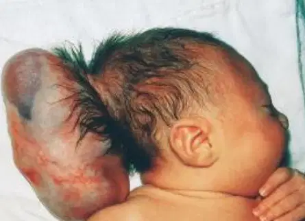
A newborn presents with a posterior midline skull mass. Imaging reveals herniation of meninges and brain tissue through a bony defect, as shown in the image. Long-term neurologic outcome in this patient most strongly depends on which variable?
Size of bony defect
Degree of CSF leakage
Presence of vertebral anomalies
Timing of surgical closure
Amount of herniated neural tissue

A child has the physical finding shown in the image but normal motor function and bowel/bladder control. Which prenatal screening result would most likely have been normal?
Maternal serum AFP
Fetal ultrasound of the spine
Vertebral segmentation
Closure of the caudal neuropore
Neural crest differentiation
Which of the following NTDs are open?
Anencephaly
Encephalocele
Meningocele
Spina Bifida Occulta
Cranioarchischisis
A patient suffers prolonged cardiac arrest with subsequent coma. MRI later reveals neuronal loss in the hippocampus and cerebellum, while other cortical regions are relatively spared.
Which property best explains the selective neuronal loss seen in this patient?
Regional variation in blood–brain barrier permeability
Differences in axonal myelination density
High metabolic demand and excitatory neurotransmitter activity
Preferential venous drainage patterns
Local microglial density
A patient presents 18 hours after severe hypoglycemia. Histology shows shrunken neurons with intensely eosinophilic cytoplasm and pyknotic nuclei.
This finding represents which pathologic process?
Apoptosis due to chronic neurodegeneration
Liquefactive necrosis with macrophage infiltration
Axonal reaction with regenerative sprouting
Earliest morphologic indicator of acute neuronal injury
Reactive gliosis
Following peripheral nerve transection, a neuron demonstrates an enlarged cell body, eccentric nucleus, and redistribution of Nissl substance.
Which cellular process is being demonstrated?
Neuronophagia
Central chromatolysis
Pseudolaminar necrosis
Wallerian degeneration
Autophagic vacuolization
A 27-year-old immunocompromised patient develops fever, seizures, and rapid neurologic decline. Brain biopsy shows neurons with intranuclear inclusions and chromatin pushed to the nuclear membrane. The patient deteriorates despite broad-spectrum antibiotics.
Which additional pathologic feature would most strongly support the underlying disease mechanism?
Progressive accumulation of α-synuclein in presynaptic terminals
Immune-mediated demyelination of periventricular white matter
Selective loss of Purkinje cells with Bergmann gliosis
Chronic astrocytic hypertrophy without neuronal loss
Lytic infection with neuronal necrosis and surrounding inflammation
An 82-year-old woman dies of pneumonia with no history of neurologic disease. Autopsy shows intracellular golden-brown granular pigment in cortical neurons, but no neuronal loss or gliosis.
Which interpretation best explains this finding?
Accumulation of indigestible oxidative byproducts over time
Marker of prior hypoxic-ischemic injury
Evidence of early neurodegenerative disease
Result of chronic microglial activation
Indicator of toxic metabolic exposure
A patient undergoes resection of a cystic brain lesion. Weeks later, imaging shows a firm, well-demarcated region surrounding the surgical cavity that limits spread of injury but contributes to seizure focus.
Which cellular response best explains both effects?
Neuronal apoptosis
Astrocytic hypertrophy and proliferation
Oligodendrocyte regeneration
Microglial phagocytosis
Endothelial fibrosis
A patient with end-stage liver disease develops confusion and asterixis. MRI shows symmetric involvement of deep gray nuclei. Symptoms improve with ammonia-lowering therapy.
Which histologic change best explains the reversibility of this condition?
Neuronal necrosis in hippocampal CA1
Demyelination of periventricular white matter
Microglial nodules with neuronophagia
Pseudolaminar cortical necrosis
Astrocytic swelling without permanent neuronal loss
After prolonged hypotension, a patient remains comatose. Early biopsy shows intensely eosinophilic neurons with shrunken nuclei. Surrounding glial cells are initially unremarkable.
Which downstream process would most likely be seen days later if the patient survives?
Recovery of affected neurons
Axonal sprouting from injured neurons
Oligodendrocyte remyelination
Astrocytic proliferation replacing necrotic tissue
Reversal of neuronal shrinkage
A patient with a decades-long history of seizures has a benign cerebellar tumor removed. Histology reveals elongated eosinophilic structures within astrocytic processes.
Which molecular feature best explains the presence of these structures?
Accumulation of misfolded synaptic proteins
Polyglucosan deposition from impaired glycogen metabolism
Stress-response protein accumulation in chronically reactive astrocytes
Neuronal cytoskeletal collapse
Immune complex deposition
A patient with untreated neurosyphilis has clusters of elongated glial nuclei surrounding foci of neuronal destruction.
Which mechanism best explains this finding?
Astrocytic scar formation
Microglial aggregation in response to focal neuronal injury
Oligodendrocyte apoptosis from ischemia
Reactive endothelial proliferation
Chronic lymphocytic infiltration
A child develops progressive ventriculomegaly weeks after viral infection of the CNS. Imaging shows irregular ventricular margins, but CSF production is normal and there is no aqueductal obstruction. Symptoms worsen when intraventricular pressure increases.
Which mechanism best explains how the original injury led to hydrocephalus?
Destruction of ependymal integrity with subependymal gliosis impairing CSF flow
Loss of cortical neurons reducing brain compliance
Microglial activation increasing CSF viscosity
Endothelial dysfunction causing increased CSF production
Astrocytic apoptosis reducing ventricular elasticity
A patient presents 12 days after a large ischemic stroke. Imaging shows decreasing edema but progressive neurologic deficits. Histology demonstrates macrophage infiltration and astrocytic proliferation at lesion margins.
Which future structural outcome is most likely if the patient survives?
Restoration of normal cortical architecture
Replacement with collagenous scar tissue
Formation of a fluid-filled cavity bordered by gliosis
Permanent cytotoxic edema
Progressive hemorrhagic expansion
Two patients suffer infarctions: one in myocardium, one in cerebral cortex. Only the cerebral lesion evolves into a cavitary defect.
Which combined property most directly accounts for this difference?
Higher vascular density and inflammatory infiltration
Lack of fibroblasts and high lipid enzymatic digestion
Increased neuronal susceptibility to hypoxia
Reduced blood–brain barrier repair
Excess astrocyte activation
A newborn has normal neural tube closure and intact early brain patterning but exhibits profound intellectual disability with a smooth cortical surface. Brainstem and cerebellum are relatively preserved.
Which embryologic process was most likely disrupted after neural tube formation?
Dorsoventral patterning by SHH
Proliferation of ventricular zone neuroblasts
Axonal pathfinding across commissures
Radial migration of neurons to the cortical plate
Synaptic pruning during postnatal life
A patient develops bilateral loss of pain and temperature sensation over the shoulders while proprioception and vibration remain intact. MRI shows a central spinal cord cavity that expands slowly.
Which fiber population is affected first, explaining the selective sensory loss?
Ipsilateral dorsal columns
Descending corticospinal tracts
Ventral horn motor neurons
Dorsal root ganglion neurons
Decussating spinothalamic fibers near the central canal
An infant presents with hydrocephalus, enlarged posterior fossa, and a cystic structure replacing the cerebellar vermis. CSF outflow from the fourth ventricle is impaired.
Which developmental failure best integrates all findings?
Failure of neural tube closure
Premature closure of cranial sutures
Degeneration of cerebellar hemispheres
Aqueductal stenosis from inflammation
Abnormal hindbrain segmentation and roof plate development
A patient survives carbon monoxide poisoning with minimal initial deficits. Two weeks later, he develops rigidity and bradykinesia.
Which mechanism best explains this delayed presentation?
Preferential injury to basal ganglia neurons with delayed cell death
Progressive hippocampal necrosis
Immune-mediated demyelination
Cortical laminar necrosis
Brainstem respiratory center injury
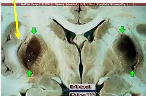
Two patients present with metabolic acidosis after ingestion of toxic alcohols. One develops visual loss with BL putamental necrosis shown in the image, while the other develops parkinsonian features.
Which structural difference best explains the divergence in neurologic findings?
Retinal ganglion cell susceptibility vs globus pallidus vulnerability
Blood–brain barrier permeability differences
Hepatic metabolism rate
CSF clearance mechanisms
Differential astrocyte response
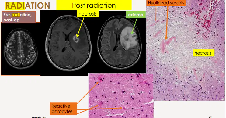
A patient treated with cranial radiation develops a mass lesion months later. Biopsy shows intramural fibrinoid necrosis, adjacent white matter edema, hyalinized vessels, and reactive astrocytes.
Which reasoning most strongly favors radiation injury rather than neoplastic recurrence?
Time course since therapy
Presence of necrosis
Vascular injury with white-matter predominance and minimal mitotic activity
Ring-enhancing lesion on MRI
Perilesional edema
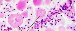
An 88-year-old woman with no neurologic disease dies of pneumonia. Autopsy reveals round, basophilic, laminated structures located predominantly in perivascular and subpial regions of the brain. There is no associated neuronal loss or gliosis. (Check structure name in explanation)
Age-related accumulation of polyglucosan material without clinical consequence
Chronic astrocytic stress response with accumulation of heat-shock proteins
Residual products of prior hypoxic-ischemic injury
Early manifestation of neurodegenerative disease
Microglial phagocytosis of apoptotic neurons
Two brain specimens show intracellular inclusions:
-Specimen A shows elongated, eosinophilic structures within astrocytic processes in a patient with a long-standing cerebellar tumor
-Specimen B shows round, laminated bodies in subpial regions of an elderly patient without neurologic disease
Which molecular distinction best differentiates the inclusions in Specimen A from those in Specimen B?
(Check structure name in explanation)
Presence of polyglucosan polymers
Association with neuronal cytoskeleton collapse
Accumulation of stress-response proteins in reactive astrocytes
Microglial origin following phagocytosis
Deposition of immune complexes
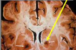
A patient experiences prolonged systemic hypotension due to CO poisoning. Days later, MRI shows ribbon-like cortical injury affecting layers III–V bilaterally, sparing deeper white matter.
Which physiologic principle best explains this injury pattern?
Preferential embolic distribution to cortical vessels
Bilateral globus pallidi necrosis
Breakdown of the blood–brain barrier at the gray–white junction
Venous infarction with cortical congestion
Immune-mediated demyelination
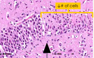
Which clinical scenario is most likely to produce pseudolaminar cortical necrosis of Sommer's sector of the hippocampus and Purkinje cells rather than a focal infarct?
Thromboembolism of the middle cerebral artery
Lacunar infarction of penetrating arteries
Hemorrhage from an arteriovenous malformation
Prolonged cardiac arrest with delayed resuscitation
Vertebral artery dissection
A patient with a slowly progressive neurodegenerative condition shows swollen axons filled with disorganized neurofilaments and organelles on histology.
Which underlying mechanism best explains this finding?
Acute neuronal necrosis
Failure of axonal transport leading to cytoskeletal accumulation
Immune-mediated myelin destruction
Reactive astrocytic proliferation
Microglial phagocytosis
A patient develops hydrocephalus after viral ventriculitis. Biopsy shows denudation of the ventricular lining with underlying astrocytic proliferation but no mitotic figures or rosette formation.
Which reasoning best excludes ependymoma?
Presence of hydrocephalus
Location near ventricles
Age of the patient
CSF flow obstruction
Absence of neoplastic cellular architecture
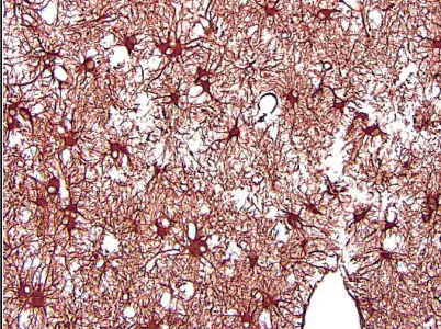
In this image, what does glial fibrillary acidic protein (GFAP) indicate?
Reactive oligodendrocytes in response to inflammation
Fibrillar eosinophilic material, indicative of axons and dendrites
Prominent nucleoli with basophilic granular cytoplasm
Stellate astrocytic processes during reactive gliosis
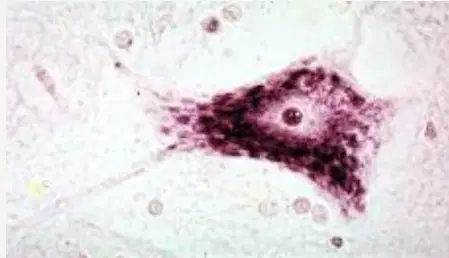
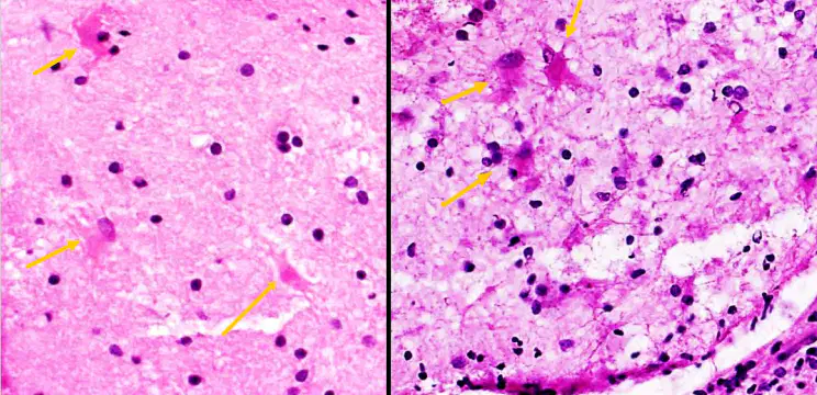
In the image shown, what histological feature(s) indicate this is a red neuron associated with an acute injury response?
Pykinosis, Nissl body loss, and eosinophilic cytoplasm
Enlarged/rounded cell body with peripheral displacement of the nucleus
Small, round, condensed nuclei with unstained cytoplasm
Large nucleus with basophilic granular cytoplasm
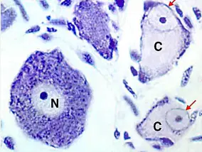
In the image shown, what process did the neurons labeled "C" undergo due to chronic injury/degeneration of the CNS?
Lipofuscin
Perikaryon vacuolization
Reactive gliosis
Central chromatolysis
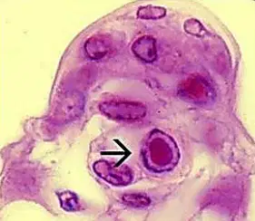
A 24-year-old man with untreated HIV presents with fever, focal seizures, and rapidly progressive confusion. MRI shows hemorrhagic necrosis involving the medial temporal lobes. Brain biopsy demonstrates intranuclear inclusions surrounded by chromatin margination in neurons and glial cells (Cowdry Type A).
Which pathophysiologic mechanism most directly explains the neuronal injury seen in this patient?
Immune-complex deposition within cerebral vessels
Lytic viral replication causing nuclear structural disruption
Retrograde axonal transport with synaptic failure
Mitochondrial dysfunction from viral toxins
Demyelination mediated by CD8⁺ T cells
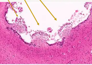
A 6-week-old infant born to a mother with no prenatal care presents with seizures, hearing loss, and petechiae. Neuroimaging shows periventricular calcifications and ventriculomegaly. Histologic examination reveals enlarged cells with a dense intranuclear inclusion surrounded by a clear halo (Owl's eye; not this image).
As shown in the image, which cell type is most directly responsible for propagation of this infection within the CNS?
Neurons of the hippocampus
Oligodendrocytes of periventricular white matter
Astrocytes involved in glutamate recycling
Ependymal and endothelial cells lining the ventricles
Microglia forming nodules
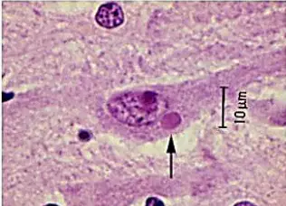
A 32-year-old man presents with agitation, hydrophobia, and autonomic instability several weeks after a bat bite. Despite intensive care, he rapidly deteriorates and dies. Postmortem examination reveals eosinophilic cytoplasmic inclusions within pyramidal neurons of the hippocampus and Purkinje cells of the cerebellum (Negri bodies).
Which viral property most directly explains the localization of these inclusions?
Retrograde axonal transport from peripheral nerves
Preferential infection of dividing cells
Hematogenous spread through fenestrated capillaries
Viral integration into host nuclear DNA
Antibody-dependent enhancement
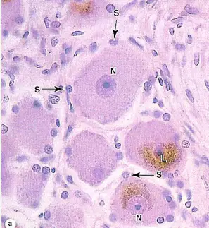
An 84-year-old man with no history of neurologic disease dies from complications of pneumonia. Autopsy of the brain reveals neurons containing coarse, yellow-brown cytoplasmic granules. There is no associated gliosis, inflammation, or neuronal loss, and surrounding tissue architecture is preserved.
Which mechanism best explains the presence of these granules without associated neurologic dysfunction?
Chronic hypoxic injury resulting in irreversible neuronal damage
Viral replication with residual inclusion formation
Immune-mediated clearance of apoptotic neurons
Failure of lysosomal degradation leading to accumulation of indigestible oxidative byproducts
Pathologic protein aggregation associated with neurodegenerative disease
A 52-year-old man presents with new-onset focal seizures several weeks after a traumatic brain injury. MRI shows a resolving contusion without evidence of tumor. A biopsy taken from the lesion margin demonstrates the cells shown in the image. Which function of the cells shown most directly explains both the patient’s seizure activity and the well-demarcated nature of the lesion?
Phagocytosis of necrotic neurons to prevent inflammation
Proliferation and hypertrophy forming a glial scar that isolates injured tissue
Remyelination of damaged axons
Production of inflammatory cytokines that promote neuronal death
Differentiation into oligodendrocytes to restore conduction
A child presents with progressive headaches and visual difficulties. Neuroimaging reveals a suprasellar mass with cystic components compressing adjacent structures in the region of the pituitary. Histologic examination shows an epithelial tumor consistent with a lesion derived from developmental remnants of the Rathke pouch rather than mature neural tissue.
Which embryologic origin best explains the location and growth pattern of this lesion?
Neuroectoderm of the diencephalon
Mesoderm of the skull base
Oral ectoderm associated with pituitary development
Neural crest cells migrating to the sella
Endodermal derivatives of the foregut
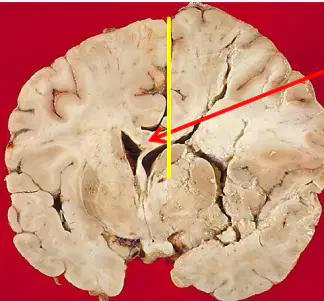
A 71-year-old man develops sudden-onset left face/arm weakness and neglect. CT head shows a large acute right hemispheric infarct with edema. Over the next 24 hours he becomes progressively somnolent and repeat imaging shows midline shift with the cingulate gyrus pushed beneath the falx. Which additional finding is most consistent with this process?
Transtenorial herniation compressing ipsilateral CN III
Subfalcine herniation with cingulate gyrus displacement across the midline
Tonsillar herniation causing medullary compression
Intraventricular hemorrhage due to rupture of Charcot-Bouchard microaneurysms
Central pontine myelinolysis
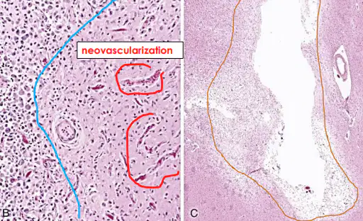
A 66-year-old woman has an ischemic stroke. Ten days later she dies from an unrelated pulmonary embolism. Histology of the infarcted region shows prominent foamy macrophages with surrounding reactive gliosis and neovascularization. Which cellular process best matches the expected timing of these histologic findings?
Neutrophilic infiltration beginning at infarct margins within hours
Formation of a collagenous scar within 3–5 days
Early “red neurons” as the best indicator of chronic neuronal injury
Caseous necrosis due to ischemia
Foamy macrophages with reactive gliosis peaking around ~10 days
Neuroimaging reveals a crescent-shaped extra-axial fluid collection over the cerebral hemisphere that is isodense with CSF and does not demonstrate blood products (hygroma). The underlying brain parenchyma appears structurally intact.
Which anatomic disruption most directly accounts for this finding?
Rupture of cortical arteries supplying the subdural space
Failure of arachnoid granulations to absorb CSF
Disruption of the arachnoid membrane allowing CSF to enter the subdural space
Liquefactive necrosis of adjacent cortex
Progressive venous congestion from bridging vein thrombosis
A neonate with trisomy 13/SHH defect is found to have incomplete separation of the cerebral hemispheres, resulting in a single forebrain structure. Which additional abnormality is associated with this diagnosis?
Developmental delay
Seizures
Dilated occipital horns
Midline facial abnormalities
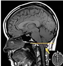
Neuroimaging demonstrates downward displacement of cerebellar structures through the foramen magnum, resulting in compression at the cervicomedullary junction.
This abnormality is best categorized as a defect of which of the following?
Forebrain cleavage
Commissural fiber development
Posterior fossa structural development
Neuronal migration
Cortical lamination
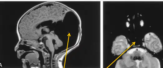
What diagnosis is associated with cerebellar vermis agenesis leading to obstructed CSF flow out of the 4th ventricle/large posterior fossa cyst (brainstem nuclei dysplasia)?
Arnold-Chiari malformation
Syringomyelia
Dandy-Walker malformation
Joubert syndrome
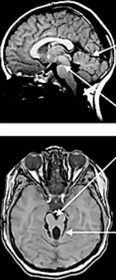
Which diagnosis is associated with vermis hypoplasia, elongated cerebellar peduncles, altered brainstem shape, and "molar tooth sign"?
Syringomyelia
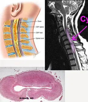
Which diagnosis is associated with central SPC enlargement due to fluid-filled cleft-like cavity (syrinx), BL pain and temp loss of UEs due to central white commissure impedance, and kyphoscoliosis?
Chiari malformation
Joubert syndrome
Syringomyelia/Hydromyelia
Dandy-Walker malformation
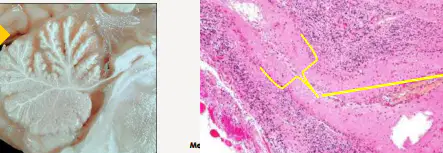
Which toxin is associated with granule cell loss in the anterior vermis (left image), cerebellar issues, and Purkinje cell loss and Bergmann gliosis between the depleted cell layer and the molecular layer (right image)?
Methanol
Ethanol
Carbon monoxide
Radiation
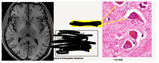
In patients with hepatic encephalopathy, which cell type is responsible for the glial response due to increased ammonia in the BG, cortex, and other subcortical gray matter regions (histo image)? Also, which area, as shown on the scan, experiences BL symmetrical hyperintensity?
Alzheimer Type I cells ; frontal lobe
Alzheimer Type II cells ; Globus Pallidi
Oligodendrocytes ; occipital lobe
Astrocytes ; Globus Pallidi
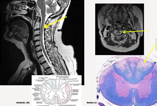
A patient with vitamin B12 deficiency begins experiencing paresthesias and BLE ataxia due to what defect affecting ascending posterior columns and descending pyramidal tracts, as shown in the images?
Myelination defect
Astrocyte defect
Defective neuronal migration
CSF flow defect
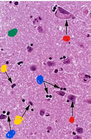
Associate the correct color with the cell type shown on the H&E stain.
Red - neurones
Blue - oligodendrocytes
Yellow - astrocytes
Green - neuropil
Red - oligodendrocytes
Blue - astrocytes
Yellow - neurones
Green - neuropil
Red - neuropil
Blue - oligodendrocytes
Yellow - astrocytes
Green - neurones
Red - astrocytes
Blue - neurones
Yellow - neuropil
Green - oligodendrocytes

