
 Worksheets
Nervous System
Total questions: 62
Worksheet time: 3590secs
This divides your brain into two hemispheres:
Cerebellum
Pons
Corpus Collosum
Medulla
The lobe that processes what I see is the
frontal
parietal
occipital
temporal
This part of my brain is related to balance and muscle coordination
brain stem
pons
midbrain
cerebellum






What do Dendrites do?
Intakes information from other neurons
Produces fatty tissue for the Myelin Sheath
Sends information to the next cell
Controls the speed of electrical signal through the axon
What do Schwann Cells do?
Intakes information from other neurons
Produces fatty tissue for the Myelin Sheath
Sends information to the next cell
Controls the speed of electrical signal through the axon

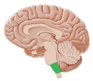
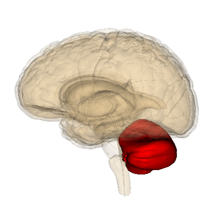
What structure is being highlighted?
cerebrum
cerebellum
pons
brain stem
Which part of the neuron hold the nucleus?
cell body
axon
neuron
axon terminal
What structures make up the Central Nervous System?
Brain
Spinal cord
nerves
Brain, spinal cord and nerves
Which structure is composed of these 3 parts: pons, midbrain, and medulla oblongata?
CNS
Brain Stem
PNS
Cerebellum
What is another name for a nerve?
nerve
brain
signal sender
neuron

If you are having trouble with your BALANCE and COORDINATION, which part of your brain should be checked?
cerebrum
cerebellum
brain stem
spinal cord
Select the VOLUNTARY action.


Select the INVOLUNTARY action.


Space between myelin sheaths
node of ranvier
Nissl body
synapse
dendrite
What are the 2 major divisions of the peripheral nervous system (PNS)?
Sensory & Motor
Peripheral & Central
Somatic & Autonomic
Brain & Spinal Cord
What is the purpose of the myelin sheath on the axon?
To maintain temperature
To improve conductivity by insulating axon
To protect the axon
To release neurotransmitters

Elevated ridges on the surface of the brain are called:
Gyrus
Fissures
Lobes
Sulcus

These supporting cells (neuroglia) contain cilia and found in the lining of brain's ventricles
astrocyte
ependymal cells
Schwann cells
satellite cells

Star-shaped glial cells that form part of the blood–brain barrier
astrocyte
ependymal cells
Schwann cells
satellite cells
Star-shaped glial cells that form part of the blood–brain barrier
astrocyte
ependymal cells
Schwann cells
satellite cells


In the figure above, what is occurring at step #4?
Neuron receives enough stimulation (15 mV), threshold is reached (-55mV)
Na+ voltage channels open allowing Na+ to diffuse inward, depolarizing the membrane
K+ voltage channels open and Na+ voltage channels open
K+ voltage channels open allowing K+ to diffuse out, repolarizing the membrane.
Hamsters. Massive amounts of hamsters



_______ is characterized by inappropriate words and inability to understand spoken language.
Grapheme-Color synesthesia
Synesthesia
Wernicke's aphasia
Broca's aphasia

