
 Worksheets
EKG CET Review #2
Total questions: 100
Worksheet time: 8hrs 20mins
What is the most common rhythm that could produce blood clots.
Atrial fibrillation
Wandering baseline
Second Degree Heart Block
None of these
On an ECG, the P wave represents __________________________.
Contraction of the Atria
Contration of the Ventricle
The pause at the AV node
The impulse traveling through the bundle branches
What is the term for rapid heartbeat?
Angina Pectoris
Fibrillations
Tachycardia
Ischemia
The most accurate way to determine the rate of any rhythm strip is the
Block Method (300, 150, 100, etc.)
6 inch method (number of complexes between 6 inches)
Counting complexes for 30 seconds and multiplying by 2
Estimation Method
When the electrical impulse originates from somewhere other than the SA node, it is said to be
blanket firing
rapid firing
ectopic firing
subdued firing
Which of these represents an entire cardiac cycle?
QRS complex
P-R complex
R-R interval
QRST complex
Which of these could cause sinus arrythmias?
An inexperienced technician
Changes in breathing
A heart block
Not winning Homecoming Queen

Which of the following best describes this strip?
Tachycardia
Normal Sinus Rhythm
Bradycardia
Asystole
Use the rule of 300 to determine approximate Heart Rate.
300/6 = 50
300/3.5 = 75
300/2 = 150
Note enough information.

Which of the following best describes this strip?
Tachycardia
Normal Sinus Rhythm
Bradycardia
Asystole

Use the rule of 300 to determine approximate Heart Rate.
300/6 = 50
300/3.5 = 75
300/1.5 = 200
Not enough information.

Which of the following best describes this strip?
Tachycardia
Normal Sinus Rhythm
Bradycardia
Asystole
If there is a flat P wave which structures in the heart are damaged?
SA/AV node
Bicuspid valve
Bundle of His
Purkinje fibers


What does depolarization result in?
relaxation
contraction
repulsion
attraction
Which node is considered the "pacemaker" of the heart?
SA
AV

Where are the Purkinje Fibers located?
A
B
C
D
Blood enters the heart from the _______?
Superior and inferior vena cava
Bicuspid and Tricuspid
aorta
pulmonary artery
The normal heart rhythm is called?
Sinus brady
ventricle fibrillation
Normal sinus rhythm
Junctional Rhytms
Normal heart rate for an adult is?
20 to 100/min
40 to 120/min
60 to 100/min
20 to 60/min
The SA Node is known as?
Pacemaker
gatekeeper
ventricle
cardiac muscle
The SA Node generates impulses at?
60-100 times per minute
60-40 times per minute
40-20 time per minute
20-100 times per minutes
which is the correct order?
SA Node, AV Node, Bundle of His, Lt and Rt Bundle Branches, Purkinje Fibers
AV Node, SA Node, Lt and Rt Bundle Branches, Bundle of His, Purkinje Fibers
Bundle of His, Lt and Rt Bundle Branches, Bundles of His, Purkinje Fibers, AV Node, SA Node
Purkinje Fibers, Bundle of His, AV Node, Lt and Rt Bundle Branches
AV Node can fire at a rate of?
60-100 times per minute
40-60 times per minute
40-20 times per minute
60-20 times per minute
The QRS complex is normally __________ seconds in duration
0.06 to 0.10
0.04 to 0.10
0.08 to 0.12
0.10 to 0.12
the AV impulse is
60-100
40-60
20-40
30-70
the impulse of SA node is
60-100
40-60
20-40
30-70
the purkinje fibers impulse is
60-100
40-60
20-40
30-70
The nurse is reviewing a patient’s electrocardiogram. Which wave should the nurse recognize as being the first positive wave in a normal cardiac cycle?
P wave
Q wave
R wave
S wave
A patient is experiencing electrocardiogram changes. Which wave should the nurse recognize as representing ventricular repolarization?
Q wave
R wave
S wave
T wave
After reviewing an electrocardiogram the nurse determines that an electrical impulse originated in a patient’s the sinoatrial node. What did the nurse see on this tracing?
An upright T wave
An inverted T wave
A positive P wave before a QRS complex
A negative P wave before a QRS complex
The nurse is reviewing cardiac anatomy with a patient scheduled for pacemaker insertion. Where should the nurse identify the location of the sinus node for the patient?
Left atrium
Right atrium
Left ventricle
Right ventricle
A patient has a normal heartbeat of 72 beats per minute. What area of the conduction system should the nurse consider as functioning normally in this patient?
Bundle of His
Sinoatrial node
Purkinje’s fibers
Atrioventricular node
The nurse is determining the functioning of a patient’s sinoatrial node. How many times per minute should the nurse expect the node to normally fire?
20 to 40
25 to 55
40 to 85
60 to 100
The nurse determines that a patient’s electrocardiogram is demonstrating normal sinus rhythm. What did the nurse observe to come to this conclusion? (Select all that apply.)
Regular rhythm
P wave before each QRS complex
Wide QRS intervals
Heart rate 83 beats per minute
The nurse is analyzing a client’s electrocardiogram tracing. Which of the following complexes is not normally seen on an electrocardiogram tracing?
P wave
QRS complex
T wave
U wave
Blood pressure is recorded as
Hydrostatic /diastolic
Diastolic/ hydrostatic
Systolic/diastolic
Diastolic/systolic
Pressure exerted on the walls of the blood vessels is termed
Hypertension
Vesicular pressure
Blood Pressure
Heart pressure
Blood pressure is used to determine
electrical current though the heart
function of coronary artery
if adequate blood is getting to tissues.
Speed of blood through veins


Normal blood pressure is...
120/80 mmHg
80/120 mmHg
140/90 mmHg
80/140 mmHg

High blood pressure is also called ...
diastolic pressure
systolic pressure
hypotension
hypertension

Person has a blood pressure of 140 mmHg/ 90 mmHg. How would you categorize their blood pressure?
Normal range
prehypertension
hypertension
hypotension
Highest pressure against blood vessels. Represented by first "thump" sound
diastolic pressure
systolic pressure
hypotension
hyperstension
lowest pressure against the blood vessels of the body. Measured when the "thump" stops
diastolic pressure
systolic pressure
hypotension
hypertension
During depolarization, the inside of the cell becomes _____.
more negative
less negative
During repolarization, the inside of the cell becomes _____.
more negative
less negative
EKG Technicians must be responsible for and to recognize or identify all of the following, EXCEPT:
Patient Assessment
Palpitations
Diabetes
Pulmonary Embolism
This individual uses the EKG as their first tool of investigation.
EKG Technician
Cardiologist
ER Physician
ICU Physician
The result is a dramatic drop in blood pressure or equal systolic/diastolic , eg. 120/115 (fatal).
Atrial Hypertrophy
Cardiac Tamponade
Abberant EKG Rhythms
Impulse Re-entry Cycles
If too high or too low, they can affect your heart's electrical impulses and contribute to arrhythmia development
Abberant EKG Rhythms
Palpitations
Cardiac Electrolytes
Ectopic Beats
A sensation that the heart is racing, pounding, fluttering, or skipping a beat, often bothersome, but hardly ever a sign of heart disease.
Ectopic Beats
Palpitations
Impulse Re-entry Cycles
Aberrant EKG Rhythms
Plays a critical role in the transmission of electrical impulses in the heart (T Wave).
Sodium
Potassium
Calcium
Magnesium
Too much may cause arrhythmias, MI, and plaque build-up.
Sodium
Potassium
Calcium
Magnesium
The monitor will show electrical activity in the heart, but the patient will have no palpable pulse.
Cardiac Tamponade
Agonal Rhythm
Asystole
PEA
Plays a critical role in the transmission of electrical impulses in the heart (QRS Complex).
Sodium
Potassium
Calcium
Magnesium
They are common and may occur for no known reason.
Aberrant EKG Rhythms
Ectopic Beats
Palpitations
Impulse Re-entry Cycles
Affects QT Interval
Sodium
Potassium
Calcium
Magnesium

Interpret this Rhythm:
3rd Degree AV Block
Agonal Rhythm
Asystole
PEA
An abnormally low concentration of oxygen in the blood.
Apnea
Dyspnea
Hypoxemia
Low Pulse Oximetry
Used to treat Torsades de Pointes
Sodium
Potassium
Calcium
Magnesium
Help trigger and conduct the electrical impulses in your heart
Impulse Re-entry Cycles
Ectopic Beats
Aberrant EKG Rhythms
Cardiac Electrolytes
A condition in which one or more arteries in the lungs become blocked by a blood clot.
PE
MI
CVA
PEA
Choose the 3rd degree heart block:
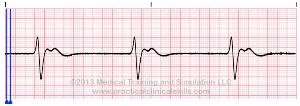
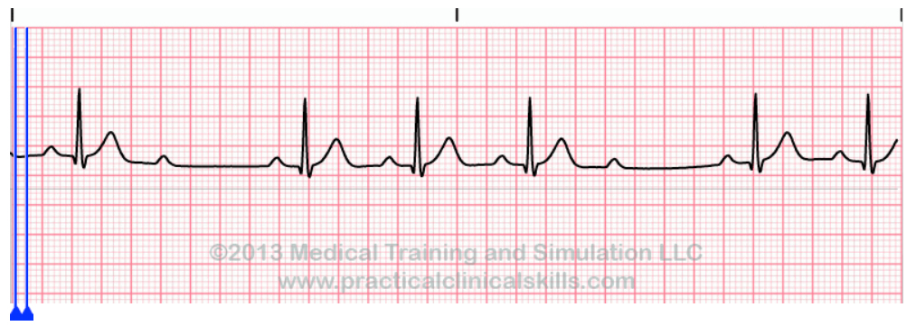
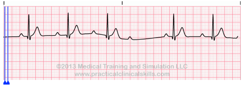
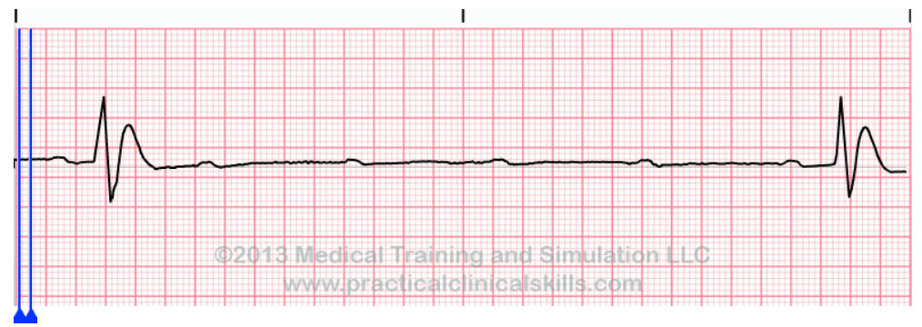
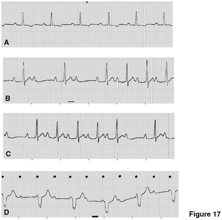
WHICH STRIP IS CONSTANT WITH SINUS RHYTHM WITH FIRST DEGREE AV BLOCK?
A
B
C
D
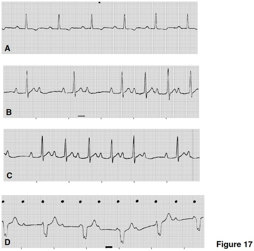
Which strip in Figure 17 is consistent with Mobitz I second-degree AV block (Wenckebach)?
A
B
C
D
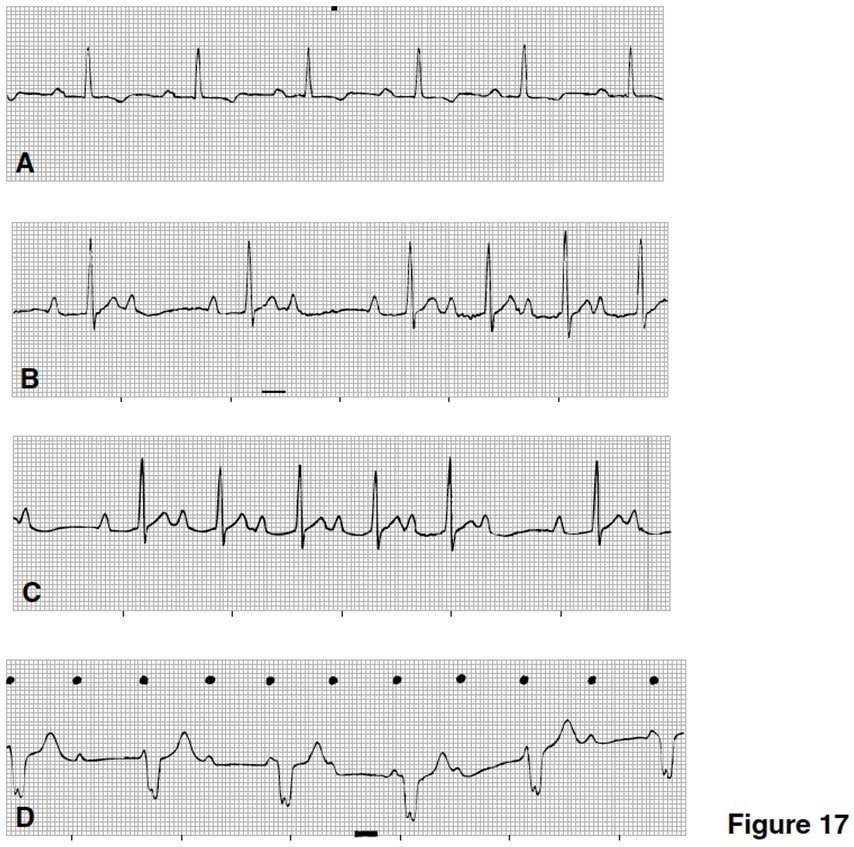
Which strip in Figure 17 is consistent with Mobitz II second-degree AV block?
A
B
C
D
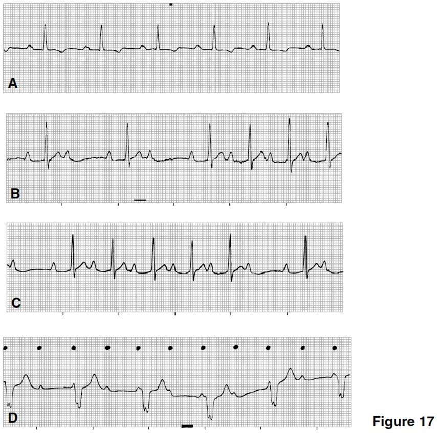
Which strip in Figure 17 is consistent with third-degree AV block?
A
B
C
D
AV dissociation is a hallmark of
first-degree AV block.
Mobitz I second-degree AV block (Wenckebach).
Mobitz II second-degree AV block.
third-degree AV block.
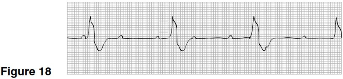
In Figure 18, which of the heart's pacemakers is controlling the ventricle?
sinus node
atrium
AV node
ventricle
Treatment for first-degree AV block involves
atropine
digitalis
electrical shock to the heart
removing the cause
Treatment for third-degree AV block usually includes
pacemaker
lidocaine
digitalis
amiodarone
All AV blocks are caused by a block at the bundle branches.
true
false
In AV blocks, the site of the block is either the AV node or the bundle branches.
true
false
In first-degree AV block, all sinus impulses do get through to the ventricles.
true
false
pacemakers treat ______________?
Mobits II, and 3rd degree AV Blocks
Mobitz I and MobitzII
Only 3rd degree blocks
Only Wenchebachs
Mobitz I can progress to 3rd degree AV block
true
false
The origin of the P waves in AV blocks is the ________
AV node
sinus node
Bundle branches
U wave
The AV block that has gradually prolonging PR intervals until a QRS is blocked is ________.
Wenckebach
First degree
3rd degree
In ______________________ all sinus impulses get through to the ventricles.
first degree AV block
3rd degree AV block
2nd degree Mobitz I
2nd degree Mobitz II

Why is this 3rd degree heart block?
because there is artial fibrillation
too much time between the p wave and qrs complex
The p, qrs and t waves show no pattern
all of the above
What type of heart block is this?
2nd degree
1st degree
3rd degree
all of the above

Why is this 1st degree heart block?
The distance between the P wave and qrs complex is too long
The distance between the P wave and qrs complex is too short
There is not regular pattern
The QRS complex is too big

What type of heart block is this?
2nd degree
1st degree
3rd degree
all of the above
What type of heart block is this?
2nd degree
1st degree
3rd degree
The paper on an ECG normally moves at:
50mm/sec
75mm/sec
25cm/sec
25mm/sec
The width of each small box represents:
0.04 seconds
0.02 seconds
0.40 seconds
0.20 seconds
What is the time frame occupied by one lead on a regular 12-lead ECG?
0.3 seconds
3 seconds
13 seconds
30 seconds
The small boxes on ECG paper measure:
1 cm by 0.20 seconds
1mm by 0.20 seconds
1cm by 0.04 seconds
1mm by 0.04 seconds
The big boxes on the ECG paper measure:
5mm by 0.20 seconds
1mm by 0.20 seconds
5mm by 0.04 seconds
1mm by 0.04 seconds
A wave that is 10 small boxes high and three small boxes wide is described as being:
1.0mm by 0.3 seconds
10mm by 0.12 seconds
12mm by 0.3 seconds
12mm by 0.10 seconds
A distance of 2 big boxes and 2 little boxes wide is described as being:
22mm wide
0.22 seconds wide
0.48 seconds wide
4.8 seconds wide

