
 Worksheets
Ch 9 Transport in animals
Total questions: 45
Worksheet time: 35mins
Which type of cell is found in the greatest numbers in blood?
Red blood cell
White blood cell
Platelets
What happens to breathing and heart rate during exercise?
Breathing rate increases and heart rate decreases
They both increase
They both decrease
Why is the left wall of the heart have a thicker layer of muscle than the right side?
To stop the blood flowing backwards
To push the blood with more force at high pressure so it goes to all body cells.
To push the blood at high pressure to the lungs.
What is the name of the liquid part of the blood?
Platelets
Plasma
Red blood cells
What do platelets do?
They stick together and make fibres that make scabs (blood clots) that stop pathogens entering the blood or stop you bleeding to death.
They form your immune system and destroy pathogens.
They carry oxygen around the body attached to haemoglobin.
What do phagocytes do?
Engulf (surround) microbes and digest them.
Produce antibodies.
Release a toxin that kills pathogens.
Why do arteries have a thick muscle layer?
To provide energy for the blood vessel.
To prevent the blood flowing backwards.
To help withstand the high pressure blood, also to contract to control blood flow.
List the structures in the circulatory system the blood travels through from vena cava to aorta.
Vena cava, lungs, pulmonary vein, left atrium, left ventricle, aorta, right atrium, right ventricle, pulmonary artery,
Vena cava, left atrium, right ventricle, pulmonary vein lungs, pulmonary artery, right atrium, left ventricle, aorta
Vena cava, right atrium, right ventricle, pulmonary artery, lungs, pulmonary vein, left atrium, left ventricle, aorta
Why do veins and the heart have valves?
To stop the backflow of blood
To contract to push the blood under high pressure
To cause the pulse
What type of blood vessel surround alveoli and other tissues in the body?
Capillaries
Arteries
Veins
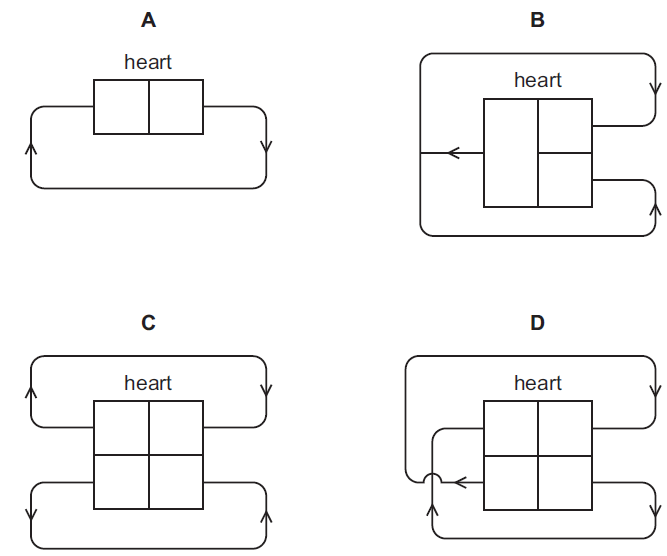
Which diagram shows the human double circulatory system?
A
B
C
D
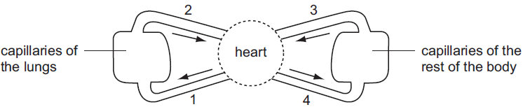
The diagram shows a double circulatory system. Which vessels carry oxygenated blood?
1 and 2
1 and 4
2 and 3
2 and 4

The diagram shows some parts from the blood of a mammal.
Which part would contain the breakdown products of bacterial cells?
A
B
C
D
Which describe the structure and function of a red blood cell?
structure: cell contents are dead
function: transport of water
structure: has a nucleus
function: produces antibodies
structure: has cilia
function: moves particles in the respiratory tract
structure: has no nucleus
function: transport of substances

The photograph shows human blood cells as seen under a microscope at high power.
Which are red cells?
P and Q
Q and R
R and S
S and P

The photograph shows two blood cells, X and Y.
What are the functions of cells X and Y?
cell X: carries out phagocytosis
cell Y: carries oxygen
cell X: carries oxygen
cell Y: produces antibodies
cell X: carries oxygen
cell Y: carries carbon dioxide
cell X: produces antibodies
cell Y: carries oxygen
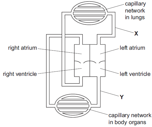
The diagram shows the circulatory system of a mammal.
What describes the blood in vessels X and Y?
X: deoxygenated
Y: deoxygenated
X: deoxygenated
Y:oxygenated
X:oxygenated
Y: deoxygenated
X: oxygenated
Y: oxygenated

The diagram shows human blood as seen through a light microscope.
A person’s blood is unable to clot.
Which component of the blood is not functioning properly?
A
B
C
D
Which chamber of the heart has the most muscular wall?
left atrium
left ventricle
right atrium
right ventricle
Which is a function of some white blood cells?
to carry glucose
to carry oxygen
to produce antibiotics
to produce antibodies
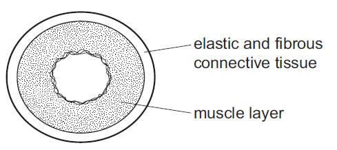
The diagram shows a cross-section through a human blood vessel.
Which type of blood vessel does the diagram show?
an artery
a capillary
a vein
a ventricle

The diagram shows human blood cells, as seen under a microscope.
What is the function of cell X?
to carry glucose
to carry oxygen
to defend against disease
to make the blood clot

The diagrams show some components of the blood of a mammal.
Which component causes the blood to start clotting?
A
B
C
D
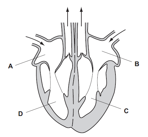
The diagram shows the directions of blood flow through a heart.
Which chamber of the heart receives blood directly from the lungs?
A
B
C
D
Name the main artery of the human body.
Name the vein that links the small intestine (ileum) to the liver.

Which of the above shows a vein? Write TWO reasons for your answer.
In the heart which ventricle has the thickest wall? Give a reason for your answer.
In which way is the pulmonary artery different from the other arteries?

A
B
C
D

A
B
C
D

A
B
C
D

A
B
C
D
Which blood vessel carries blood at the highest pressure?
Arteries
Veins
Capillaries
Which blood vessel contains valves?
Arteries
Veins
Capillaries
Which blood vessel carries blood away from the heart?
Arteries
Veins
Capillaries
Which blood vessel carries blood towards the heart?
Arteries
Veins
Capillaries
Which component of blood contains phagocytes and lymphocytes?
Red Blood Cells
White Blood Cells
Platelets
Plasma
Which component of blood is responsible for transporting oxygen?
Red Blood Cells
White Blood Cells
Platelets
Plasma
What is the function of phagocytes?
Transport oxygen around the body
Engulf and digest pathogens
Produce antibodies which destroy pathogens
Clot together to prevent bleeding
What blood vessel transports deoxygenated blood from the body to the heart?
Vena Cava
Aorta
Pulmonary Vein
Pulmonary Artery
What blood vessel provides the heart with its own blood supply?
Vena Cava
Aorta
Coronary Artery
Pulmonary Artery
Which chamber of the heart receives blood from the vena cava?
Right Atrium
Right Ventricle
Left Atrium
Left Ventricle

A
B
C
D
Which chamber of the heart sends blood to the lungs?
Right Atrium
Right Ventricle
Left Atrium
Left Ventricle

