
NEW
Font size
 Worksheets
Integumentary System
Total questions: 70
Worksheet time: 53mins

Identify the structure indicated
Hair shaft
Hair root
Hair folicle
Hair papilla

Identify the structure indicated
Hair shaft
Hair root
Hair folicle
Hair papilla

Identify the structure indicated
Hair shaft
Hair root
Hair folicle
Hair papilla

Identify the structure indicated by the red arrow
Blood vessels
Dermal Papillae
Hair Papilla
Adipose tissue

Identify the structure indicated by the red arrow
Sebaceous Gland
Hair follicle
Arrector pili muscle
Sudoriferous gland

Identify the structure indicated by the red arrow
blood vessels
sebaceous glands
sudoriferous glands
nerves

Identify the structure indicated by the red arrow
blood vessels
sebaceous glands
sudoriferous glands
nerves

Identify the structure indicated by the red arrow
blood vessels
sebaceous glands
sudoriferous glands
nerves

Identify the structure indicated by the red arrow
blood vessels
sebaceous glands
sudoriferous glands
nerves

Identify the structure indicated by the red arrow
epidermis
dermis
dermal papillae
hypodermis/subcutaneous

Identify the structure indicated by the red arrow
epidermis
dermis
dermal papillae
hypodermis/subcutaneous

Identify the structure indicated by the red arrow
epidermis
dermis
dermal papillae
hypodermis/subcutaneous

Identify the structure indicated by the red arrow
epidermis
dermis
dermal papillae
hypodermis/subcutaneous

The protective dead layer of cells of the outer epidermis is called the:
stratum spinosum
stratum granulosum
stratum corneum
stratum basale
Melanocytes lie primarily within the _________________ of the skin
stratum corneum
dermis
stratum lucidum
stratum basale

What is one important function for melanin?
cause the skin to be cooler
increase the amount of blood vessels
strengthen the skin or eyes
absorb harmful radiation

The epidermal ridges or fingerprints are probably caused by the _____.
genes in the epidermis
dermal papillae
smooth muscle in dermis
amount of keratin in cells
The strength of the dermis would mainly be due to _____.
the amount of blood vessels
the distribution of elastic fibers
the presence of collagen fibers
the thickness of the layers
It is the body's __________________ glands that secretes an oily liquid that naturally moisturizes the skin.
sweat gland
eccrine
sebaceous gland
apocrine

The layer that binds skin to deep tissue layers.
Stratum Lucidum
Dermis
Stratum Granulosum
Reticular Layer
What skin disease is caused by too much exposure to the sun's ultra-violet radiation?
acne
dermatitis
skin cancer
rash
The epidermis is composed of what kind of cells?
Simple squamous epithelial
Stratified cuboidal epithelial
Stratified squamous epithelial
Transitional epithelial
What types of cells found in the epidermis contain nerve endings that are touch receptors?
Keratinocytes
Langerhans cells
Melanocytes
Merkel cells
What layer of the skin contains sweat glands?
Dermis
Epidermis
Hypodermis
Subcutaneous
What part of a hair can be seen?
Follicle
Matrix
Root
Shaft
What part of the hair provides cells for new growth?
Bulb
Matrix
Papilla
Root
Concerning skin color, which of these statements is NOT correctly matched?
Skin appears yellow—carotene present
Skin appears blue (cyanosis)—oxygenated blood
No skin pigmentation (albinism)—genetic disorder
Skin tans—increased melanin production
Skin color is due to a pigment called _____.
The "clear layer" of the epidermis that is only present in certain areas of the body (palms of the hands and soles of the feet) is called:
Stratum granulosum
Stratum spinosum
Stratum corneum
Stratum lucidum

Which type of cell is found in the hypodermis?
adipose tissue
keratinocytes
Langerhans (dendritic) cells
melanocytes
Merkel cells
Full thickness burns involve damage to the ______.
epidermis, only
epidermis, dermis, and hypodermis
epidermis and dermis, only
muscle, only
Psoriasis is a chronic inflammatory skin disease. It is most characteristic of a ___________ plaque.
raised
silvery
hardened
red
Which type of sweat gland is found in the arm pits and produces a foul odor due to bacteria decomposing organic wastes?
merocrine
apocrine
sebaceous
holocrine
Dermal tissue is made of areolar & dense connective tissue to give skin _________& _________.
Hairiness & oil
Rigidity & tensile strength
Sweat & oil
Toughness & elasticity
The pigment carotene can give a person a _________.
Black color
Red color
Orange/Yellow color
Pink color
Using the acronym ABCDE as it relates to skin cancer, A refers to
Atypical
Alright
Asymmetrical
Absence
Tiny openings at the skin’s surface are called ---
epidermis
pores
dermis
oil gland

How does sweating actually keep us cooler?
Through condensation
It's colder than the air outside
Through sublimation
Though evaporation
The part of the integumentary system that is found all over the body except for the palms, soles of the feet and lips
hair
muscles
skin
nails
A contagious fungal infection that affects the skin on the feet. It can also spread to the toenails and the hands.
sunburn
albinism
athlete's foot
warts
A group of inherited disorders where there is little or no production of the pigment melanin.
herpes
psoriasis
albinism
athlete's foot
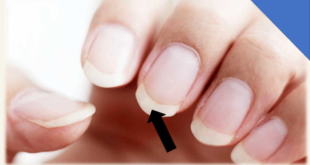
This is part of the nail plate that extends past the fingertip. It is referred as “distal edge”, that is shaped or trimmed in the nail care service.
Lunula
Free edge
Nail Groove
Cuticle
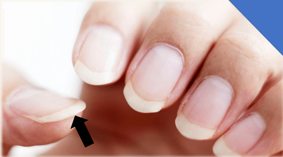
It is found at the end of the fingertip. This serves as protective seal or barrier from harmful bacteria and it is considered a sensitive part.
Hyponychium
Nail Groove
Mantle
Nail Wall
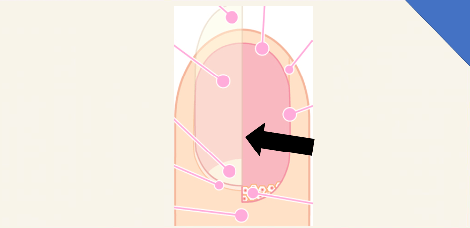
This part of the nail where the nail plate rests on. It is also a continuation of the matrix and it is abundantly supplied with blood vessels and nerves. This makes our nails appear pinkish.
Cuticle
Nail Groove
Free Edge
Nail Bed
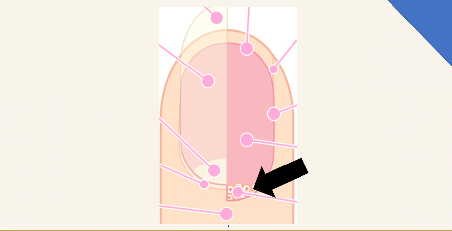
This is referred also as “root of the nail”. It is situated beneath the cuticle. It is where the keratin cells are manufactured forming our nail plate. It contains blood vessels and nerves. When it is damaged the nail will be deformed as it grows.
Nail Plate
Nail Wall
Matrix
Lunula
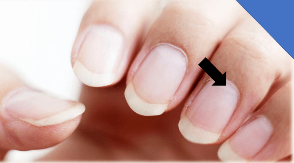
It is referred to as “moon” which is found at the base of the nail, a front end of the matrix. It is pearly in color because of the keratin cells that haven’t been flattened yet. As this keratin cells lose viability, they separate becoming transparent and forming our nail plate.
Lunula
Nail wall
Free edge
Hyponychium
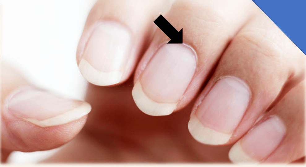
It is the overlapping epidermis around the nail found at the base. It serves as a protection from harmful bacteria.
Mantle
Cuticle
Lunula
Hyponychium

A
Hair Papilla
Melanocytes
Hair Matrix
Fibrous Sheath
Epithelial Root Sheath
B
Hair Papilla
Melanocytes
Hair Matrix
Fibrous Sheath
Epithelial Root Sheath

c
Hair Papilla
Melanocytes
Hair Matrix
Fibrous Sheath
Epithelial Root Sheath

c
Hair Papilla
Hair Follicle
Hair Matrix
Fibrous Sheath
Epithelial Root Sheath

A
Cuticle
Hair Follicle
Hair Bulb
Cortex
Hair Root

B
Cuticle
Hair Follicle
Hair Bulb
Cortex
Medulla
C
Cuticle
Hair Follicle
Hair Bulb
Cortex
Medulla

A
Hair Shaft
Hair Root
Hair Follicle
Arrector Pili Muscle
Hair Bulb

