
 Worksheets
Cardiovascular review A&P
Total questions: 65
Worksheet time: 22mins
Valve between the left atrium and the left ventricle.
tricuspid valve
mitral valve
pulmonary valve
aortic valve
Thickest walled chamber of the heart.
left atrium
right ventricle
left ventricle
right atrium
Valve between the left ventricle and the aorta.
aortic valve
pulmonary valve
tricuspid valve
mitral valve
These vessels carry blood to each lung from the right ventricle.
superior vena cava
pulmonary arteries
pulmonary veins
aorta
Valve between the right atrium and the right ventricle.
mitral valve
pulmonary valve
aortic valve
tricuspid valve
Blood in the right atrium:
Oxygenated
Deoxygenated
Blood in the left ventricle:
Oxygenated
Deoxygenated
Blood in the pulmonary artery:
Oxygenated
Deoxygenated
Blood in the pulmonary veins:
Oxygenated
Deoxygenated
Blood in the aorta:
Oxygenated
Deoxygenated
Blood in the left atrium:
Oxygenated
Deoxygenated
Blood in the Inferior Vena Cava:
Oxygenated
Deoxygenated
How many chambers does the heart have?
4
3
2
1
What is the upper chamber of the heart that receives blood from the body?
Aorta
Right atrium
Right ventricle
Left atrium
What is the main function of valves in the circulatory system?
To pump blood throughout the body
To prevent blood from moving in the wrong direction
To prevent platelets from clotting the blood
To keep the body stable

#2
Septum
Right ventricle
Left ventricle
Right Atrium

Pink color....
Lungs
Septum
Pulmonary artery
Superior vena cava

The relaxing of the heart is known as
Diastole
Systole
Heart Rate
Cardiac Arrest
The contraction of the heart is known as?
Stroke volume
Diastole
Cardiac cycle
Systole
This initiates the action potential that causes the heart to beat
Inferior vena cava
Atrioventricular node (AV node)
Superior vena cava
Sinoatrial node (SA node)
This is responsible for distributing electrical impulses through the heart
Atrioventricular node
Sinoatrial node
Inferior vena cava
Superior vena cava
These are the smallest blood vessels with the thinnest walls in order to maximize diffusion of nutrients in and out of the interstitial fluid
arteries
veins
capillaries
aorta
These carry blood away from the heart
Arteries
Veins
Blood traveling through the systemic circuit goes through the _____ side of the heart and out to the body
right
left
What causes atherosclerosis?
high blood pressure
an increase in blood viscosity
accumulation of fatty materials
an increase in blood volume
Blood returning to the heart from the pulmonary circuit first enters the
right atrium.
right ventricle
left atrium
left ventricle
The right ventricle pumps blood to the
right and left lungs
left ventricle.
left atrium.
aorta.
What color do Eosinophils stain?
Red
Purple
Light Pink/Clear
A heart rate below 60 beats per minute (bpm) is called:
Tachycardia
Bradycardia
Fibrillation
Heart Murmer
The pacemaker is also known as the SA node. What does SA mean?
Sinoatrial
Sensory Arteriosis
Stimulus Apheresis
Still Alive...
What is the function of white blood cells?
carry oxygen
fight infection
carry nutrients
clot blood
Which WBC is most numerous?
basophil
eosinophil
neutrophil
monocytes
What is the average life span of an erythrocyte?
10 days
12 days
120 days
8000 days
What type of tissue is blood?
Epithelial
Connective
Muscle
Nerve
If your WBC count is raised, it could be a sign of _____.
infection
leukemia
leukocytosis
all of the above
Which WBC kills parasites?
neutrophils
eosinophils
monocytes
basophils
Which type of WBC is the "first responder", most mobile & active?
Neutrophils
Eosinophils
Basophils
Lymphocytes

What is blood made of?
oxygen and plasma
red blood cells and white blood cells
plasma, red blood cells, white blood cells, and platelets.
plasma and platelets

What is the type leukocyte shown in the image?
Lymphocyte
Monocyte
Neutrophil
Eosinophil
Basophil

What is the type leukocyte in your RIGHT SIDE of image?
Lymphocyte
Monocyte
Neutrophil
Eosinophil
Basophil
What is the type leukocyte in your LEFT SIDE of image?
Lymphocyte
Monocyte
Neutrophil
Eosinophil
Basophil
What is CBC
Comprehensive blood count
Complete biology count
Collaborative blood count
Complete blood count
In anemia blood doesn't have sufficient amount of
Protein
RBC
Cholesterol
Carbohydrate
This blood type contains ONLY A and Rh anitgens?
B-
B+
O+
A+
A-

The proteins found on the surface of RBCs that allow us to type blood as A, B, O or AB are called
antigens
antibodies
aggluntates
Rh factors
Platelets are best described as
giant multinucleated cells
immature leukocytes
fragments of cells
lymphoid cells
Blood cell formation occurs in the red bone marrow, a process called ____.
fibrinolysis
syneresis
hematopoiesis
erythrocytosis
What happens to cells that are going through Leukemia?
cells start to disappear
abnormal white blood cell growth
cells become easily infected
abnormal red blood cell growth
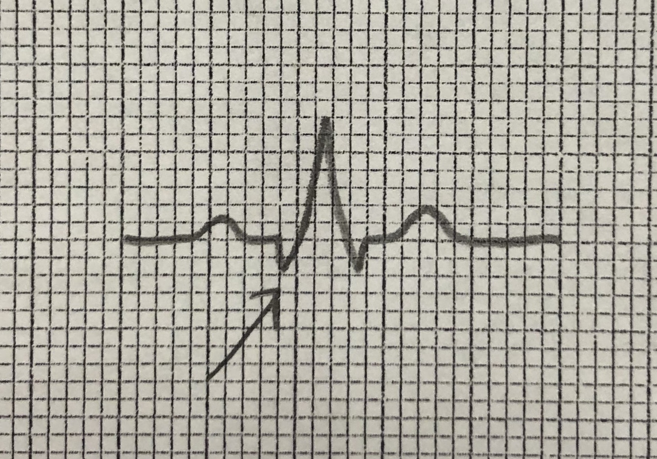
Name this part of the ekg tracing.
P
R
Q
T
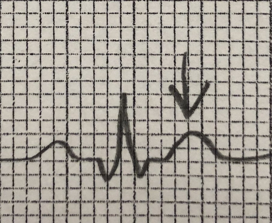
Name this part of the ekg tracing.
Q
R
S
T
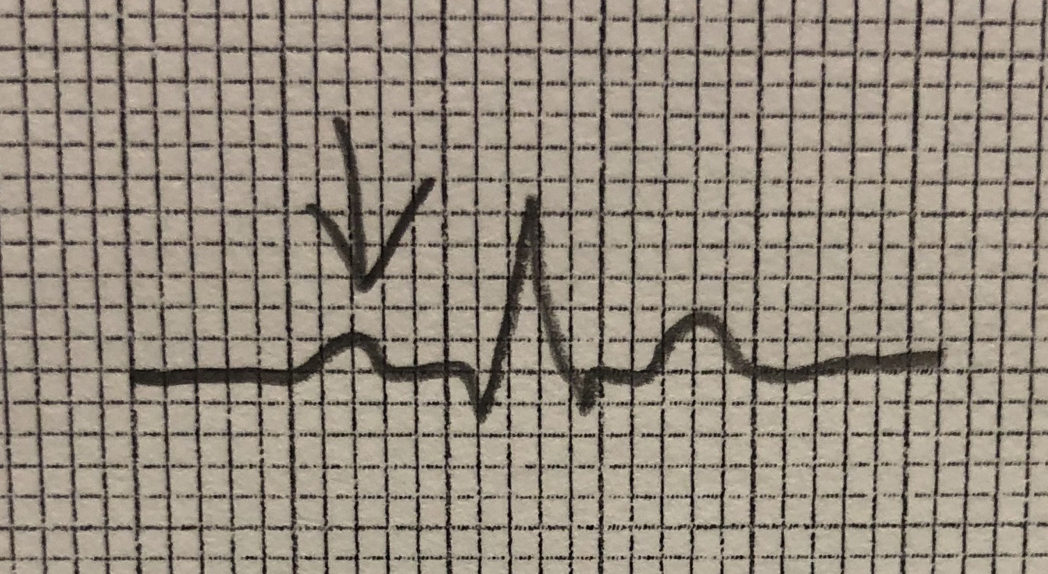
Name this part of the ekg tracing.
P
Q
R
S

Identify this rhythm
Supra-ventricular tachycardia
Atrial fibrillation
Sinus tachycardia
Atrial flutter
The QR complex occurs:
When the ventricles contract.
When the heart rest after a contraction.
When the atria contract.
When the heart begins to beat from the pacemaker.
What does the T wave show?
The contraction of the ventricles.
The contraction of the atria.
If the rhythm is normal.
The relaxation of the heart.
fragments of cells that are used in blood vessel repair
RBCs
WBCs
Platelets
(SELECT ALL CORRECT ANSWERS) What antigens are present on the surface of their red blood cells if someone has type A+ blood?
A antigen
B antigen
Rh antigen
Neither A nor B antigen
(SELECT ALL CORRECT ANSWERS) What antigens are present on the surface of their red blood cells if someone has type O- blood?
A antigen
B antigen
Rh antigen
Neither A nor B antigen

