
 Worksheets
Unit 3 Final Review
Total questions: 87
Worksheet time: 53mins

Number 1
epimysium
fascicle
blood vessel
bone

number 2
tendon
epimysium
fascicle
perimysium

number 3
fascicle
epimysium
bone
blood vessel

number 4
myofibril
perimysium
bone
tendon

number 5
bone
fascicle
endomysium
myofibril

number 6
fascilcle
myofibril
endomysium
tendon

number 7
tendon
myofibril
bonne
blood vessel

number 9
tendon
blood vessel
fascicle
sarcolemma

number 10
sarcolemma
muscle fiber
endomysium
bone
What is the main function of the muscular system?
Movement
Blood filtration
Excretion of waste
Calcium homeostasis
How do muscles produce movement?
Muscles contract by pushing bones
Muscles contract by pulling bones
Muscles contract by pushing and pulling bones
Muscles do not produce movement
Which of the following is NOT a function of the muscular system?
Maintain posture
Stabilize joints
Generate heat
Filter toxins and excess waste from blood
Which of the following is NOT a type of muscle tissue?
Smooth
Rough
Cardiac
Skeletal
Which of the following is the correct order of muscle structures going from largest to smallest
Muscle --> Muscle fascicles --> Muscle fibers --> Myofibrils --> Sarcomeres
Sarcomeres --> Myofibrils --> Muscle fibers --> Muscle fascicles --> Muscles
Muscle --> Muscle fibers --> Muscle fascicles --> Sarcomeres --> Myofibrils
Muscle fibers --> Muscle fascicles --> Muscle --> Myofibrils --> Sarcomeres
Which connective tissue wraps around the entire outside of each muscle fiber?
Perimysium
Epimysium
Endomysium
A bundle of muscle fibers
muscle fiber
fascicle
sarcolemma
aponeurosis
agonist
The delicate membrane surrounding each striated muscle fiber
muscle fiber
fascicle
sarcolemma
aponeurosis
agonist
Role played by a skeletal muscle to cause a movement
agonist
antagonist
contractility
extensibility
irritability
Role played by a skeletal muscle acting to slow or stop a movement
agonist
antagonist
contractility
extensibility
irritability

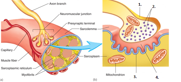
The point at which the nerve cell reaches the sarcolemma of the muscle fiber is the _____.
Neurotransmitter
Acetylcholine (ACh)
Synaptic cleft
Neuromuscular Junction (NMJ)
Once calcium is pumped into the axon terminal what happens?
The muscle contracts
ACh is released into the synaptic cleft.
Sodium pumps into the sarcolemma.
The muscle contracts.
Once ACh binds to receptor cells on the sarcolemma, what takes place?
Sarcolemma becomes temporarily permeable to sodium.
The muscle contracts.
Calcium is pumped into the axon terminal.
Synaptic vesicles move to the end of the terminal.
Once sodium is pumped into the sarcolemma, what happens?
Action potential is transferred to the sarcolemma to start the process of contraction.
The process ends and ACh breaks down.
Calcium is pumped into the axon terminal.
ACh is released into the synaptic cleft.
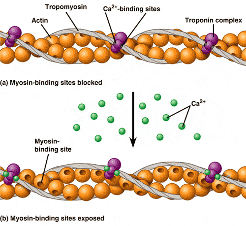
As calcium binds to actin the myosin heads are allowed to bind to the actin forming a cross-bridge and begin to slide over mysoin.
True
False
When ATP binds to the myosin head the myosin head then _____________.
completes a power stroke
attaches to actin
rotates
detaches from actin

Looking at the figure what molecule will allow the myosin head to detach from actin.
ADP
Myosin
ATP
Creatine Phosphate
What causes the muscle to stop contracting?
Lack of effort
Myosin-Heads fail to attach
Calcium Ions are pumped away
Muscle fibre tears
Calcium binds to ___of the troponin-tropomyosin complex.
troponin
tropomyosin
actin
myosin
Once sodium is pumped into the sarcoplasm, what happens?
Voltage of the cell changes to become more positive.
The process ends and ACh breaks down.
Calcium is pumped out of the sarcoplasmic reticulum
Acetylcholine is released into the synaptic cleft.
Once acetylcholine binds to receptor cells on the sarcolemma, what takes place?
Ion channels open and sodium rushes inside the cell
The muscle contracts.
Calcium is pumped out of the sarcoplasmic reticulum.
Synaptic vesicles release acetylcholine into the synapse
When ion channels in the sarcolemma first open to allow ions in/out of the muscle cell, changing the voltage of the cell, how and why does the voltage change?
Becomes more positive because sodium ions rush inside the cell
Becomes more positive because sodium ions rush out of the cell
Becomes more negative because sodium ions rush inside the cell
Becomes more negative because sodium ions rush out of the cell
When ion channels in the sarcolemma first open to allow ions in/out of the muscle cell, changing the voltage of the cell, what happens next?
Other ion channels are triggered to open in a cascade down the sarcolemma
Acetylcholine is released from the synaptic vesicles
Acetylcholine binds to the receptors on the sarcolemma
The sarcoplasmic reticulum is triggered by the voltage change to release calcium ions into the sarcoplasm
What triggers the first few ion channels to open in the sarcolemma?
The binding of acetylcholine to receptors
The release of acetylcholine into the synapse
A voltage change nearby
The action potential traveling through the motor neuron
After the initial few ion channels are triggered to open by the acetylcholine, what causes the rest of the ion channels to open one after the other in a cascade?
Changes in voltage
The binding of acetylcholine to receptors
The release of acetylcholine into the synapse
The sarcoplasmic reticulum releases calcium ions

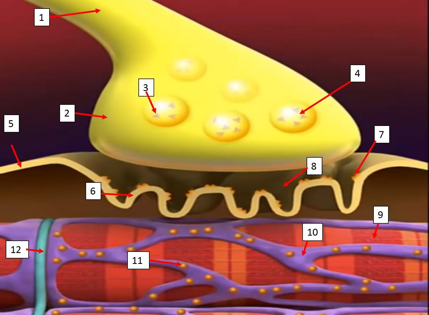
What is structure B?
motor neuron
muscle cell
acetylcholine
sarcolemma
What is structure E?
sodium ion
acetylcholine
calcium ion
synapse
What is structure F?
ion channel
acetylcholine receptor
sarcolemma
sarcoplasmic reticulum
What is structure H?
transverse tubule
muscle fiber
sarcolemma
sarcoplasmic reticulum

What is structure K?
sarcoplasmic reticulum
sarcoplasm
sodium ion
calcium ion
What is structure M?
sarcoplasmic reticulum
sarcoplasm
sodium ion
calcium ion
What is structure L?
ion channel
sarcoplasm
sarcoplasmic reticulum
acetylcholine receptor
What is the opposing muscle group for the biceps?
abdominals
triceps
deltoids
hamstrings
What is the opposing muscle group for the quadriceps?
biceps
lower back
hamstrings
obliques
What is the agonist and what is the antagonist in a bicep curl?
biceps are the agonist, triceps are the antagonist
triceps are the agonist, biceps are the antagonist
The agonist and antagonist work in __________ to create movement
tandem
random
opposition
synchronization
_________ carries the nerve impulse deep into the myofibrils.
T-Tubules
Synapses
Sarcoplasm
Neurotransmitters
True or False: Calcium Ions bind to Tropomyosin to change the shape of Troponin.
True
False
Which proteins make up the thin filament? Select all that apply.
Actin, myosin, and troponin
Actin, troponin, and tropomyosin
Troponin, myosin, and tropomyosin
Myosin, actin, and tropomyosin
What triggers tropomyosin to shift and expose the actin binding sites during a muscle contraction?
Calcium binding to troponin
Troponin binding to myosin
Myosin head forming a cross bridge
Acetylcholine attaching to receptors on the sarcolemma
Ca2+ released by sarcoplasmic reticulum will bind to
Actin
Troponin
Myosin
Tropomyosin

Identify the pink structures? It is a globular protein complex, occuring with tropomyosin in the thin filaments of muscle tissue. Binds to calcium ions, moving tropomyosin away from the myosin binding sites on actin filaments.
actin
myosin
tropomyosin
troponin
cross bridge

Identify X? A protein that covers the mysoin binding sites on the actin filament in a relaxed muscle, preventing contraction.
actin
myosin
tropomyosin
troponin
cross bridge

Identify the blue structures? a protein that forms (together with myosin) the contractile filaments of muscle cells.
actin
myosin
tropomyosin
troponin
cross bridge

What muscle is the arrow pointing toward?
biceps
traps
pecs
triceps

What muscle is the arrow pointing to?
Pectoralis (Pecs)
Biceps
Triceps
Lats

What muscle is the arrow pointing to?
Biceps
Triceps
Lats
Quads


What type of contraction is happening here??
isostabilize
isotonic- eccentric
isotonic- concentric
isometric
How glucose and aerobic respiration are related.
Glucose is broken down into ATP in the presence of oxygen
Glucose is broken down into ATP in the absence of oxygen
When ATP is created, glucose is released
For every ATP, a glucose is created
Muscle fatigue is...
when the muscle is unable to contract
when you are too tired to exercise
when the muscle is contracted more than 30 secondsd
when O2 comes from direct phosphorylation
What is the correlation between muscle fatigue and oxygen debt?
When O2 decreases, muscle fatigue increases
When O2 increases, muscle fatigue decreases
When O2 is obtained, muscle fatigue decreases
The lesser the oxygen debt, the lesser the muscle fatige
What disorder causes the impairment of motor function due to a disruption in brain development?
ALS
Fibromyalgia
Muscular dystrophy
The disorder the causes widespread pain in the muscles and joints and causes fatigue.
fibromylagia
cerebral palsy
muscular dystrophy
myasthenia gravis
ALS
Muscular Dystrophy
Myasthenia gravis

