
 Worksheets
Anatomy Midterm Study Practice
Total questions: 129
Worksheet time: 1hrs 12mins
What are the four types of tissues in the human body? (Select all that apply)
Epithelial
Connective
Muscle
Nervous
How do we classify epithelial tissue?
Shape
Shape and number of layers
Counting the number of individual cells
Squamous

Skeletal
Smooth
Cardiac
Dense regular

Dense Regular
Cardiac
Skeletal
Smooth
What is the most abundant type of tissue in the human body?
Nervous
Muscle
Connective
Epithelial
Striations can be seen in which type of muscle tissues?
Smooth and cardiac
smooth and skeletal
skeletal and cardiac
Cardiac only
What are the two main cells that make up nervous tissue?
neurons
myelin
axon
neuroglia


The head is superior to the chest
True
False

Are fingers proximal or distal to to the heart?
Proximal
Distal
What body position is considered the “starting point” for describing body positions?
SOFT Planes
Anatomical Position
Quadrants
Cavities
Sagittal Planes divide the body
into right v left
into front v back
into unsymmetrical parts
into top v bottom
Transverse planes divide
into right v left
into top v bottom
into unsymmetrical parts
into front v back
The foot is ______ to the ankle.
Lateral
Posterior
Superior
Inferior
Which cavity contains the brain?
Oral
Cranial
Orbital
Abdominopelvic
The posterior body cavity contains:
Thoracic and Abdominal
Thoracic and Spinal
Pelvic, Abdominal, and Thoracic
Spinal and Cranial
Buccal means _______ and carpal means _____
Cheek of face; wrist
Cheek of face; toes
Groin; wrist
Groin; toes
Crural means ______; fibular means ______
Lateral, lower leg; lower leg
Lower leg; wrist
wrist; lower leg
Lower leg; lateral, lower leg
Frontal means ______; Mental means _____
Chin; forehead
Forehead; Chin
Eyes; Chin
Chin; brain

What is this bone?
Tibia
Tarsals
Patella
Sternum

What is this bone?
Ulna
Radius
Humerus
Metacarpal

What is this muscle?
Anterior deltoids
Quadriceps
Pectoralis Major
Latissimus Dorsi

What is this muscle?
Latisimus Dorsi
Teres Major
Rhomboids
Biceps brachii
Which is the bottom layer of the epidermis?
Stratum basale
Stratum lucidum
Stratum spinosum
Stratum corneum
Stratum granulosum
Which is the top layer of the epidermis?
Stratum basale
Stratum lucidum
Stratum spinosum
Stratum corneum
Stratum granulosum
What does the arrector pili muscle do?
Push sweat out
Thermoregulation
Make hair stick up
Waterproofing
Which type of sweat gland is found in the armpit?
Sebaceous
Apocrine
Eccrine
The integumentary system is made up of the:
lymph nodes, interstitial fluid, and lungs
skin, the oil and sweat glands, sensory receptors, hair, and nails
the heart, arteries, and white blood cells
skin, nails, bladder, and anus
Voluntary muscles are .
Striated
non straited
cardiac
myologic
This system includes the kidneys and the bladder:
circulatory
exretory
endocrine
integumentary
The uppermost and largest bone of the arm, extending from the elbow to the shoulder is the __________
Humerus
metacarpus
radius
phlanges
What are the main layers of the skin from superficial to deep?
epidermis, dermis, hypodermis
dermis, hypodermis, epidermis
dermis, epidermis, hypodermis
hypodermis, dermis, epidermis
Which branch of science specifically studies tissues?
Anatomy
Physiology
Cytology
Histology
What is the primary function of the endocrine system?
Produces & secretes hormones
Defend the body from & fight infection
Collect, process, & respond to sensory information
Gas exchange & nutrient transport
The skin is included in which organ system?
Integumentary System
Lymphatic System
Muscular System
Reproductive System
The axial portion of the body includes all of the following regions except for...
Head
Neck
Torso
Arms
Which of the following anatomical descriptions is FALSE?
The calves are posterior to the shins
The abdomen is anterior to the spine
The nose is dorsal to the mouth
The eyes are ventral to the ears
Which of the following is the correct anatomical descriptor of the elbows and fingers?
The elbows are intermediate between the fingers and toes
The elbows are lateral to the fingers
The elbows are distal to the toes
The elbows are proximal to the fingers
Which body cavity holds the heart & lungs?
Thoracic Cavity
Pleural Cavity
Pericardial Cavity
Abdominopelvic Cavity
Joint cavities are called...
Synovial Cavities
Orbital Cavities
Pleural Cavities
Ventral Cavities
Cells that secrete products are called...
Glands
Squamous Cells
Cartilage Cells
Adipose Cells
Which of the following is NOT a classification of epithelial tissue?
Squamous
Cuboidal
Columnar
Dense
Which of the following is NOT a type of connective tissue?
Bone
Cartilage
Blood
Muscle
Which type of muscle tissues propel substances along internal pathways?
Skeletal
Smooth
Cardiac
Loose

Which letter refers to the hypodermis?
A
B
C
D
E
Which letter refers to the papillary dermis?
A
B
C
D
E

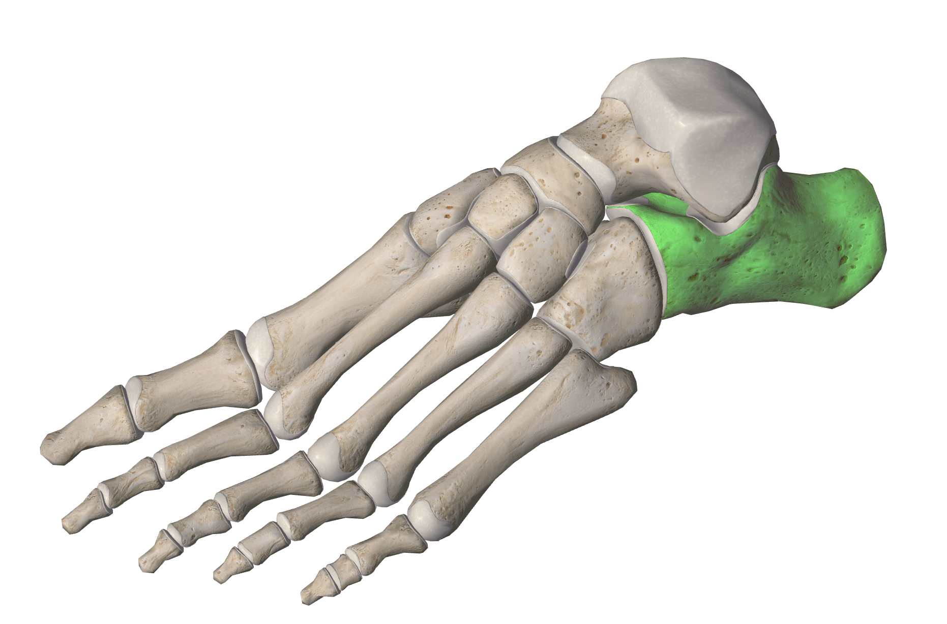
What bones is highlighted?
Femur
Humerous
Skull
Shin


How many bones are in an adult human skeleton?
206
268
300
365
Calcium is stored in _ bone marrow.
yellow
red
spongy
compact
Which of these is a type of long bone?
carpals
tarsals
skull
femur
The sternum is an example of a _ bone.
flat
long
sesamoid
irregular

Which term describes #2?
lamella
central canal
osteon
periosteum
What is hematopoiesis?
Building bones
Blood cell formation
Destroying blood cells
Swelling caused by blood

Structure 7 is the outer covering of the bone made up of fibrous connect tissue called
Medullary Cavity
Compact Bone
Yellow Marrow
Periosteum


Identify bone "F"
Temporal
Parietal
Sphenoid
Zygomatic

Identify Bone "H"
Zygomatic
Maxilla
Sphenoid
Lacrimal


temporal
sphenoid
zygomatic
occipital

occipital foramen
vertebral foramen
foramen magnum
occipital arch

What are the structures in the picture?
foramen magnum
occipital condyles
external auditory meatus
zygomatic process
sella turcica

What is the structure in the picture?
foramen magnum
occipital condyles
external auditory meatus
zygomatic process
sella turcica

What is the structure in the picture?
foramen magnum
occipital condyles
external auditory meatus
zygomatic process
sella turcica

What part of the ethmoid bone is B?
cribiform plate
crista galli
perpendicular plate
sella turcica

What is the structure in the picture?
foramen magnum
occipital condyles
external auditory meatus
zygomatic process
mastoid process

What is the structure in the picture?
foramen magnum
occipital condyles
styloid process
zygomatic process
mastoid process

What is the structure in this picture?
infraorbital foramen
palatine process
zygomatic arch
horizontal plate
mental foramen

What type of suture is this?
coronal
squamosal
lamboidal
sagittal

What is the structure on this picture?
infraorbital foramen
palatine process
zygomatic arch
mental foramen

The depressions on the temporal bones (within the red ovals) are called...
mandibular notch
occipital crest
mandibular fossae
zygomatic arch
Among of the classification of bone markings below which is not an openings
Foramen
Sinus
Meatus
Fossa
Which region of the vertebral column contains the most vertebrae
Cervical
Thoracic
Lumbar
Sacrum
The ribs that are not attached to the sternum are called
Floating ribs
False ribs
True ribs
None of the above

What classification of bone marking is the red area
Epicondyle
Fossa
Condyle
Facet
What classification of bone marking is the green area
Epicondyle
Fossa
Condyle
Facet
Which does not belong to that axial skeleton
Skull
Vertebral Column
Thoracic cage
Pelvic bone
It is any protuberance or projection of a bone
Bone marking
Process
Features
Ramus

What is the bone labeled 1?
ischium
pubis
ilium
acetabulum

What feature is labeled 6?
obturator foramen
acetabulum
greater sciatic notch
pubis symphysis

The iliac crest is at which number?
7
9
4
5
What bone articulates with the coxal bone at the acetabulum?
Tibia
Fibula
Femur
Humerus
The head of the humerus articulates with the
Tarsals
Acetabulum
Glenoid cavity
Trochlear notch
The head of the radius articulates with the
Carpals
Capitulum
Trochlea
Styloid process

Name the outside portion of the tibia shown in blue.
Medial malleolus
Lateral malleolus
Acetabulum
Talus


Metacarpals articulate with the (a) proximally.


Identify the structure marked with the X: This cartilaginous joint joins the two pubic bones together.
pubic symphysis
obturator foramen
iliac crest
acetabulum

E
LCL
medial meniscus
MCL
lateral meniscus
D
LCL
ACL
MCL
IT band
N
LCL
ACL
MCL
patellar tendon
J
LCL
ACL
MCL
PCL
M
LCL
ACL
MCL
PCL

What is the name of this bony feature?
Acromion process
Lesser Tubercle
Coracoacromial Arch
Coracoid process

What is the name of this joint?
Acromioclavicular joint
Coracoclavicular joint
Humeroclavicular joint
Sternoclavicular joint
Which small muscles in the shoulder all work in conjunction to STABILIZE the humeral head?
Rotator cuff muscles
Deltoid (all 3 heads)
Pectoralis (both major and minor)
Biceps Brachii (both heads)

What rotator cuff muscle fits in this section of the scapula?
Supraspinatus
Teres Minor
Infraspinatus
Subscapularis

What muscle is shown in the image?
(a)

What muscle is shown in the image?
(a)

What muscle is shown in the image?
(a)

What muscle is shown in the image?
(a)

What muscle is shown in the image?
(a)

Which label is the muscle fiber?
A
B
C
D
Which type of muscle is involuntary and striated?
Cardiac
Smooth
Skeletal
What is the scientific name for a muscle fiber bundle?
Tendon
Ligament
Fascicle
Perimysium

Which label is pointing to the epimysium?
A
B
C
D

