
 Worksheets
Cardiovascular Test
Total questions: 81
Worksheet time: 4hrs 3mins
Which heart chambers are thicker: atria or ventricles? Why?
ventricles; they pump blood, atria just receive.
atria; they pump blood, ventricles just receive.
atria; because they are on top.
ventricles; because they are on the bottom.
Which ventricle is thicker: left or right? Why?
left: pumps blood to the body, while the right goes to lungs.
left; ventricles are just thicker.
right; the left lung is smaller.
right: pumps blood to the body, while the left goes to lungs.

What structure is indicated by Letter A ?
pulmonary trunk
aorta
inferior vena cava
pulmonary veins
What structure is indicated by Letter F ?
pulmonary artery
aorta
inferior vena cava
pulmonary veins
What structure is indicated by Letter H ?
pulmonary artery
superior vena cava
inferior vena cava
pulmonary veins
What structure is indicated by Letter K ?
ventricles
septum
superior vena cava
pulmonary veins
What structure is indicated by Letter C ?
left ventricle
right ventricle
left atrium
right atrium
What structure is indicated by Letter L ?
left pulmonary veins
right pulmonary veins
right pulmonary artery
left pulmonary artery
What structure is indicated by Letter J ?
left pulmonary veins
right pulmonary veins
right pulmonary artery
left pulmonary artery
The heart chamber that receives blood coming back from the body is the _____________.
left ventricle
right ventricle
left atrium
right atrium
The heart chamber that pumps blood out to the body is the _____________.
left ventricle
right ventricle
left atrium
right atrium
T/F: The pulmonary veins carry blood back to the heart from the body.
True
False; pulmonary veins
False; aorta
False; vena cavae
The largest blood vessel in the body is the _________.
aorta
superior vena cava
inferior vena cava
femoral artery
The medical term for a heart attack is ___________.
myocardial infarction
cardiac arrest
angina pectoris
cardiomyopathy
Bradycardia is _____________.
condition of a slow heartbeat
surgical removal of a clot
condition of a rapid heartbeat
hardening of a vein
The best procedure to see the anatomy of the heart is a/an ___________________.
echocardiogram
complete blood count
heart transplant
electrocardiogram
Veins always carry ___________________.
blood toward the heart
blood away from the heart.
oxygen-poor blood
oxygen-rich blood
What is another name for the circulatory system?
Blood System
Heart Operations
Cardiovascular System
Cardiorespiratory System
Arteries carry what type of blood?
Oxygenated
Deoxygenated
Veins carry what type of blood?
Oxygenated
Deoxygenated
In which direction do arteries carry blood?
To the body
From the heart
To the heart
From the body
Which is located in the upper two chambers of the heart?
Atrium
Ventricle
Which is located in the lower two chambers of the heart?
Atrium
Ventricle
Roughly how many quarts of blood are in the adult human body?
1-2
4-6
8-10
10-12
Which of the following is NOT a component of blood?
Water
Plasma
Red Blood Cells
Nitrogen
What takes up the most portion in a vial of blood?
Red Blood Cells
White Blood Cells
Plasma
Platelets
What are erythrocytes?
White Blood Cells
Plasma
Platelets
Red Blood Cells
What are leukocytes?
White Blood Cells
Plasma
Platelets
Red Blood Cells
Cardi
Heart
Blood
Blood Vessels
Veins
Hem
Blood
Plasma
Platelet
Protein
Sound/feeling of blood against the walls of the arteries.
Heart pulse
Pulse
Blood Pressure
None of the above
Major pulse point located in the neck.
Brachial
Femoral
Radial
Carotid
Major pulse point located in the wrist.
Brachial
Femoral
Radial
Carotid
Major pulse point located in the elbow.
Brachial
Femoral
Radial
Carotid
Major pulse point located in the upper leg.
Brachial
Femoral
Radial
Carotid
What finger must you never take a pulse with?
Ring
Middle
Pinky
Thumb
Top number in blood pressure.
Diastolic
Systolic
Bottom number in blood pressure.
Diastolic
Systolic
Pressure when the heart is relaxing.
Systolic
Diastolic
What is a normal blood pressure reading?
110/70
120/80
125/95
130/90
A condition in which red blood cells are shaped like bananas.
Anemia
Sickle Cell Anemia
Leukemia
Heart Attack
When there is an inadequate number of red blood cells.
Sickle Cell Anemia
Anemia
Leukemia
Erythrodemia


The heart is located here
mediastinum
ventral cavity
pleural cavity
thoracic cavity
Separates the atria
interventricular septum
interventricular sulcus
interatrial septum
interatrial sulcus


Highest pressure against blood vessels. Represented by first heat sound
diastolic pressure
systolic pressure
hypotension
hyperstension

Loos fitting sac around the heart
myocardium
endocardium
epicardium
parietal pericardium
Mrs. Smith has been diagnosed with hypertension. This means that the upper number in her blood pressure reading is over 140mmHg. The upper number in a blood pressure reading is called __________.
pulse pressure
systolic
pulse
diastolic
On an EKG the contractions of the ventricles is represented by the ______________.
T wave
PR interval
QRS complex
P wave
The hardening and narrowing of arteries caused by a build-up of cholesterol plaque is called ___________.
venosclerosis
arteriosclerosis
phlebomegaly
arteriomegaly
Type of circulation that refers to blood flow from the heart to the lungs and back to the heart again?
Systemic circulation
Renal circulation
Portal circulation
Pulmonary circulation
Type of circulation that refers to the blood flowing through the entire body?
Systemic circulation
Renal circulation
Portal circulation
Pulmonary circulation

Instrument used to measure blood pressure?
AED
ECG
Sphygmomanometer
ultrasound
The _________________arteries carry blood and oxygen to all parts of the heart.
coronet
coronary
corridor
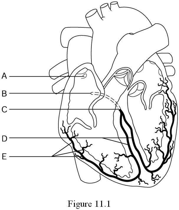
The part of the cardiac conduction system that goes down the interventricular septum is the:
AV node
SA node
Bundle of His
Purkinje Fibers
The area of the heart that divides the right and left ventricles is called the:
Chordae tendinae
Papillary muscles
Interventricular septum
Foramen ovale

The string like feature that attaches to and helps close the AV valves is the:
Chordae tendinae
Papillary muscles
Interventricular septum
Foramen ovale
The correct sequence of the three events in ECG
P wave , T wave , QRS complex
QRS complex , P wave , T wave
T wave , QRS complex , P wave
P wave, QRS complex, T wave
Why is a myocardial infarction life threatening?
A damaged heart muscle cannot pump blood efficiently.
Swollen veins lose elasticity, which decreases blood flow to vital organs.
A minor bump or fall could lead to internal bleediing.
It inhibits the exchange of oxygen and carbon dioxide in body cells.

