
 Worksheets
A&P Lab
Total questions: 114
Worksheet time: 3hrs 51mins
Standing arms at sides, palms and head forward
Standard Anatomical position
Supine
Prone
anterior surface facing up
Standard anatomical position
Supine
Prone
anterior surface facing down
Standard anatomical position
Supine
Prone
Anterior or ventral
Front/Belly side
Back side
Posterior or dorsal
Front/Belly side
Back side
Superior
closer to top of head
farther away from top of head
closer to surface
farther from surface
Inferior
closer to top of head
farther away from top of head
closer to surface
farther from surface
Medial
toward midline
away from midline
closer to surface
nearest to point of origin
Lateral
toward midline
away from midline
farther from point of origin
farther from surface
Proximal
nearest to point of origin
closer to surface
toward midline
closer to top of head
Distal
farther from point of origin
farther away from top of head
farther from surface
away from midline
Superficial
closer to surface
nearest to point of origin
toward midline
closer to top of head
Deep
farther from surface
away from midline
farther from point of origin
farther away from top of head
Appendicular
arms and legs
head, neck, trunk
soles of feet
armpit
Axial
head, neck, trunk
arms and legs
lower trunk
chest
Axillary
armpit
arms and legs
arm
wrist
Brachial
arm
armpit
arms and legs
elbow
Carpal
wrist
elbow
arm
armpit
Cubital
elbow
wrist
arm
armpit
Inguinal
groin
lower trunk
knee
thigh
Pelvic
lower trunk
groin
belly
thigh
Palmar
palms of hands
wrist
arm
soles of feet
Patellar
knee
lower trunk
ankle
soles of feet
Thoracic
chest
navel
belly
thigh
Umbilical
navel
belly
chest
thigh
Cranial (Cephalic)
head
neck
groin
face
Cervical
neck
head
groin
belly
Facial
face
neck
head
Abdominal
belly
chest
navel
lower trunk
Femoral
thigh
ankle
knee
lower trunk
Plantar
soles of feet
ankle
palms of hands
arms and legs
Tarsal
ankle
soles of feet
palms of hands
wrist
separates right from left
Sagittal plane
Transverse (horizontal) plane
Frontal (coronal) plane
separates top from bottom
Sagittal plane
Transverse (horizontal) plane
Frontal (coronal) plane
separates front from back
Sagittal plane
Transverse (horizontal) plane
Frontal (coronal) plane
Serous Membranes
one membrane forms two layers surrounding an organ
two membrane forms two layers surrounding an organ
three membrane forms two layers surrounding an organ
four membrane forms two layers surrounding an organ
Serous membranes: Visceral
in direct contact with organ
not in contact with organ
Serous Membranes: Parietal
in direct contact with organ
not in contact with organ
Pericardial membranes
heart
lungs
abdominopelvic organs
Pleural membranes
heart
lungs
abdominopelvic organs
Peritoneal membranes
heart
lungs
abdominopelvic organs
Name four types of tissue
(a)
Causes stage to move upward or downward
ADJUSTMENT KNOB
CONDENSER
FIELD OF VIEW
STAGE
Serves as a handle for carrying the microscope
ARM
CONDENSER
ADJUSTMENT KNOB
FIELD OF VIEW
Contains a lens at the top of the body tube
EYEPIECE (OCULAR)
FIELD OF VIEW
IRIS DIAPHRAGM
OBJECTIVE LENS SYSTEM
Circular area see through the eyepiece
FIELD OF VIEW
EYEPIECE (OCULAR)
STAGE
IRIS DIAPHRAGM
Increases or decreases the light intensity.
IRIS DIAPHRAGM
OBJECTIVE LENS SYSTEM
EYEPIECE (OCULAR)
STAGE
Part to which the objective lenses are attached.
NOSEPIECE
OBJECTIVE LENS SYSTEM
STAGE
CONDENSER
After light passes through the specimen, it next enters this lens system.
OBJECTIVE LENS SYSTEM
IRIS DIAPHRAGM
FIELD OF VIEW
CONDENSER
Platform that supports the microscope slide.
STAGE
ARM
CONDENSER
ADJUSTMENT KNOB
Holds a microscope slide in position.
STAGE CLIP
CONDENSER
ARM
OBJECTIVE LENS SYSTEM
9 Body Regions
R&L Hypochondriac
Epigastric & Hypogastric
Umbilical
R&L Iliac
R&L Hypachondriac
Body Cavities
Abdominal cavity
Abdominopelvic cavity
Pelvic cavity
Thoracic cavity
Cranial cavity Vertebral cavity
Columnar cavity
Squamous cavity
Cuboidal cavity
Pleural Cavity
Visceral Pleura Cavity
Parietal Pleura Cavity
loosely coiled fibers containing protein and DNA within nucleus.
Chromatin
Cytoplasm
Endoplasmic reticulum
Golgi Apparatus
occupies space between plasma membrane and nucleus.
Cytoplasm
Chromatin
Endoplasmic reticulum
Golgi Apparatus
composed of membrane-bound canals for tubular transport throughout the cytoplasm
Endoplasmic reticulum
Cytoplasm
Chromatin
Golgi Apparatus
flattened membraneous sacs that package a secretion
Golgi Apparatus
Endoplasmic reticulum
Cytoplasm
Chromatin
membraneous sac that contains digestive enzymes
Lysosome
Microtubule
Mitochondrion
slender tubes that provide movement in cilia and flagella
Microtubule
Mitochondrion
Nuclear envelope
Nucleolus
location of ATP production from digested food molecules
Mitochondrion
Microtubule
Nuclear envelope
Nucleolus
separates nuclear contents from cytoplasm
Nuclear envelope
Nucleolus
Mitochondrion
Microtubule
dense body of RNA within the nucleus
Nucleolus
Nuclear envelope
Mitochondrion
Microtubule
Small RNA containing particles for the synthesis of proteins.
Ribosome
Vesicle
Membraneous sac formed by the pinching off of pieces of cell membrane.
Vesicle
Ribosome
The structure and function of the serous membrane is
smooth membrane
rough membrane
serous membrane
The structure and function of the serous membrane is smooth membrane, consisting of a thin layer of cells which secrete
serous fluid
membrane fluid
visceral fluid
The structure and function of the serous membrane is smooth membrane, consisting of a thin layer of cells that secrete serous fluid and a thin connective tissue layer between the
parietal layer
visceral layer
The structure and function of the serous membrane is smooth membrane, consisting of a thin layer of cells that secrete serous fluid and a thin connective tissue layer between the parietal layer and the
visceral layer
membrane layer
Lines the walls of the body cavity
parietal layer
visceral layer
Covers the organs
visceral layer
parietal layer
For total magnification you multiply the 4x, 10x, 40x, and 100x lens by
10x
100x
40x
A solution with a higher concentration of solutes than the cell
Hypertonic
Hypotonic
Isotonic
A solution with a lower concentration of solutes than the cell
Hypertonic
Hypotonic
Isotonic
A solution with the same concentration of solutes as the cell
Hypertonic
Hypotonic
Isotonic
4 main tissue types
Connective
Epithelial
Muscle
Nervous
Skeletal
Supports other tissues and binds them together
(Ex: bone, blood, and lymph tissue)
Connective tissue
Epithelial tissue
Muscle tissue
Nervous tissue
Provides covering (Ex: skin and the linings of various passages inside the body)
Connective tissue
Epithelial tissue
Muscle tissue
Nervous tissue
Includes striated (voluntary) muscles that move the skeleton and smooth muscle, such as the muscle that surround the stomach
Connective tissue
Epithelial tissue
Muscle tissue
Nervous tissue
Made up of nerve cells (neurons) and is used to carry "messages" to and from parts of the body
Connective tissue
Epithelial tissue
Muscle tissue
Nervous tissue
Tissue is a group of cells that have
similar structure and that function together as a unit
opposite structure and that function separately
3 main types of fibers secreted by fiber blast
Collagen fibers
Elastic fibers
Reticular fibers
Lucidum fibers
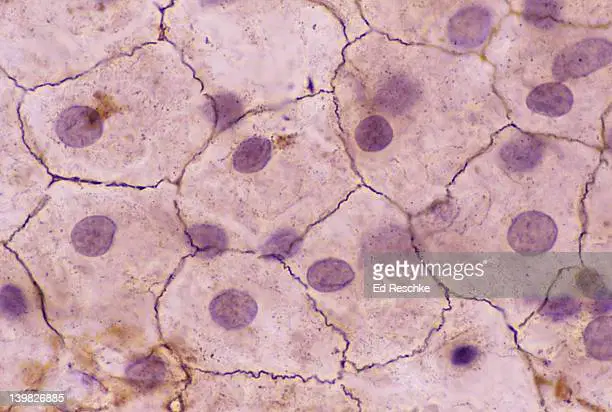
A single layer of thin flattened cells
Simple Squamous
Simple Cuboidal
Simple Columnar

A single layer of cube shaped cells
Simple Squamous
Simple Cubiodal
Simple Columnar

A single layer of column-shaped cells
Simple Squamous
Simple Cuboidal
Simple Columnar
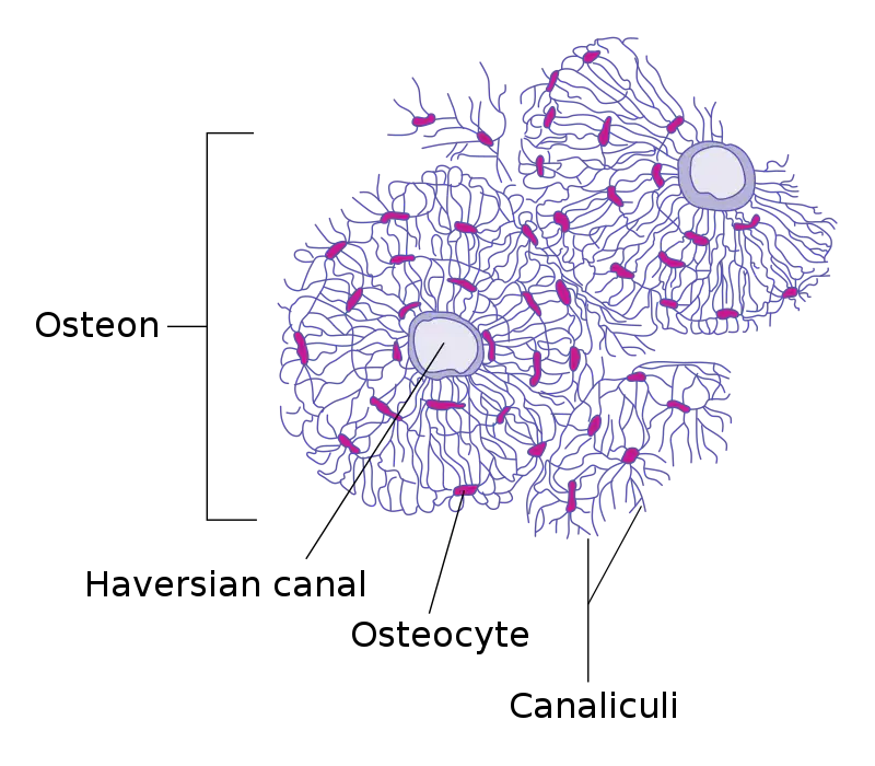
The huge circle of the osteon is called the
Haversian canal
Osteocyte
Canaliculi

The bone cells/osteocytes (small black dots) are called
Lacunae
Haversian canal
Canaliculi
The rounded portion found at the end of the bones
Epiphysis
Metaphysis
Diaphysis
The widest part of a long bone
Epiphysis
Metaphysis
Diaphysis
The central shaft running between proximal and distal ends of the bone
Epiphysis
Metaphysis
Diaphysis
True Ribs (Direct connection)
1-7
8-10
11-12
False Ribs (coastal margin, no direct connection)
1-7
8-10
11-12
Floating
1-7
8-10
11-12
Held together by dense connective tissue
Fibrous joints
Cartilaginous joints
Synovial joints
Held together by cartilage
Fibrous joints
Cartilaginous joints
Synovial joints
Free-moving and enclosed in a capsule
Fibrous joints
Cartilaginous joints
Synovial joints
Immovable joint
Synarthrosis
Amphiarthrosis
Diathrosis
Slightly movable joint
Synarthrosis
Amphiarthrosis
Diathrosis
Freely moveable joint
Synarthrosis
Amphiarthrosis
Diathrosis

Smooth Muscle
Cardiac Muscle
Skeletal Muscle

Smooth Muscle
Cardiac Muscle
Skeletal Muscle
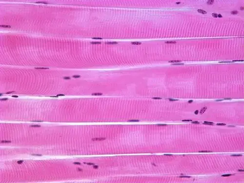
Smooth Muscle
Cardiac Muscle
Skeletal Muscle
Involuntary, found in the stomach/intestines. It forms organs like the stomach and bladder
Smooth Muscle
Cardiac Muscles
Skeletal Muscle
Involuntary, found in the heart. It contracts the heart to pump blood
Smooth Muscle
Cardiac Muscle
Skeletal Muscle
Voluntary, found in the shoulder, abdominals, and hamstrings (etc). They move bones and other structures.
Smooth Muscle
Cardiac Muscle
Skeletal Muscle
Made up of the brain and spinal cord
Central Nervous System (CNS)
Peripheral Nervous System (PNS)
Made up of nerves that branch off from the spinal cord and extend to all parts of the body
Central Nervous System (CNS)
Peripheral Nervous System (PNS)
Neuroglial Cell Types in CNS
Astrocytes
Ependymal Cells
Oligodendrocytes
Microglia
Schwann Cells
Neuroglial Cell Types in PNS
Astrocytes
Microglia
Ependymal
Schwann Cells
Satellite Cells
Inner layer of brain
Pia mater
Arachnoid
Dura mater
Middle layer of brain
Pia mater
Arachnoid
Dura mater
Outer layer of brain
Pia mater
Arachnoid
Dura mater
The cerebral lobe is responsible for
vision
hearing
taste
touch
There is ___ cranial nerve pairs that exists in the human body
8
12
24
What separates the Thoracic cavity and the Abdominal cavity
Diaphragm
Abdominalpelvic
Heart

