
 Worksheets
A&P - 6.1-6.6 - 2026 - Skeletal histology & physiology
Total questions: 70
Worksheet time: 1hrs 10mins
Blood cell formation is called ___.
hematocrit
homozygous
heterozygous
hematopoiesis
Which means “to break?”
-blast
-clast
osteo-
-phage

What are small needle-like pieces of bone with lots of open spaces in spongy bone?
mesenchyme
lamellae
lacuna
trabeculae
Match bone shapes
parietal & temporal skull bones
flat bones
cube-like bone shape
short bones
patella
sesamoid bone
vertebra
irregular bone
femur & radius
long bones
Where in bone does hematopoiesis occur?
central medullary cavity
red bone marrow
diaphysis
periosteum
Match PTH & Calcitonin
PTH is secreted from the parathyroid gland
when blood calcium levels are too ____.
low
Calcitonin is secreted from the thyroid gland when blood calcium levels are too ____.
high
PTH & Calcitonin are ___.
hormones
What is the end of bone nearest to the torso?
proximal epiphysis
distal epiphysis
Which is a rare disorder caused by any one of a number of faulty genes that results in either too little collagen formation or a poor quality of collagen and causes the bone matrix to have decreased flexibility and be more easily broken than normal bone?
osteogenesis imperfecta
rickets
scurvy
osteoporosis
What are mature bone cells derived from osteoblasts?
osteoclasts
osteocytes
adipocytes
erythrocytes
Reorder the process of making bone cells
osteoprogenitor cells
osteoblast
osteocyte
Which are ultramicroscopic links that connect the lacunae to one another and the central canal?
canaliculi
osteocyte
linkaculi
lamellae

What is also called a perforating canal, connect bones cells with one another, and links the central canals of osteons?
Haversian canal
Volkmann's canal
What is primarily collagen (90% of bone protein), which gives bone its high tensile strength?
osteoid
osteocyte
osteoblast
osteoclast
Anatomical Position= standing (a) with the face directed (b) , the arms hanging to the side,
and the palms facing forward.

Interpret the diagram
A
osteoblast
B
osteocyte
osteoprogenitor cell
developed from stem cell
categorize bone types
humerus
carpals
skull
ribs

Identify the BIG red arrow's structure
diaphysis
epiphysis

Identify "A"
lamellae
lacuna
osteocyte
canaliculi

Identify "C"
lamellae
lacuna
canaliculi
epiphysis
Match bone structure & composition

Haversian canal

periosteum

lamellae
The typical adult skeleton has how many bones?
155
206
250
433
600
Which of the following is NOT a classification term for bones?
Long
Short
Irregular
Flat
Symmetrical

Which ossification produces flat bones?
Endochondral ossification
Intramembranous ossification
Appositional growth is when the bone
thickens
elongates
The process of bone formation is called
nitrification
ossification
ammonification
calcification

In long bones, bone tissue first begins to replace hyaline cartilage in the center of the diaphysis or the...
(a) . After this, blood vessels infiltrate the epiphyses and bone tissue forms at the (b) . This process is called (c)

In a fetus, which bones ossify first?
carpals
skull
Match the following Fractures

Oblique

Spiral

Impaction

Greenstick
Select all the apply
Select the 4 cells that are associated with bone health & remodeling
Osteoprogenitor
Lacunae
Osteocyte
Osteoblast
Osteoclast
In a fetus, flat bones ossify first.
How is this an adaptation?
*Adaptations are helpful
protection of internal organs
ability to walk
synthesis of vitamin D
filtering waste and producing bile
A baby has _____ bones at birth.
65
about 300
206
about 6,200
% of bone calcium is exchanged each year
3-5 %
75-77 %
85 - 90 %
50-60 %
What is the formation of blood cells called?
Hematoma
Hematocrit
Hippopotamus
Hematopoiesis
What are the five functions of the skeletal system?
Support
Protection
Insulation
Transmitting Signals
Breathing
Support
Protection
Movement
Storage
Manufacturing
Regulating Temp
Digestion
Support
Protection
Circulation
Heat Production
Movement
Storage
Immunity
Manufacturing

"A" = ?
process
facet
Match general features of bone

main part

constriction between head & body

smooth round articular surface

branch off body

What does "C" of ACL stand for?
Cruciate
Collateral
Connective
Condyle
Match the following joints

suture

ball & socket

hinge
Match joint classifications
Synarthroses
immovable
Amphiarthroses
slightly movable
Diarthroses
movable
Osteoid (the organic material of bone)
-Provides flexibility (the "glue")
Mineral salts (inorganic material of bone)
Provides bone strength and hardness (the stuff that sticks to the "glue")
Hormones that maintain calcium levels
Calcitonin and parathyroid
What are the 4 things that affect bone growth and development?
**Select FOUR
nutrition
sunlight
exercise
hormones
metamorphic

Hormones affect bone growth. The effects of estrogen on the epiphyseal plate is somewhat _____ than that of testosterone.
stronger
weaker

The prepatellar bursa is inflamed in the image on the right, shown in red.
What is inflammation of bursa?
suturitis
patellitis
mellitus
bursitis

What are discs of fibrocartilage between articulating surfaces of some synovial joints?
menisci
bursa
sutures
tibia

Match the following parts of the synovial joint
A
synovial cavity
B
bursa
C
tendon
D
articular cartilage
E
ligament
Examples of a Ball & Socket Joint are found in the ___ and ___.
*Select 2 answers.
Shoulder
Knee
Hip
Ankle

process

foramen

meatus

sinus
Match bone structure openings & projection
process
prominent projection
foramen
hole
meatus
canal, tunnel
sinus
cavity

What term best describes the movement in the picture?
Adduction
Abduction
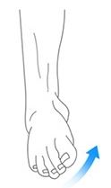

Identify the joint action being performed.
Inversion
Eversion


Which movement is shown in the picture?
circumduction
rotation
Hip Extension


Joint movements?
Hip Flexion
Hip Extension
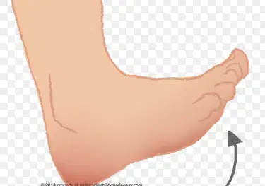
dorsiflexion
pointing toes toward the ground
dorsiflexion
plantarflexion
Match bone joint terms
Palm turns from posterior to anterior
Palm turns from anterior to posterior
ACL
anterior cruciate ligament
MCL
medial collateral ligament
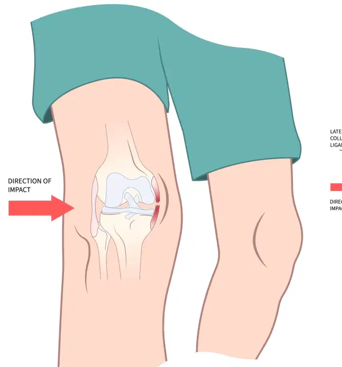
What knee ligament is torn in this image?
It is located toward the center of the body. It is opposite the LCL.
**It is highlighted in pinkish/red.
ACL
MCL
PCL

Which ligament is torn in this image?
It attaches the femur and fibula.
It is located towards the outside of the body, opposite the MCL.
ACL
LCL
PCL
Match joint movement terms.
origin
less movable end
insertion
more movable end
elevation
raising (shrugging) the shoulders
depression
lowering (drooping) the shoulders
Match bone ridges
line
low ridge
crest (crista)
prominent ridge
spine
very high ridge
Match bone structure openings & projection
process
prominent projection
foramen
hole
meatus
canal, tunnel
sinus
cavity

