
 Worksheets
PATH 184 LO2
Total questions: 70
Worksheet time: 52mins
A patient that has ascites would require an increase in kVp
True
False
A patient that has a small or large bowel obstruction would require an increase in kVp
True
False
The process of digestion begins in the
stomach
small bowel
mouth
large bowel
The rhythmic smooth muscle contractions that help move food through the digestive tract is called
peristalsis
deglutition
mastication
emulsification
The greatest amount of digestion occurs in the
transverse colon
stomach
sigmoid colon
duodenum
This organ is responsible for secreting an alkaline solution to neutralize the acid carried into the small intestine from the stomach.
liver
gall bladder
pancreas
stomach
Tracheoesophageal Fistulas can be congenital or acquired
True
False
The type of congenital Tracheoesophageal Fistula that may go undetected into adulthood.
Type 1
Type 3
Type 4
Type 2
The most common cause of acute esophagitis
medications
infections
reflux
physical injury
DOES NOT tend to decrease the pressure of the esophageal sphincter, causing reflux.
Water
Coffee
Alcohol
Chocolate
On a double-contrast study, looks like superficial ulcerations that appear as streaks or dots of barium superimposed on the flat mucosa of the distal esophagus.
Esophageal Cancer
Esophageal Diverticula
Esophageal Varices
Esophagitis
Severe reflux leads to normal squamous lining being replaced by columnar epithelium
Barrett Esophagus
Esophageal Cancer
Esophageal Diverticula
Esophageal Varices
Ulcerations tend to develop at a distance from the hiatal hernia and tend to result in a smooth, tapered stricture.
Esophageal Diverticula
Esophageal Cancer
Barrett Esophagus
Esophagitis
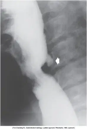
Name this pathology
Esophageal Diverticula
Esophagitis
Barrett Esophagus
Esophageal Cancer

Name this pathology
Esophageal Cancer
Esophagitis
Barrett Esophagus
Esophageal Diverticula
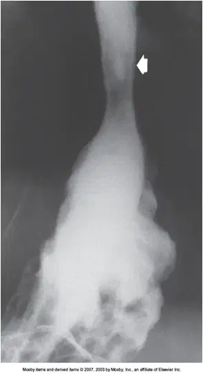
Name this pathology
Barrett Esophagus
Esophagitis
Esophageal Cancer
Esophageal Diverticula

Name this pathology
Esophageal Cancer
Esophagitis
Esophageal Diverticula
Barrett Esophagus
Progressive dysphagia in a person older than 40 should be assumed to be this condition until demonstrated otherwise.
Esophageal Cancer
Esophagitis
Barrett Esophagus
Esophageal Diverticula
This modality is a major method of staging patients with Esophageal Cancer
MRI
CT
US
Plain Radiography
Zenker Diverticula arise from this area of the esophagus
upper anterior
upper posterior
lower anterior
lower posterior
Dilated veins in the wall of the esophagus usually a result of cirrhosis of the liver.
Esophagitis
Esophageal Varices
Barrett Esophagus
Esophageal Diverticula

Name the pathology shown here. "Rosary Beads"
Esophagitis
Esophageal Cancer
Esophageal Varices
Barrett Esophagus
Symptoms associated with hiatal hernia include all except ...
Dysphagia
Esophageal Stenosis
Esophageal Ulcer
Esopahagitis
A functional obstruction of the distal section of the esophagus with proximal dilation.
Hiatal Hernia
Achalasia
Esophageal Varices
Esophagitis
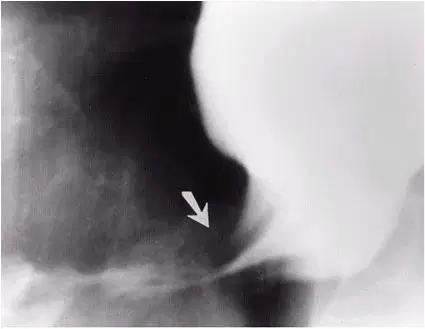
Name the pathology seen here.
Achalasia
Esophagitis
Esophageal Cancer
Hiatal Hernia
Appears as a widened mediastinum on the right side of the cardiac shadow on the PA Chest projection and a posterior widening on the Lateral Chest projection.
Barrett Esophagus
Hiatal Hernia
Esophagitis
Achalasia
Most common cause of Esophageal Perforation in an otherwise healthy esophagus.
Esophagitis
severe vomiting
external trauma
Peptic Ulcer
A complete or almost complete obstruction, preventing food from entering the duodenum.
Duodenal Stenosis
Duodenal Ulcer
Peptic Ulcer
Pyloric Stenosis
The most common manifestation of Peptic Ulcer Disease
Pyloric Stenosis
Duodenal Ulcer
Gastric Ulcer
Stomach Cancer
The most common cause of upper GI bleeding.
Stomach Cancer
Peptic Ulcer
Disease
Pyloric Stenosis
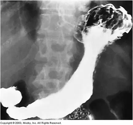
Name this pathology.
Peptic Ulcer Disease
Gastric Ulcer
Pyloric Stenosis
Stomach Cancer
This disorder usually involves the terminal ileum
Crohn Disease
Adynamic Ileus
Intussusception
Malabsorption Disorder
Small bowel obstruction and fistulas are a common result of this condition.
Malabsorption Disorder
Crohn Disease
Adynamic Ileus
Intussusception
Mostly caused from previous surgery.
Small Bowel Obstruction
Intussusception
Malabsorption Disorder
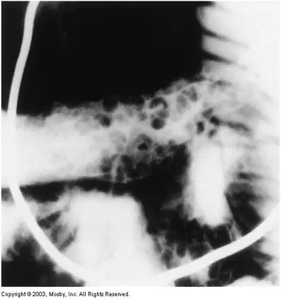
Name this pathology. "Cobblestone"
Small Bowel Obstruction
Adynamic Ileus
Crohn Disease
Intussusception
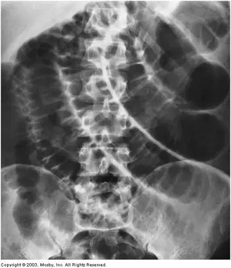
Name the pathology seen here. "Step-ladder"
Malabsorption Syndrome
Adynamic Ileus
Crohn Disease
Small Bowel Obstruction
In order to distinguish small bowel obstruction from large bowel obstruction, it is crucial to look at the positioning of the dilated loops of bowel.
False
True
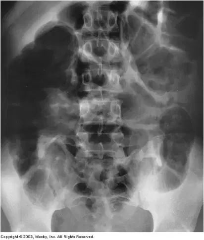
Name the pathology seen here. Dilated loops of small and large bowel with no indication for a point of obstruction.
Adynamic Ileus
Intussusception
Small Bowel Obstruction
Crohn Disease
This condition is most common in children and usually involves the ileocecal valve.
Adynamic Ileus
Small Bowel Obstruction
Intussusception
Malabsorption Syndrome
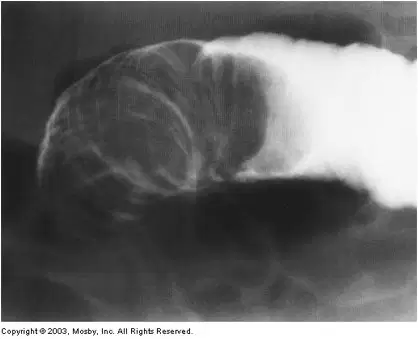
Name the pathology demonstrated here. "Coiled-Spring"
Intussesception
Adynamic Ileus
Small Bowel Obstruction
Crohn Disease

Name the pathology seen here. "Double Bubble"
Intussusception
Small Bowel Obstruction
Malabsorption Syndrome
Duodenal Atresia
Diagnosis for this pathology does not rely on radiographic findings, but clinical symptoms and lab results.
Ischemic Colitis
Irritable Bowel Syndrome
Acute Appendicitis
Ulcerative Colitis
Outpouchings through the muscular layers at points of weakness in the bowel wall.
Diverticulitis
Volvulus
Diverticulosis
Polyps
Most common area for diverticula to occur in the colon.
Ascending
Sigmoid
Cecum
Descending
A common complication of Diverticulitis is fistulas.
True
False
A complication of this condition is toxic megacolon where a segment of the bowel appears extremely dilated.
Large Bowel Obstruction
Ulcerative Colitis
Diverticulitis
Irritable Bowel Syndrome

This pathology appears as a "lead pipe" colon, with the loss of normal haustral markings.
Colon Cancer
Large Bowel Obstruction
Ulcerative Colitis
Diverticulosis
The proximal portion of the colon is most frequently involved in this pathology.
Ulcerative Colitis
Crohn Colitis
Diverticulitis
Appendicitis
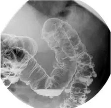
Name the pathology seen here.
Crohn Colitis
Ulcerative Colitis
Diverticulosis
Ischemic Colitis
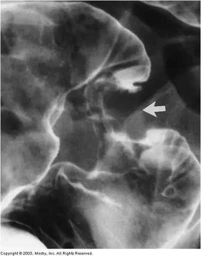
Name the pathology seen here. "Apple core"
Diverticulosis
Ulcerative Colitis
Crohn Colitis
Colon Cancer
This pathology presents as a sudden onset of lower abdominal pain and rectal bleeding.
Crohn Colitis
Ulcerative Colitis
Ischemic Colitis
Appendicitis
Patients with this condition present with alternating bouts of constipation and diarrhea.
Ulcerative Colitis
Crohn Colitis
Irritable Bowel Syndrome
Ischemic Colitis
Ulcerative Colitis and Polyps make a person predisposed to the development of this pathology.
Colon Cancer
Crohn Colitis
Diverticulitis
Irritable Bowel Syndrome
This pathology is most often caused by colon cancer.
Irritable Bowel Syndrome
Large Bowel Obstruction
Diverticulitis
Crohn Colitis
The major concern with Large Bowel Obstruction is perforation of this area.
Splenic Flexure
Hepatic Flexure
Cecum
Sigmoid
Volvulus tends to occur in the transverse colon.
True
False
You are predisposed to this pathology if you are female, fluffy, and forty.
Gallstones
Appendicitis
Ulcerative Colitis
Diverticulosis
Most common result of an obstructed cystic duct caused by an impacted gallstone.
Acute Cholecystitis
Emphysematous Cholecystitis
Vaccine is required by the childhood immunization process due to the high incidence.
Hepatitis A
Hepatitis B
Hepatitis C
Hepatitis E
A condition where fibrous connective tissue replaces destroyed liver cells with cells that have no liver cell function.
Hepatic Metastases
Hepatitis C
Hepatitis B
Cirrhosis
The liver appears darker on CT in Cirrhosis.
True
False
Cirrhosis of the liver can lead to all EXCEPT this pathology
Splenomegaly
Ascites
Esophageal Varices
Pleural Effusion
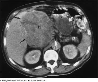
Name the pathology shown here.
Cirrhosis
Hepatocellular Carcinoma
Ascites
Hepatic Metastases
The modality of choice for Hepatocellular Carcinoma and Hepatic Metastases
Fluoroscopy
CT
Plain Radiography
US
The elevation of this enzyme will confirm the diagnosis of Acute Pancreatitis.
Lipase
Amylase
Protease
Esterase
In Chronic Pancreatitis, calcifications are most frequently seen in this area of the pancreas.
Head
Body
Tail

Name the pathology seen here.
Acute Pancreatitis
Chronic Pancreatitis
Pancreatic Cancer
Pancreatic Pseudocyst
Percutaneous Biliary Drainage is a method of treatment for patients with this condition.
Acute Pancreatitis
Chronic Pancreatitis
Pancreatic Cancer
Pancreatic Pseudocyst

Name the pathology seen here.
Ascites
Pneumothorax
Pneumoperitoneum
Pleural Effusion
Most often caused by trauma.
Cirrhosis of the liver
Appendicitis
Spleen Rupture
Pneumoperitoneum

