
 Worksheets
Anatomy II Exam 3
Total questions: 84
Worksheet time: 55mins
The 2 structural divisions of the Respiratory System are:
Upper Respiratory Tract & Respiratory Zone
Upper Respiratory Tract & Lower Respiratory Tract
Conducting Zone & Respiratory Zone
Conducting Zone and Lower Respiratory Tract
What organs belong in the Upper Respiratory Tract and the Lower Respiratory Tract?
Upper: nose , nasal cavity, pharynx, larynx
Lower: larynx, trachea, bronchi, lungs
Upper: trachea, bronchi, lungs, larynx
Lower: bronchi, bronchioles, terminal bronchioles, nose, pharynx, larynx, trachea
Upper: bronchi, bronchioles, terminal bronchioles, nose, pharynx, larynx, trachea
Lower: larynx, trachea, bronchi, lungs
What happens in the Upper Respiratory Tract?
involved with bringing air to the site of the external respiration
involved with gas exchange (alveoli)
air entering is filtered, warmed, and humidified
lined spaces in facial bones
What happens in the Lower Respiratory Tract?
involved with bringing air to the site of external respiration
air entering is filtered, warmed, and humidified
increase the surface area of the nasal cavities to provide warming and humidification of air as it passes to the lungs
It is involved with gas exchange (alveoli)
The 2 functional divisions of the respiratory system are:
Conducting Zone & Respiratory Zone
Upper Respiratory Tract & Lower Respiratory Tract
Upper Respiratory Tract & Respiratory Zone
Lower Respiratory Tract & Conducting Zone
What organs belong in the Conducting and Respiratory Zone?
Conducting: respiratory bronchioles, alveolar ducts, alveolar sacs, and alveoli
Respiratory: nose, pharynx, larynx, trachea, bronchi, bronchioles, and terminal bronchioles
Conducting: larynx, trachea, bronchi, lungs
Respiratory: nose, nasal cavity, pharynx, larynx
Conducting: nose, pharynx, larynx, trachea, bronchi, bronchioles, and terminal bronchioles
Respiratory: respiratory bronchioles, alveolar ducts, alveolar sacs, and alveoli
What is the function of the Conducting Zone?
it is the main site of gas exchange
it is involved with bringing air to the site of external respiration
air entering is filtered, warmed, and humidified
it is involved with gas exchange (alveoli)
What happens in the Respiratory Zone?
it is involved with gas exchange (alveoli)
air entering is filtered, warmed, and humidified
it is the main site of gas exchange
involved with bringing air to the site of external respiration
What 3 components make up the nasal septum?
vomer, septal cartilage, ethmoid
nasopharynx, oropharynx, laryngopharynx
Submucosa, Hyaline cartilage, adventitia
Apex, Base, Cardia Notch
What are paranasal sinuses?
mucous membrane-lined spaces in facial bones
Entrance or opening of glottis
Segment of lung tissue that each segmental bronchus supplies
allow for exchange of gases with the pulmonary capillaries
What is the function of paranasal sinuses?
to adhere tightly to the surface of the lungs
to increase the surface area of the nasal cavities to provide warming and humidification of air as it passes to the lungs
to support vocal folds and lateral aspects of epiglottis
to increase surface area of nasal mucosa
What is the function of the nasal conchae?
to increase surface area of nasal mucosa
to increase the surface area of the nasal cavities to provide warming and humidification of air as it passes to the lungs.
to supports vocal folds and lateral aspects of epiglottis
to provide flexibility to tracheal pipe
What are the 3 regions of the pharynx?
vomer, septal cartilage, ethmoid
Nasopharynx, oropharynx, laryngopharynx
Thyroid cartilage, cricoid cartilage, arytenoid cartilage
Submucosa, hyaline cartilage, adventitia
What is the location of the Nasopharynx?
posterior to larynx
posterior to oral cavity and extends from soft palate to hyoid bone
posterior to nasal cavity and extends to soft palate
anterior lateral surface of larynx
What structures are in the Nasopharynx?
Thyroid cartilage, cricoid cartilage, arytenoid cartilage
vomer, septal cartilage, ethmoid
palatine tonsils and small lingual tonsil
Pharyngeal tonsils and openings of the Eustachian tubes
What is the location of the Oropharynx?
posterior to nasal cavity and extends to soft palate
posterior to larynx
posterior to oral cavity and extends from soft palate to hyoid bone
anterior lateral surface of larynx
What structures are in the Oropharynx?
thyroid cartilage, cricoid cartilage, cuneiform cartilage
pharyngeal tonsils and openings of the Eustachian tubes
palatine tonsils and small lingual tonsil
vomer, septal cartilage, ethmoid
Where is the Laryngopharynx located?
posterior to larynx
posterior to oral cavity and extends from soft palate to hyoid bone
anterior lateral surface of larynx
posterior to nasal cavity and extends to soft palate
How many pieces of cartilage make up the larynx?
3
5
11
9
What are the 9 cartilages of the larynx?
Thyroid, cricoid, arytenoid, corniculate, cuneiform, epiglottis
carina, alveoli, rima glottidis, vocal folds, primary bronchi, secondary bronchi, tertiary bronchi, apex, cardia notch
Parietal pleura, Visceral pleura, Pleural cavity, Pleural fluid
C-rings, alveolar wall, alveolar sacs, pulmonary capillaries, capillary basement membrane
Which 3 cartilages are paired?
epiglottis, thyroid, cricoid
corniculate, cricoid, epiglottis
cuneiform, cricoid, corniculate
Arytenoid, corniculate, cuneiform
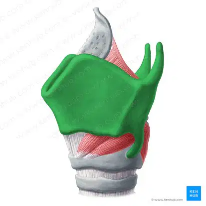
What is the function of the thyroid cartilage and its location?
inferior wall of larynx or base, connects larynx and trachea
anterior lateral surface of larynx, to protect the vocal cords
attached to root of the tongue, prevents food from entering the larynx
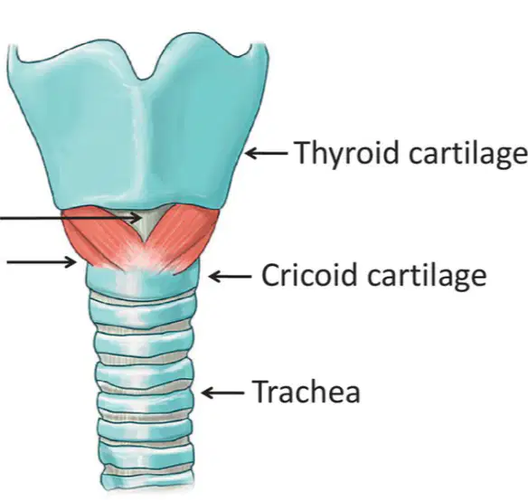
What is the function of the cricoid cartilage and where is it located?
anterior lateral surface of larynx, to protect the vocal cords
attached to root of the tongue, prevents food from entering the larynx
inferior wall of larynx or base, connects larynx and trachea
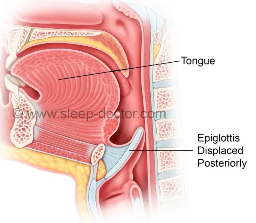
What is the function of the epiglottis and where is it located?
located at posterior, influence changes in position and tension of vocal folds
inferior wall of larynx or base, connects larynx and trachea
attached to root of the tongue, prevents food from entering the larynx
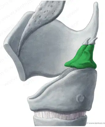
What is the function of the arytenoid cartilage and where is it located?
attached to root of the tongue, prevents food from entering the larynx
anterior lateral surface of larynx, to protect the vocal cords
located at posterior, influence changes in position and tension of vocal folds
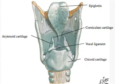
What is the function of the corniculate cartilage and where is it located?
located at apex of arytenoid cartilage, connects the cricoid cartilage and the thyroid cartilage
attached to root of the tongue, prevents food from entering the larynx
located at posterior, influence changes in position and tension of vocal folds
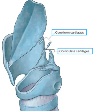
What is the function of the cuneiform cartilage and where is it located?
located at apex of arytenoid cartilage, connects the cricoid cartilage and the thyroid cartilage
inferior wall of larynx or base, connects larynx and trachea
supports vocal folds and lateral aspects of epiglottis
What is the rima glottidis?
the space between the vocal folds and the pair of mucous membrane folds
Entrance or opening of glottis
What is the rima glottis?
Entrance or opening of glottis
the space between the vocal folds and the pair of mucous membrane folds
Where are the vocal cords located?
Entrance or opening of glottis
anterior lateral surface of larynx
High in the larynx
located at posterior
How is pitch controlled?
by tension on the vocal folds
with the epiglottis
Air path flow chart
Nasal cavity > Pharynx > Larynx > Parietal Pleura > Visceral Pleura > Pleural cavity > Alveoli
Nasal cavity > Pharynx > Larynx > bronchioles > alveolar ducts > alveolar sacs > alveoli
Nasal cavity > Pharynx > Larynx > Trachea > Primary bronchi > Secondary bronchi > Tertiary bronchi > Bronchioles > Alveoli
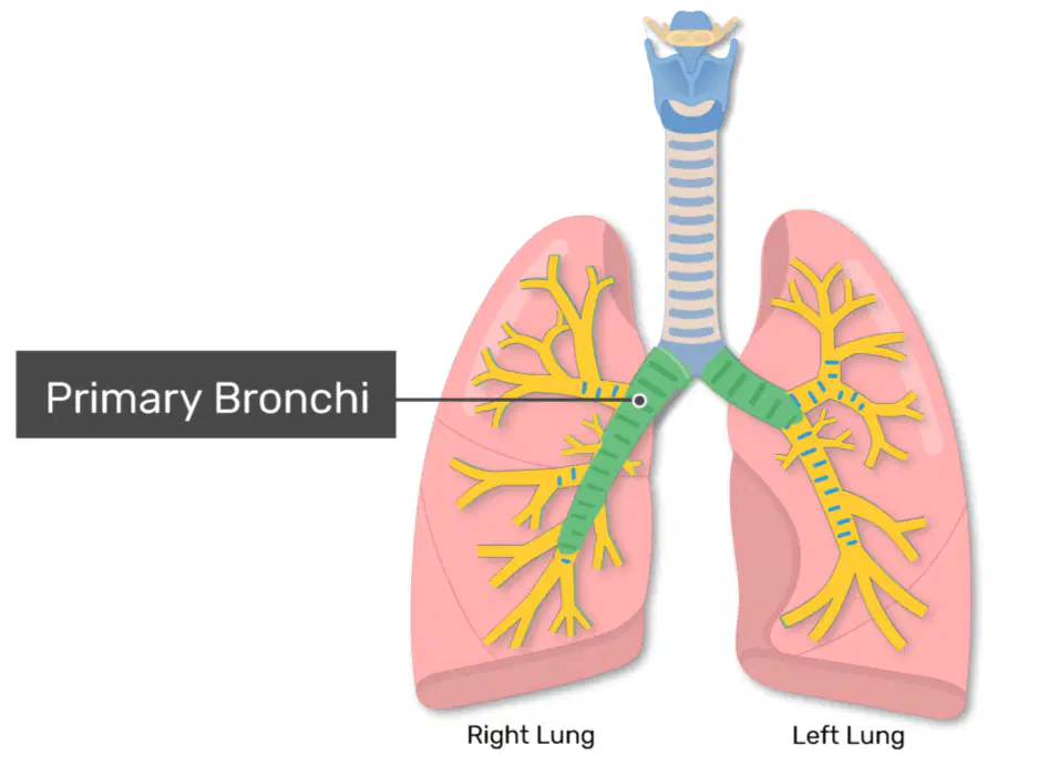
Primary bronchi (also called the mainstem bronchi branches) is
the left and right main bronchi in the upper portion of your lungs
the middle of your lungs
the edge of your lungs
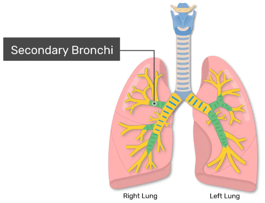
Secondary bronchi is:
the left and right main bronchi in the upper portion of your lungs
the edge of your lungs
the middle of your lungs
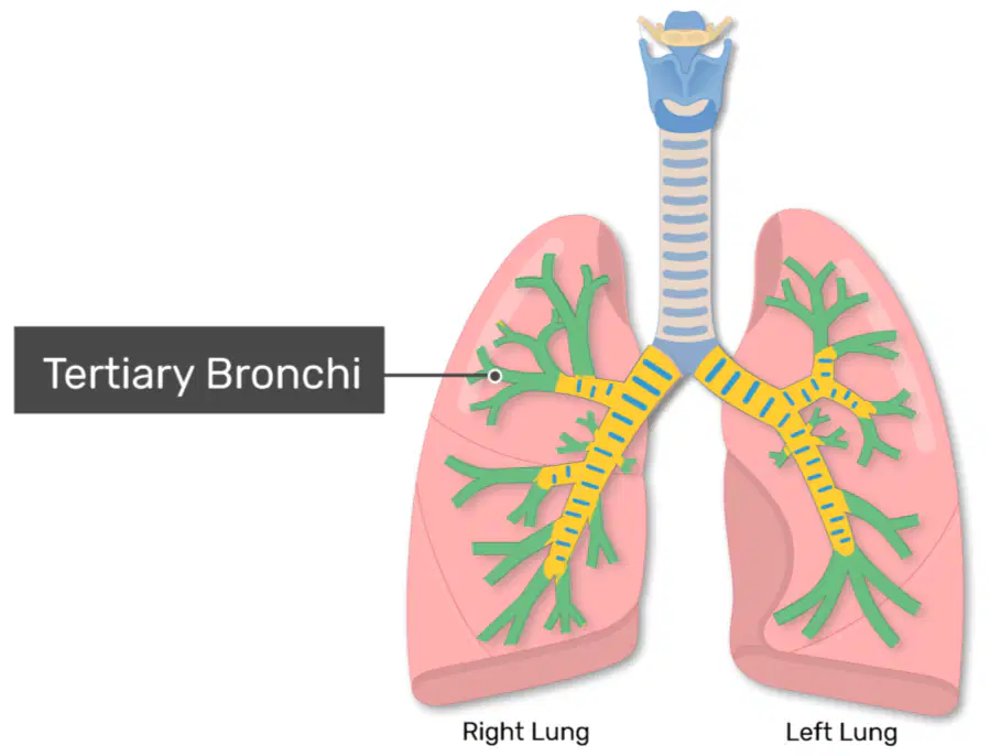
Tertiary bronchi is:
the edge of your lungs
the left and right main bronchi in the upper portion of your lungs
the middle of your lungs
What are the 4 layers of the trachea?
Parietal pleura, visceral pleura, pleural cavity, pleural fluid
Mucosa, submucosa, hyaline cartilage, adventitia
Apex, base, cardia notch, alveolar wall
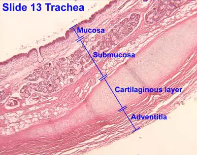
The mucosa is:
provide flexibility to tracheal pipe
loose CT, abundant blood and lymph vessels
binds trachea to adjacent structures
mucous secreting epithelium
The submucosa is:
mucous secreting epithelium
binds trachea to adjacent structures
provide flexibility to tracheal pipe
loose CT, abundant blood and lymph vessels
The hyaline cartilage is:
loose CT, abundant blood and lymph vessels
provide flexibility to tracheal pipe
mucous secreting epithelium
binds trachea to adjacent structures
The adventitia is:
mucous secreting epithelium
provide flexibility to tracheal pipe
binds trachea to adjacent structures
loose CT, abundant blood and lymph vessels
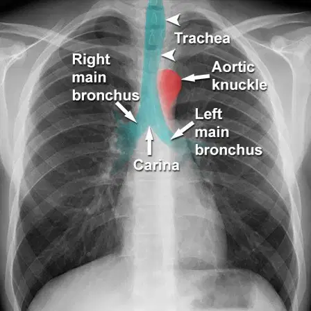
What is the carina?
Entrance or opening of glottis
an internal ridge located at the junction of the two mainstem bronchi area for triggering the cough reflex
Segment of lung tissue that each segmental bronchus supplies

Bronchial tree flow chart
Trachea > Primary bronchi > Secondary bronchi > Tertiary bronchi > Bronchioles > Terminal bronchioles
Nasal cavity > Pharynx > Larynx > Trachea > Primary bronchi > Secondary bronchi > Tertiary bronchi > Bronchioles > Alveoli
Respiratory bronchioles > Alveolar ducts > Alveolar saccules > Pulmonary alveoli
What changes happen as we move from the bronchi to the bronchioles? (multiple answers)
(Take your time)
Mucous membrane changes from ciliated pseudostratified epithelium to non-ciliated cuboidal epithelium
C-rings of the trachea are replaced by plates of cartilage in the bronchi
As the amount of cartilage decreases, the amount of smooth muscle increases
secrete substance called surfactant that prevents collapse of the alveoli
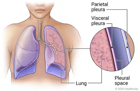
What is the Parietal Pleura?
adhere tightly to the surface of the lungs
space between the parietal and visceral pleural layers
line the walls of the thoracic cavity
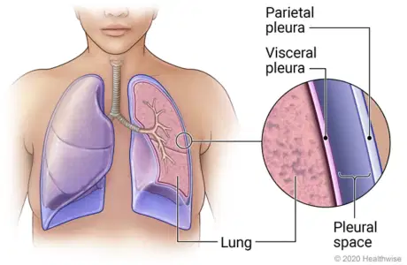
What is the visceral pleura?
space between the parietal and visceral pleural layers
adhere tightly to the surface of the lungs
line the walls of thoracic cavity
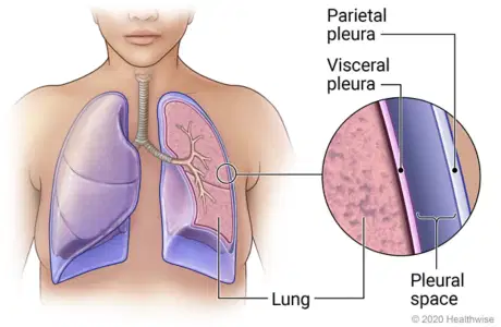
What is the Pleural Cavity/Space?
adhere tightly to the surface of the lungs
line the walls of thoracic cavity
space between the parietal and visceral pleural layers
What is the function of the Pleural Fluid?
to reduce friction
to trigger the cough reflex
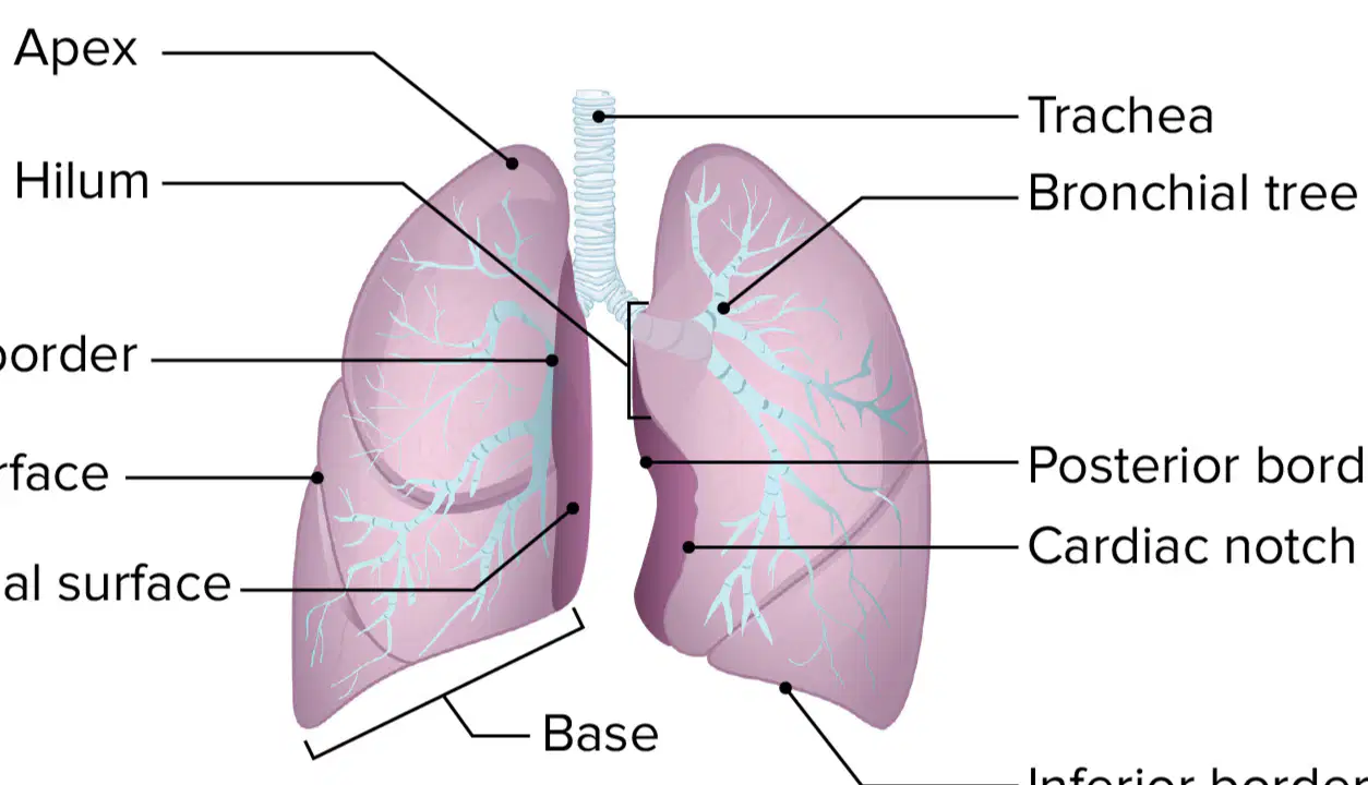
Where are the lung structures (apex, base & cardiac notch) located?
Apex - superior, slightly above the clavicles
Base - rests on the diaphragm
Cardiac Notch - makes left lung 10% smaller than the right lung
Apex - rests on diaphragm
Base - superior, slightly above the clavicles
Cardiac Notch - makes left lung 10% smaller than the right lung
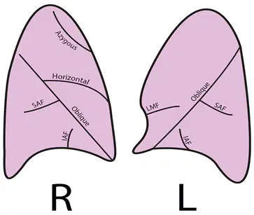
What fissures divide the right and left lung?
Right: Oblique fissure
Left: Oblique & horizontal fissure
Right: Oblique & horizontal fissure
Left: Oblique fissure
What is the bronchopulmonary segment?
an internal ridge located at the junction of the two mainstem bronchi area for triggering the cough reflex
Segment of lung tissue that each segmental bronchus supplies
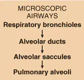
Microscopic airway flow chart
Trachea > Primary bronchi > Secondary bronchi > Tertiary bronchi > Bronchioles > Terminal bronchioles
Respiratory bronchioles > Alveolar ducts > Alveolar saccules > Pulmonary alveoli
Nasal cavity > Pharynx > Larynx > Trachea > Primary bronchi > Secondary bronchi > Tertiary bronchi > Bronchioles > Alveoli
What is the function of type I and type II alveolar cells?
Type I: allow for gas exchange with pulmonary capillaries
Type II: secrete substance called surfactant that prevents collapse of the alveoli during exhalation
Type I: secrete substance called surfactant that prevents collapse of the alveoli during exhalation
Type II: allow for gas exchange with pulmonary capillaries
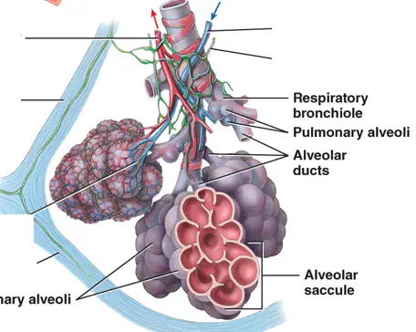
What structures make up the pulmonary lobule?
Parietal pleura, visceral pleura, pleural cavity
Apex, base, cardiac notch
Bronchiole, alveolar ducts, sacs, and alveoli
What 4 structures make up the respiratory membrane?
Bronchiole, alveolar ducts, sacs, and alveoli
Alveolar wall, Epithelial basement membrane, Capillary basement membrane, Capillary endothelium
Parietal pleura, visceral pleura, pleural cavity, pleural fluid
How does gas flow, in relation to pressure?
High to low pressure
Low to high pressure
During inhalation lungs expand which ____ lung volume and ____ lung pressure.
increases, decreases
increases, increases
decreases, increases
During exhalation lungs shrink which ____ lung volume and ____ lung pressure.
increases, decreases
increases, increases
decreases, increases
For exhalation to occur alveolar pressure must be ____ than atmospheric pressure
lower
higher
For inhalation to occur alveolar pressure must be ____ than atmospheric pressure
lower
higher
What is Boyle's Law?
increasing the partial pressure of a gas “over” a solution will result in more of the gas dissolving into the solution
volume and pressure are inversely related
the pressure of each gas is directly proportional to the percentage of that gas in the total mixture
What is Dalton's Law?
increasing the partial pressure of a gas “over” a solution will result in more of the gas dissolving into the solution
volume and pressure are inversely related
the pressure of each gas is directly proportional to the percentage of that gas in the total mixture
What is Henry's Law?
increasing the partial pressure of a gas “over” a solution will result in more of the gas dissolving into the solution
volume and pressure are inversely related
the pressure of each gas is directly proportional to the percentage of that gas in the total mixture
What is pulmonary ventilation?
exchange of gases between blood in systemic capillaries and tissue cells
exchange of gases between alveoli of lungs and blood in pulmonary capillaries across respiratory membrane
process of inhaling and exhaling and involves exchange of air between atmosphere and alveoli of lungs
What is external respiration?
exchange of gases between blood in systemic capillaries and tissue cells
exchange of gases between alveoli of lungs and blood in pulmonary capillaries across respiratory membrane
process of inhaling and exhaling and involves exchange of air between atmosphere and alveoli of lungs
What is internal respiration?
exchange of gases between blood in systemic capillaries and tissue cells
exchange of gases between alveoli of lungs and blood in pulmonary capillaries across respiratory membrane
process of inhaling and exhaling and involves exchange of air between atmosphere and alveoli of lungs
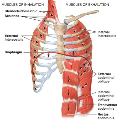
What muscles are involved in normal quiet inhalation and exhalation?
Quiet inhalation: Internal intercostals, external abdominal oblique, transverse abdominis and rectus abdominis muscles
Quiet exhalation: Diaphragm and external intercostals
Quiet inhalation: Diaphragm and external intercostals
Quiet exhalation: Internal intercostals, external abdominal oblique, transverse abdominis and rectus abdominis muscles
What additional muscles are involved in deep/forceful inhalation and exhalation?
Deep inhalation: scales and sternocleidomastoid
Deep exhalation: abdominal and internal intercostals
Deep inhalation: abdominal and internal intercostals
Deep exhalation: scales and sternocleidomastoid
What is eupnea, costal breathing and diaphragmatic breathing?
Eupnea: normal pattern of quiet breathing
Costal breathing: pattern of deep breathing (abdominal)
Diaphragmatic breathing: pattern of shallow breathing (chest)
Eupnea: normal pattern of quiet breathing
Costal breathing: pattern of shallow breathing (chest)
Diaphragmatic breathing: pattern of deep breathing (abdominal)
Eupnea: pattern of deep breathing (abdominal)
Costal breathing: pattern of shallow breathing (chest)
Diaphragmatic breathing: normal pattern of quiet breathing
What is tidal volume (Vt)?
volume of one breath (500 mL) inhaled
number of breaths a person takes per minute (12 breaths per minute)
total volume of air inhaled and exhaled each minute = respiratory x tidal volume = 12 breaths x 500 ml = 6,000 ml/min
What is respiratory rate?
volume of one breath (500 mL) inhaled
number of breaths a person takes per minute (12 breaths per minute)
total volume of air inhaled and exhaled each minute = respiratory x tidal volume = 12 breaths x 500 ml = 6,000 ml/min
What is minute volume (MV)?
volume of one breath (500 mL) inhaled
number of breaths a person takes per minute (12 breaths per minute)
total volume of air inhaled and exhaled each minute = respiratory x tidal volume = 12 breaths x 500 ml = 6,000 ml/min
What is the anatomical dead space and how does it affect minute ventilation?
it is 30% of the remaining tidal volume in the conducting zone
Instead of 500 mL only 350 mL of gases will be exchanged at the alveoli = 500 mL x 0.70 = 350
it is volume of air/min that actually reaches the alveoli = 12 breaths/min x 350 ml = 4,200 ml/min
What is alveolar ventilation?
30% of the remaining tidal volume in the conducting zone
volume of air/min that actually reaches the alveoli = 12 breaths/min x 350 ml = 4,200 ml/min
What is inspiratory and expiratory reserve volume?
Inspiratory: additional air over the 500 ml average {M=3100 ml, F=1900 ml}
Expiratory: extra volume of air exhaled {M= 1200 ml, F= 700 mL}
Inspiratory: extra volume of air exhaled {M= 1200 ml, F= 700 mL}
Expiratory: additional air over the 500 ml average {M=3100 ml, F=1900 ml}
What is residual and minimal volume?
Residual volume: air remaining after residual volume is forced out
Minimal volume: air still present in the lungs after a force exhalation
Residual volume: air still present in the lungs after a force exhalation
Minimal volume: air remaining after residual volume is forced out
What is the inspiratory, vital, and total lung capacity?
Inspiratory: maximum volume of air that can be inspired after reaching end of normal, quiet expiration = tidal volume + inspiratory reserve volume
Vital: all the air that can be exhaled after maximum inspiration = inspiratory reserve + tidal volume + expiratory reserve
Total lung: maximum amount of air that can fill lungs, vital capacity + residual volume
Inspiratory: maximum volume of air that can be inspired after reaching end of normal, quiet expiration = tidal volume + inspiratory reserve volume
Vital: maximum amount of air that can fill lungs, vital capacity + residual volume
Total lung: all the air that can be exhaled after maximum inspiration = inspiratory reserve + tidal volume + expiratory reserve
As blood acidity increases, does the affinity of hemoglobin for oxygen increase or decrease?
Acidity increases, pH decreases = hemoglobin affinity for oxygen decreases
pH increases, acidity decreases = hemoglobin affinity for oxygen increases
If blood pH increases (alkaline), does the affinity of hemoglobin for oxygen increase or decrease?
Acidity increases, pH decreases = hemoglobin affinity for oxygen decreases
pH increases, acidity decreases = hemoglobin affinity for oxygen increases
As temperature increases, what happens to the oxygen on the hemoglobin?
Less oxygen is released
More oxygen is released
The medullary respiratory center:
is made of 2 collection of neurons
generate impulses to diaphragm via phrenic nerves and external intercostals muscles via the intercostal nerves
become activated in forceful breathing (exercise)
The Dorsal Respiratory Group (DRG):
is made of 2 collection of neurons
becomes activated in forceful breathing (exercise)
generate impulses to diaphragm via phrenic nerves and external intercostals muscles via the intercostal nerves
The Ventral Respiratory Group (VRG):
is made of 2 collection of neurons
become activated in forceful breathing (exercise)
generate impulses to diaphragm via phrenic nerves and external intercostals muscles via the intercostal nerves
What is the Pre-Botzinger complex?
cluster of neurons located in VRG that act in generation of rhythm of breathing
modifying basic rhythm of breathing generated by VRG
How does the pons influence respiration?
cluster of neurons located in VRG that act in generation of rhythm of breathing
modifying basic rhythm of breathing generated by VRG

