
 Worksheets
Lab 3 Practice Quiz
Total questions: 20
Worksheet time: 30mins

Observe this image of an angiosperm stem, and select all correct statements.
A indicates metaxylem.
A indicates vessel elements.
B indicates vessel elements.
B indicates metaxylem.

What stain was applied to the tissue in this section?
(a)

This is a section taken from a gymnosperm. What cell type is indicated by the blue and orange circle?
(a)

What tissue is indicated at C?
(a)
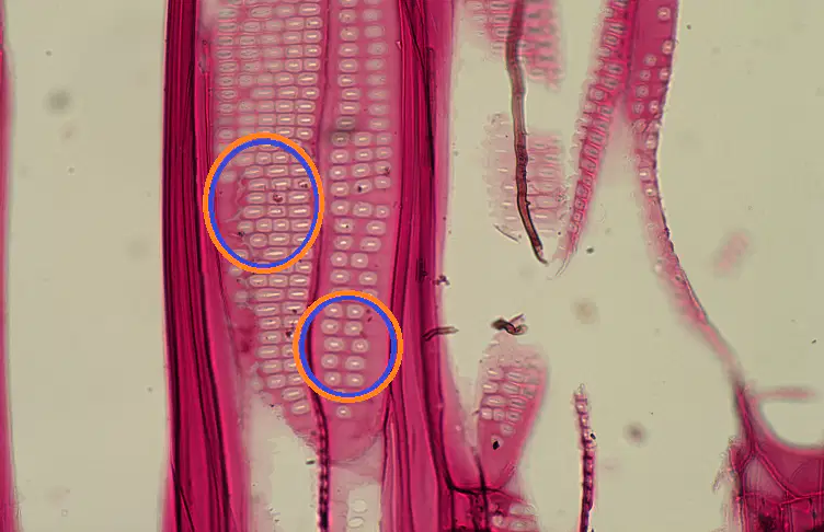
What structures are indicated by the circles?
(a)
True or False: Pits have a true membrane.
True
False
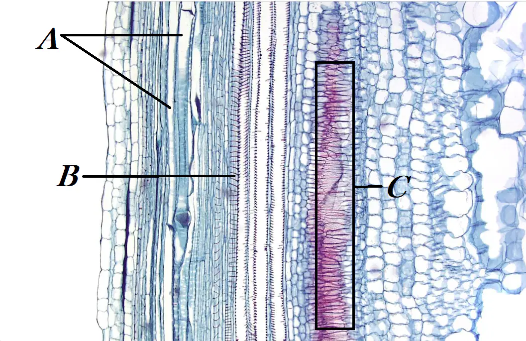
What cell type is indicated at A?
(a)
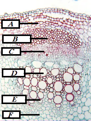
What cell type is indicated at F?
(a)
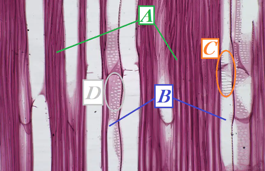
What cell type is indicated at B?
(a)

What structure is indicated by the arrow?
(a)

What cell type is indicated by B?
(a)

True or false: in this section taken from a pine tree (Pinus, a gymnosperm), the large opening indicated by the arrow is a vessel element.
True
False

What tissue is indicated by both A & B?
(a)

What cell type is indicated at B?
(a)

Which type of primary xylem is indicated by the box at C?
(a)

Which type of primary xylem indicated at E?
(a)
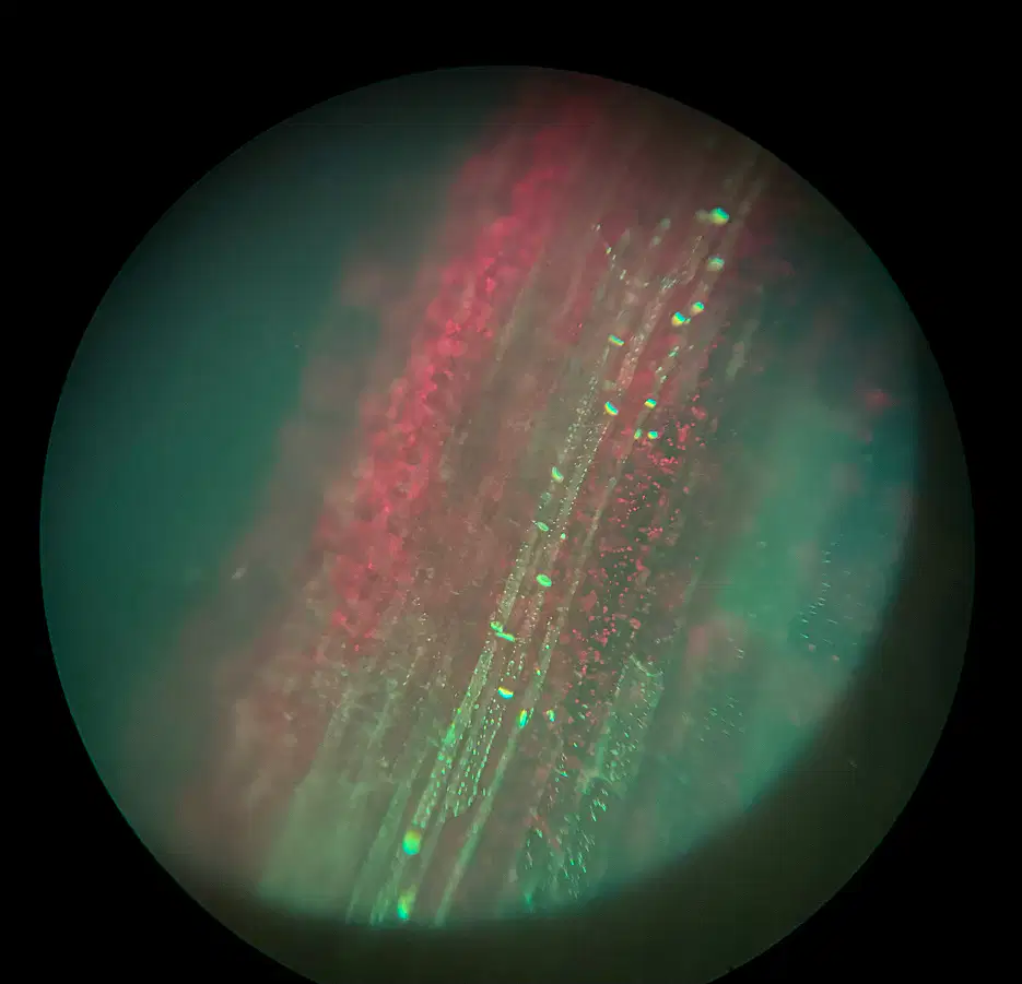
Observe the image, and select all correct statements.
A fluorescent microscope was used to generate this image.
Acid fuschin was applied to this section.
The stain is interacting with lignin in the xylem.
The bright yellow-green spots are "slime" plugs.
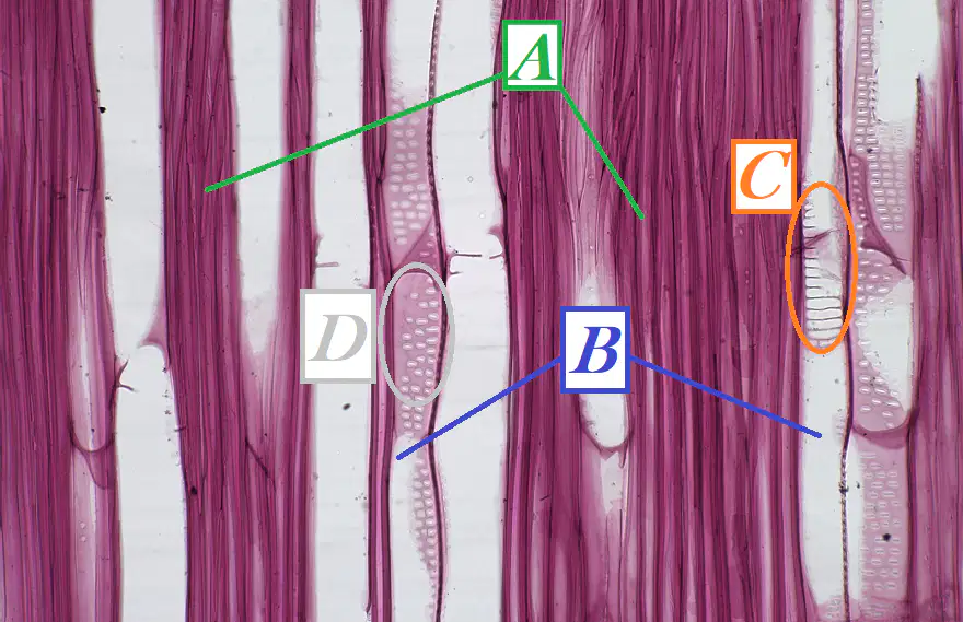
What structure is circled at C?
(a)

Observe the image, and select all correct statements.
The vascular bundle is bicollateral.
The phloem cells have thick, secondary cell walls.
The vascular bundle lacks xylem.
The xylem is surrounding the phloem on both sides.
Phloem is present on either side of the xylem.

Select the statement that best describes this image.
Aniline blue was used as a stain.
This image is of primary cell walls.
This image is of secondary cell walls.
This image is of helical secondary wall thickenings.

