
 Worksheets
1.1.3- 1.1.5 HBS TEST REVIEW
Total questions: 45
Worksheet time: 23mins

The highlighted bones are the:
Humerus
Radius
Ulna
Scapula

What are these bones (blue) called?
Tarsals
Carpals
Metatarsals
Metacarpals

The highlighted bones are known as:
Humerus
Radius
Ulna
Fibula

The highlighted bones of the body are the:
Thigh bones
Radius
Tibia
Femur

The bones (green) are known as the:
Metacarpals
Carpals
Phalanges
Digits

The bone (green) is known as the:
Ulna
Patella
Tibia
Hyoid

This irregular bones (blue on skeleton) make up the:
Hips don't lie...
Pelvic Girdle
Iliac Crest
Pubis

The bone (in green) is the:
Clavicle
Humerus
Spatula
Scapula

The bone (in red) is known as the:
Clavicle
Collarbone
Scapula
Sternum

The thicker, weight-bearing bone of the lower leg:
Femur
Tibia
Fibula
Fibia

This lower leg bone (red) is known as:
Femur
Tibia
Fibula
Tibula

What is the correct name of this bone (in red):
Hyoid
Thymus
Sternum
Breastbone

What is the name of the highlighted bone?
Humerus
Radius
Scapula
Ulna

The highlighted bones are called:
Tarsals
Metatarsals
Phalanges
Feets

These are known as:
Vertebra/Vertebrae
Spinal Cord
Spinal Bones
Pelvic Girdle
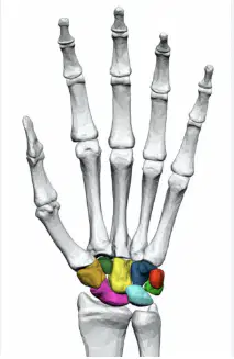
What bone is highlighted in rainbow?
Carpals
Metacarpals
Tarsals
Metatarsals
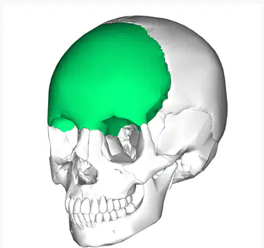
What bone is highlighted in green?
Frontal Bone
Occipital Bone
Temporal Bone
Parietal Bone
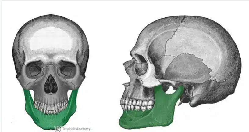
What bone is highlighted in green?
Maxilla
Mandible
Patella
Metatarsals
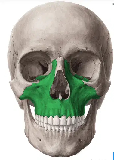
What bone is highlighted in green?
Maxilla
Mandible
Occipital
Sphenoid
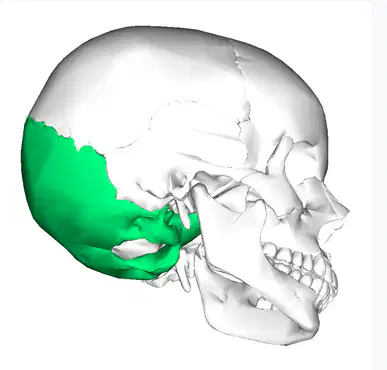
What bone is highlighted in green?
frontal bone
parietal bone
temporal bone
occipital bone
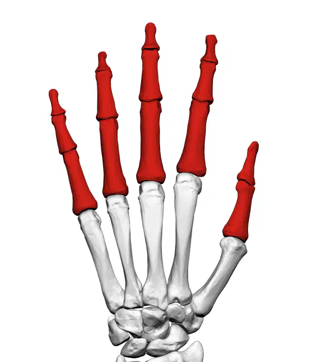
What bone is highlighted in red?
Metacarpals
Carpals
Phalanges
Tarsals
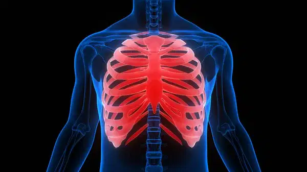
What bone is shown in the image?
Rib cage
Sphenoid
Temporal
Zygomatic
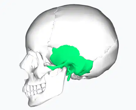
What bone is highlighted in green?
frontal bone
parietal bone
temporal bone
occipital bone
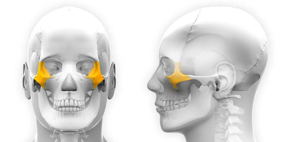
What bone is highlighted in yellow?
Frontal bone
Mandible
Maxilla
Zygomatic
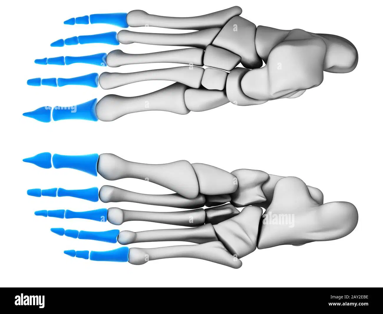
What bone is highlighted in blue?
Carpals
Tarsals
Metarsals
Phalanges
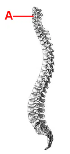
What is letter A showing in the image?
thoracic vertebra
cervical vertebra
sacral vertebra
lumbar vertebra
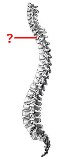
What is the diagram pointing to?
thoracic vertebra
cervical vertebra
coccygeal vertebra
lumbar vertebra
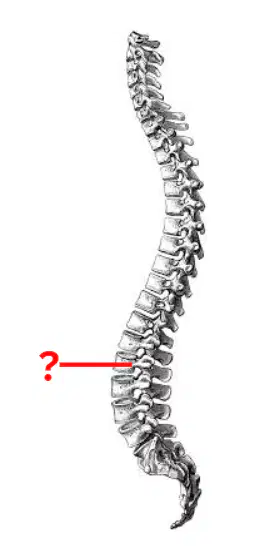
what is the diagram pointing to?
cervical vertebrae
lumbar vertebra
thoracic vertebra
sacral vertebra
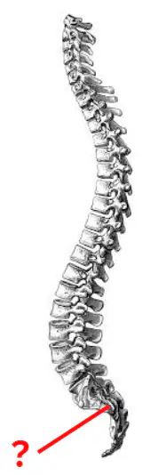
what is the diagram pointing to?
cervical vertebra
coccygeal vertebra
sacral vertebra
lumbar vertebra
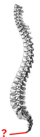
what is the diagram pointing to?
coccygeal vertebra
sacral vertebra
lumbar vertebra
thoracic vertebra
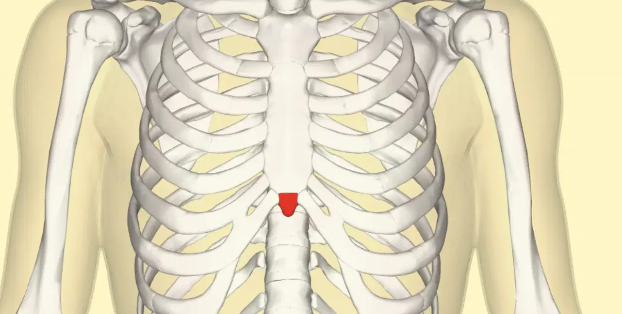
What structure is highlighted in red?
sternal body
manubrium
xiphoid process
mandible
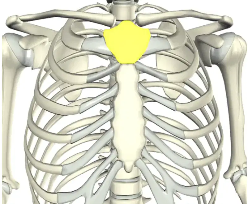
What part is highlighted in yellow?
xiphoid process
manubrium
sternal body
mandible
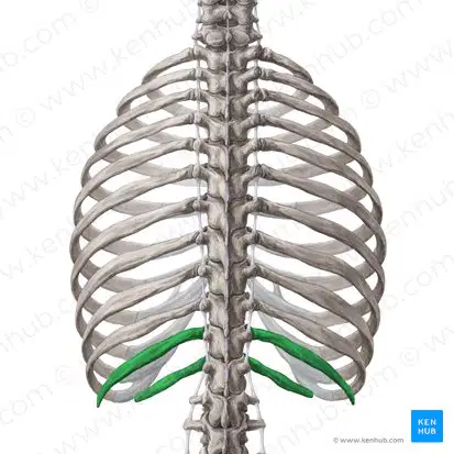
What kind of ribs are shown in the image?
false ribs
true ribs
floating ribs
unattached ribs
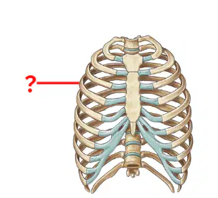
What kind of rib is the diagram pointing to?
false rib
true rib
floating rib
unattached rib
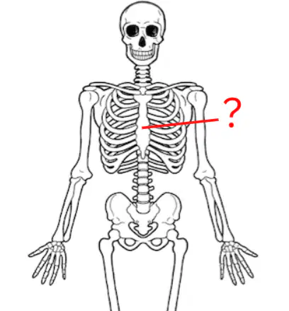
What bone?
Manubrium
Sternal Body
Xiphoid Process
Scapula
Identify which of the following is NOT correct.
Nervous Tissues- sends and receives signals
Epithelial Tissue- Protects with linings and coverings
Connective Tissue- Lines organs in the digestive system
Muscular Tissue- allows for movement in the body
You place a microscope slide on the microscope. What adjustment knob would you use FIRST to help bring the specimen into focus?
Fine adjustment knob
Coarse adjustment knob
Neither
Both
You pucker your lips to take a drink out of a straw. What muscle are you using?
Orbicularis Oris
Orbicularis Oculi
Temporalis
Intercostals
Someone acts like they are going to slap your face and you close your eyes. What muscle are you using?
Orbicularis Oris
Orbicularis Oculi
Temporalis
Intercostals
You are chewing a huge bite of hamburger. What muscle are you using?
Orbicularis Oris
Orbicularis Oculi
Temporalis
Intercostals
What is the smallest level of organization?
Cells
Tissues
Organs
Organ System
The stomach is an example of a-
Cell
Tissue
Organ
Organ system
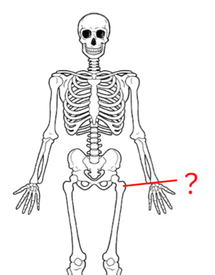
The diagram is pointing at the-
Proximal epiphysis of the femur
Distal epiphysis of the femur
Diaphysis of the femur
Periosteum of the femur
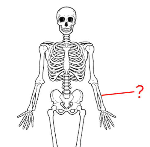
The diagram is pointing at the-
Proximal epiphysis of the radius
Distal epiphysis of the radius
Diaphysis of the radius
Periosteum of the radius
Which of the following is an example of connective tissue?
Spinal cord
Skeletal muscle
Ligaments
Lining of blood vessel

