
 Worksheets
X-ray Interpretation
Total questions: 22
Worksheet time: 18mins
What is the primary purpose of a chest X-ray?
To measure bone density in the chest area.
To assess blood flow in the brain.
To diagnose conditions related to the chest, such as lung infections or heart problems.
To evaluate digestive issues related to the stomach.
What anatomical structures are typically visible on a chest X-ray?
Kidneys
Stomach
Lungs, heart, ribs, diaphragm, major blood vessels, trachea, bronchi.
Brain
Describe the difference between AP and PA views in chest X-rays.
AP view is taken from the side; PA view is taken from the front.
AP view is taken from front to back; PA view is taken from back to front.
AP view is used for patients standing; PA view is used for patients lying down.
AP view shows the heart in more detail; PA view shows the lungs better.
What is the significance of the diaphragm's position on a chest X-ray?
The diaphragm's position is irrelevant to lung health.
The diaphragm's position only indicates heart size.
The diaphragm's position indicates lung health and potential abnormalities.
The diaphragm's position shows the age of the patient.
How can you identify the presence of fluid in the pleural space on a chest X-ray?
Presence of a solid mass in the lung field.
Normal appearance of the pleural space without any signs.
Blunting of costophrenic angles and meniscus sign on chest X-ray.
Increased lung markings and hyperinflation on chest X-ray.
What are the common signs of pneumonia on a chest X-ray?
Common signs of pneumonia on a chest X-ray include consolidation, pleural effusion, and air bronchograms.
Thickened bronchial walls
Presence of nodules
Increased lung volume
How does patient positioning affect the quality of a chest X-ray?
Patient positioning affects the quality of a chest X-ray by ensuring proper visualization of anatomical structures and minimizing distortion.
X-ray quality is solely determined by the machine settings.
Proper positioning is only important for MRI scans.
Patient positioning has no impact on X-ray quality.
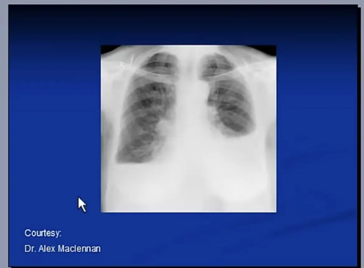
What is the the main finding?
Pleural effusion
Left lower lobe atelactasis
Pneumothorax
Left lower lobe consolidation
One of the main sign of detection of pleural effusion in chest xray is:
Cardiomegaly
Shift of the trachea toward the same side
Unclear costophrenic angles
Enlarged lung

What is the main finding?
Bilateral collapse
Bilateral consolidation
Bilateral pleural effusion
Pulmonary edema
Consolidation or infiltration is the term used when the alveoli are filled with
Puss
Fluid
Blood
All of the above
To ensure that the patient was not rotated in the x-ray image we should check:
Clavicles and spinous process
Intervertebral disc should be clear
Count of the ribs
The diaghram
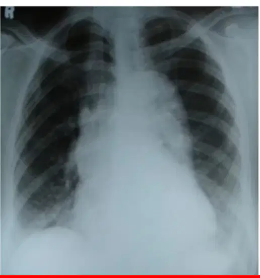
What is the main technical problem of this chest xray?
Exposure
Rotation
Count of the ribs
A and B

The arrow points to
clavicle
glenoid
corocoid process
acromion

There is a fracture of the
4th Metacarpal
5th Metacarpal
4th Metatarsal
2nd Metacarpal

The is a fracture of the
Distal radius
Ulna shaft
Humerus
Scaphoid

Labelled 3 is the
Azygous vein
Costophrenic angle
Carina
Cardiophrenic angle
Labelled 4 is the
Carina
Left main bronchus
Arotic arch
Hilum

This image shows
No abnormality detected
Bilateral effusion
Tension pneumothorax
Pneumothorax

This image shows
No abnormality detected
Left kidney stone
Foreign body
Gall stones

