
 Worksheets
ECG110 Exam #1 Review
Total questions: 52
Worksheet time: 51mins
Calculate the RVSP using these values: IVC 2.3 & collapses >50%, TR Vmax 2.8. Remember to include units.
(a)
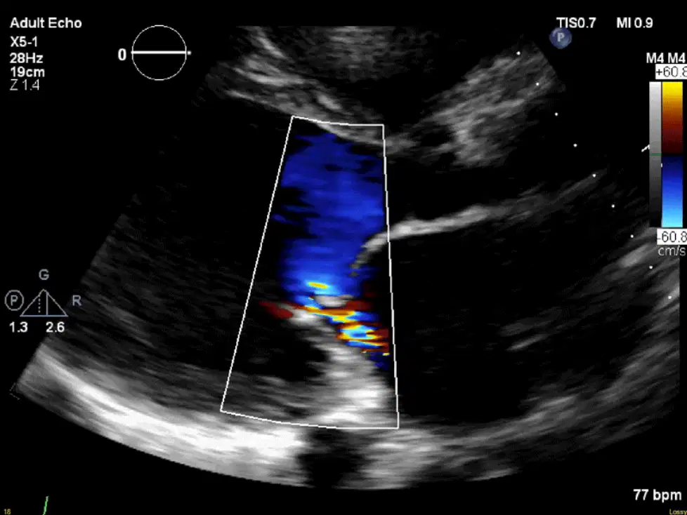
What does the mosaic "blue" flow represent in this image?
TR
AI
PI
MR
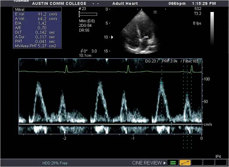
What part of the cardiac cycle does the E-wave correspond to on the MV spectral Doppler?
A. atrial contraction
B. rapid early filling
C. diastasis
D. both B & C
When acquiring LAVi measurements, which of the following would be considered a normal LAVi?
(a)
26
35 mL/m2
48 mL
What is the normal IVC diameter?
1.5-2.0 cm
2.0-3.0 cm
less than 2.0 cm
1.2-2.1 cm
Tissue Doppler Imaging...
measures the velocity of the myocardial movement as it passes through the sample gate toward the PWD cursor.
is usually a mirror image of the MV inflow waveform
is used to calculate E/e'
all of the above
As a person ages, their E wave is expected to (a)
, while the A wave is expected to (b) .
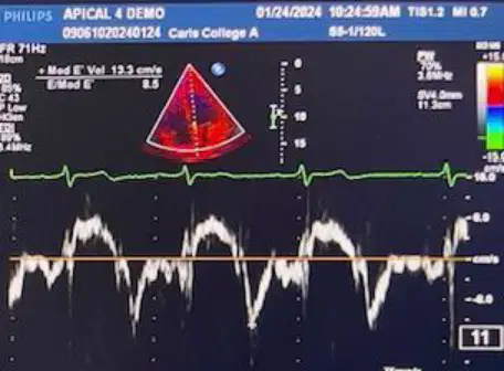
The septal annulus e' measurements from MV TDI can sometimes be contraindicated. Select 3 examples of contraindications.
Abnormal EF
Hypertrophic Cardiomyopathy
MAC
Body Habitus
Prosthetic valves
If a patient is suspect for Pseudonormal, Grade II diastolic dysfunction, the sonographer should repeat the MV Inflow PW Doppler with the Valsalva Maneuver. If the patient has truly normal filling patterns & diastolic function, what will happen to the MV Inflow waveform pattern?
It will stay the same --Big E, Little A
It will reverse -- Little E, Big A
It will equalize -- E & A will be the same size
It will disappear -- E & A will flatline
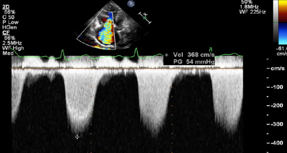
What type of Doppler is shown in this spectral Doppler tracing?
Pedoff
High PRF
PW
CW
When we estimate Right Atrial Pressure, we use the 3/8/15 method. What two pieces of information do we need to make a proper estimation?
TR VMax
IVC Diameter
IVC Collapse
Grade of PHTN
SPAP
RV dilatation is reported if the basal diameter is <41 mm and the mid diameter is <35 mm.
True
False
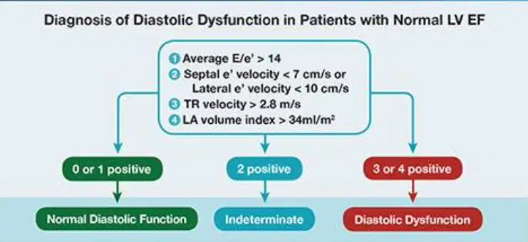
If the patient has normal LVEF, TDI e' (lateral wall) = 15 cm/s, E/e' (average lateral and septal wall) = 10, LAVi = 28 mL/m², and peak TRV = 2.4 m/s, report (a) .
insufficient data
abnormal diastolic function
inconclusive diastolic function
The diastolic function parameters include
(a)
.
CWD of the mitral inflow
LA linear dimension
Draw what a laminar RVOT waveform of normal PV flow looks like using the zero baseline given.

Increased LAVi predicts patient outcome, usually reflects increased LAP, and is a sign of (a) .
cumulative effect of decreased LV filling pressure
diastolic dysfunction
right atrial pressure severity and chronicity
nothing, it is not very accurate
The pulmonary venous flow into the left atrium is dependent upon the pressure difference between the pulmonary veins and the left atrium.
true
false
Flow traveling toward the transducer should appear ____ in color and _______ the spectral Doppler baseline.
blue, below
red, below
red, above
blue, above
What is a normal LVIDd size?
0.6-1.0 cm
3.8-5.8 cm
2.7-3.7 cm
2-3 cm
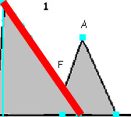
What is true about the measurement represented by the red line?
Normal value is 160-240ms
Normal value is 1.0-1.5
it is a measure of the slope, not the time
it is a measure of time, not slope
Color Doppler gain can be adjusted by the sonographer.
True
False
Draw the angle for measuring 2D structures.

The optimal angle for 2D and MMode is 90 degrees.
True
False
On what type of graph is a spectral Doppler waveform displayed?
time-depth
time-velocity
speed-time
velocity-depth
CW Doppler utilizes how many crystals to pulse a signal out and wait for it to return?
1
2
we don't know
none of these
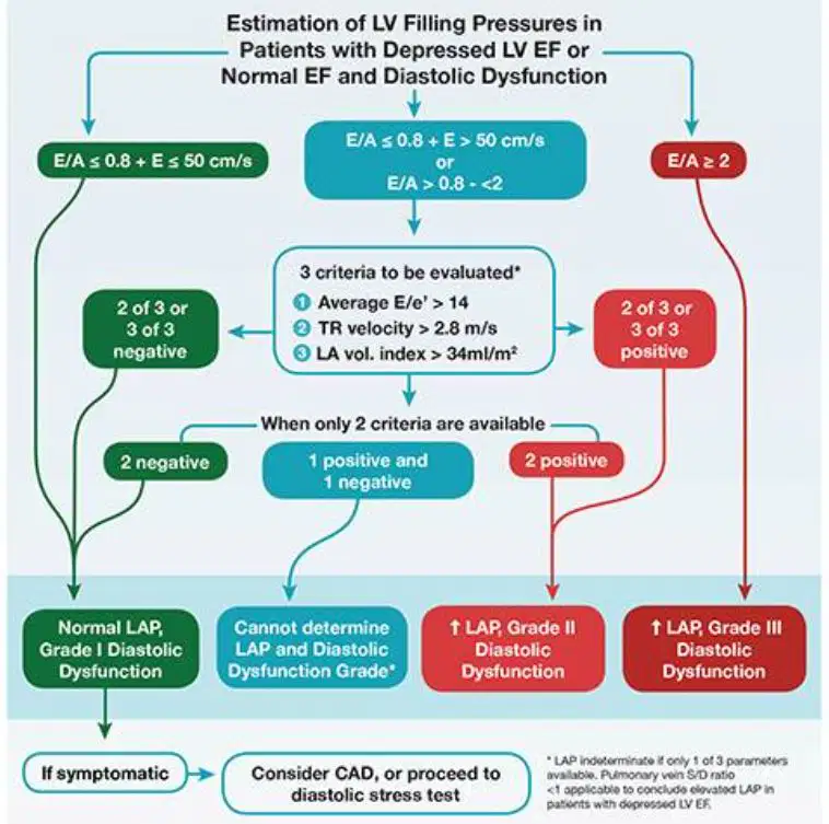
In a patient with reduced LVEF, e' (lateral wall) = 5 cm/s. E/e' = 20, LAVi = 45 mL/m², and peak TRV is 3.2 m/s, report (a) .
normal diastolic function
CAD
inconclusive diastolic function
A normal LVOT diameter is:
A. 18-22 mm
B. 1.8-2.2 mm
C. 1.8-2.2 cm
D. Both A & C are correct
CW Doppler is able to detect both high and low velocities
True
False
The non-imaging continuous wave doppler transducer is called the ______.
Pencil or Pedoff probe
Linear probe
Curved array probe
Phased array probe
When measuring PLAX structures, we take our measurements when the ventricle is at its largest. What part of the cardiac cycle is this?
end-systole
mid-diastole
end-diastole
systole
What is essential for 2D measurements in echocardiography?
Accuracy
Speed
Consistency
Depth
Work out the cardiac output of an athlete with a heartbeat of 170 bpm and a stroke volume of 70 ml.
(a)
10.9 liters per minute or 10,900ml per minute
13.9 liters per minute or 13,900ml per minute
9.9 liters per minute or 9,900ml per minute
Select the area on the EKG where end-diastole occurs.
What are 3 common indications for an echo?
Label the wall segments in this view.
The primary right ventricular diastolic function parameters include _______.
LAVi and LAP
linear dimensions
TDI of PV annulus
TV inflow E/A & decel time
Label the structures with the correct name.
LVPW
RVAW
RV
IVS
LV
Draw a line on the image showing the measurement of the Asc Ao as outlined in class.

Point to the area of the EKG where end-systole occurs.
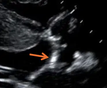
Which cusp is the arrow pointing to in the image?
RCC
NCC
LCC
PCC
Point to the Interventricular Septum on the image.
How should the Sinotubular Junction (STJ) be measured?
Leading edge to leading edge during end diastole
Inner to inner during end diastole
Leading edge to leading edge during peak systole
Inner to inner during peak systole
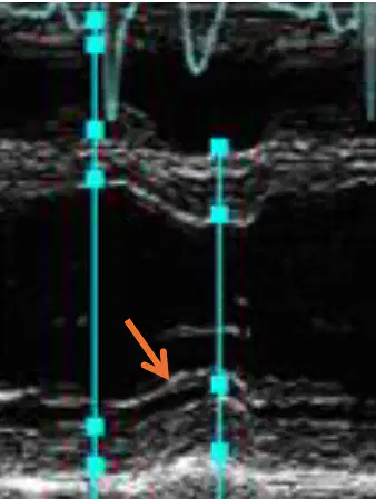
The structure indicated by the arrow is the (a)
Converting Measurements in the Metric System
When converting lengths in the metric system, you need to know the basic units. For example, 1 meter is equal to 100 centimeters or 1000 millimeters. To convert between different units, you simply move the decimal point to the right or left based on the conversion factor.
Volume conversion in the metric system involves units like liters and milliliters. For instance, 1 liter is equal to 1000 milliliters. To convert from one unit to another, you can use the conversion factor to determine the equivalent volume in the desired unit.
Unit prefixes play a crucial role in the metric system, indicating the magnitude of the measurement. Common prefixes include kilo (k), centi (c), and milli (m). Understanding these prefixes helps in converting measurements efficiently and accurately.
What are some basic units essential for length conversions in echocardiography?
Kilometers, decimeters, and centimeters
Centimeters and millimeters
Inches, feet, and yards
Pounds, ounces, and tons
Why is knowledge of unit prefixes like kilo, centi, and milli crucial for converting measurements in the metric system?
To make the measurements more accurate
To make the measurements more complicated
To make the measurements less accurate
To make the measurements impossible to understand
Label the 2D Parameters on the Echo Image Display:
Thermal & Mechanical Indices
Focal Zone (focus)
Frame Rate
Select all statements that are true regarding Color Flow Doppler (CFD):
Converts recorded flow frequencies into different colors
Superimposed on the 2D echo or M-mode image
Should be used in every view regardless of Doppler angle
Should only be used when parallel to flow
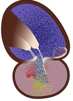
What is the term for abnormal, backward flow through a closed valve?
Stenosis
Shunt
Regurgitation
Normal flow
Label the color parameters on the image.
Color Scale
Color Box
Frame Rate
According to Bernoulli's Principle, what will Bernoulli's Equation do?
Turn velocities into pressures
Turn pressures into velocities
Turn velocities into flow
Turn pressures into flow

Explain why you wouldn't perform Spectral Doppler on this image.
Select the Doppler image displaying the highest sweep speed.
Label the structures

