
 Worksheets
Intro & Structure of Skeletal Muscle
Total questions: 50
Worksheet time: 43mins
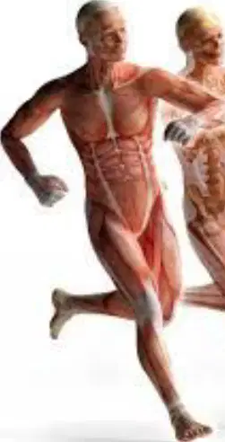
Various outcomes of muscle actions include:
Movement, posture maintenance, heat production
Only movement
Only posture maintenance
Only heat production
Identify the major parts of a skeletal muscle fiber.
Myofibrils, Sarcoplasmic Reticulum, T-tubules
Nucleus, Mitochondria, Ribosomes
Cell Membrane, Cytoplasm, Nucleus
Endoplasmic Reticulum, Golgi Apparatus, Lysosomes
Nerve stimulation of a skeletal muscle involves:
the release of neurotransmitters at the neuromuscular junction.
the contraction of cardiac muscle fibers.
the inhibition of nerve impulses.
the stimulation of smooth muscle tissue.
What are muscles classified as in the body?
Fill in the blank: The 3 types of muscle tissue in the body are (a) , smooth, and cardiac muscle.
How many skeletal muscles does the body contain?
600
500
700
800
How many muscles are in the face, and how many are used to frown and smile?
There are 43 muscles in the face, 17 are used to smile and 43 to frown.
There are 50 muscles in the face, 10 are used to smile and 40 to frown.
There are 30 muscles in the face, 5 are used to smile and 25 to frown.
There are 60 muscles in the face, 20 are used to smile and 40 to frown.
What is the smallest muscle in the body?
Stapedius
Biceps
Quadriceps
Deltoid
Which muscle is the body's largest muscle?
The gluteus maximus is the body's largest muscle.
The biceps brachii is the body's largest muscle.
The deltoid is the body's largest muscle.
The rectus abdominis is the body's largest muscle.
Where is the sartorius muscle found, and what is its average length?
The sartorius muscle is found in the thigh, and its average length is approximately 38 to 40 centimeters.
The sartorius muscle is found in the arm, and its average length is approximately 20 to 25 centimeters.
The sartorius muscle is found in the abdomen, and its average length is approximately 15 to 20 centimeters.
The sartorius muscle is found in the calf, and its average length is approximately 25 to 30 centimeters.
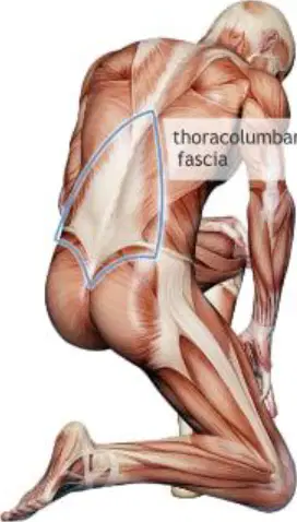
What is the name of the dense connective tissue that surrounds and separates each muscle?
A) Tendons
B) Fascia
C) Aponeuroses
D) Periosteum
This connective tissue extends beyond the ends of the muscle and gives rise to ______ that are fused to the periosteum of bones.
tendons.
ligaments.
cartilage.
fascia.
Sometimes muscles are connected to each other by broad sheets of connective tissue called ______.
aponeuroses.
ligaments.
tendons.
fasciae.
What is the layer of connective tissue around each skeletal muscle called?
A) Perimysium
B) Epimysium
C) Endomysium
D) Fascia
The perimysium extends inward from the ________; it surrounds bundles of skeletal muscle fibers, called fascicles, within each muscle.
epimysium.
endomysium.
sarcolemma.
myofibril.
Each muscle cell (fiber) is covered by a connective tissue layer called ________.
endomysium.
perimysium.
epimysium.
fascia.
What is the outermost layer of connective tissue surrounding a skeletal muscle?
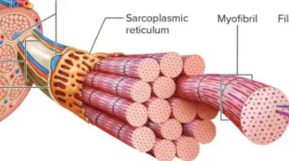
The function of the sarcoplasmic reticulum in muscle fibers is to:
store calcium ions
transmit nerve impulses
provide structural support
synthesize proteins
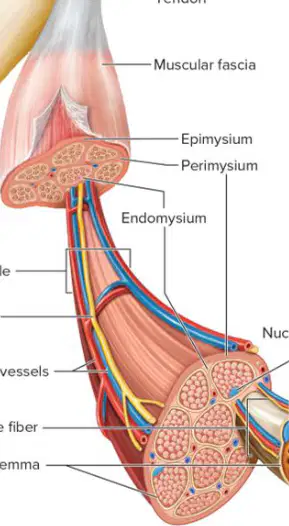
Which part of the muscle fiber contains the nucleus?
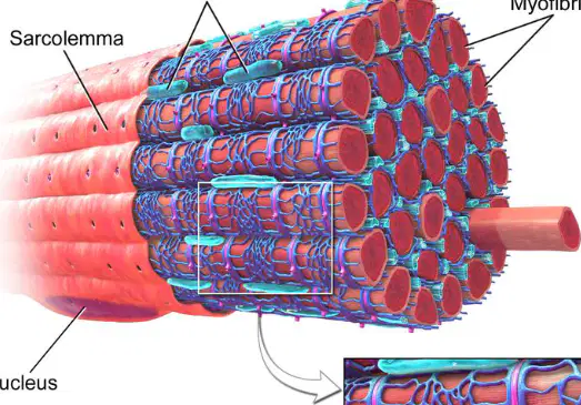
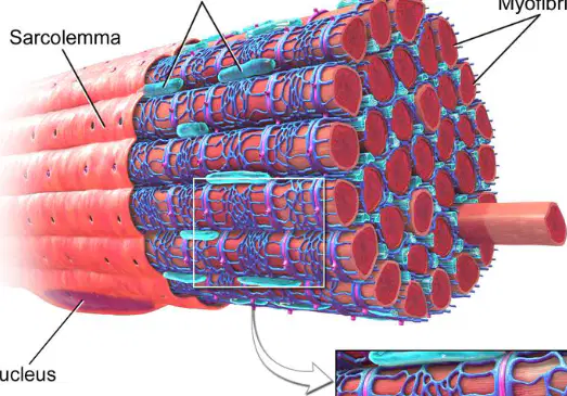
What is the cell membrane of a muscle fiber called?
Sarcolemma
Myofibril
Endomysium
Perimysium
What is the function of muscle fibers in response to stimulation?
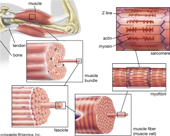
Thick filaments in myofibrils consist of the protein ________.
myosin
actin
tropomyosin
troponin
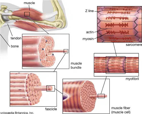
Thin filaments in myofibrils are mainly composed of the protein actin, but also contain ________ and ________.
the proteins tropomyosin and troponin.
the proteins myosin and dystrophin.
the proteins keratin and elastin.
the proteins collagen and fibrin.
What are myofibrils made up of?
Sarcomeres
Z lines
Actin filaments
Thick filaments
A sarcomere extends from one ______ to the next.
Z line
M line
A band
I band
Striations consist of an alternating pattern of ______ and ______ bands.
(a)
I bands are made up of ______ filaments, which are anchored to the ______ lines.
actin, Z
myosin, M
titin, A
nebulin, H
A bands are made up of overlapping ______ and ______ filaments.
thick, thin
long, thick
long, short
thin, long
In the center of the A band is the ______, which consists of myosin filaments only.
H zone
I band
Z line
M line
What does the M line consist of?
Actin filaments
Proteins that hold the myosin filaments in place
Myosin filaments only
Z disc
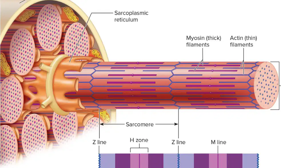
Fill in the blank: The 'Z line' is part of the _______ in the diagram.
The 'Z line' is part of the sarcomere in the diagram.
The 'Z line' is part of the neuron in the diagram.
The 'Z line' is part of the mitochondrion in the diagram.
The 'Z line' is part of the chloroplast in the diagram.
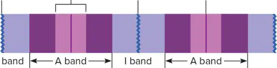
What is the function of the 'A band' as shown in the diagram?
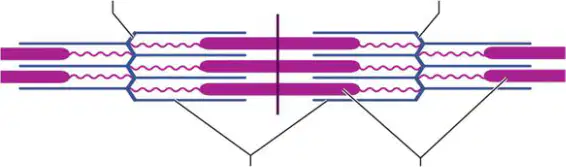
What are the two types of filaments shown in the sarcomere diagram?
What lies beneath the sarcolemma of a muscle fiber?
Sarcoplasmic reticulum (SR)
Transverse (T) tubules
Cisternae
Sarcoplasm
Each T tubule lies between how many cisternae of the sarcoplasmic reticulum?
2
1
3
4
What activates the muscle contraction mechanism when the fiber is stimulated?
Sarcoplasmic reticulum and transverse tubules
Sarcolemma and cisternae
Muscle fiber and sarcoplasm
T tubules and sarcolemma

The function of the sarcoplasmic reticulum in muscle cells is to:
store calcium ions
produce energy
synthesize proteins
transmit nerve impulses
The role of mitochondria in muscle cells is to:
produce energy through cellular respiration
store calcium ions
synthesize proteins
transport oxygen
The function of the sarcolemma in muscle cells is to:
protect the muscle cell
store calcium ions
transmit electrical signals
produce energy
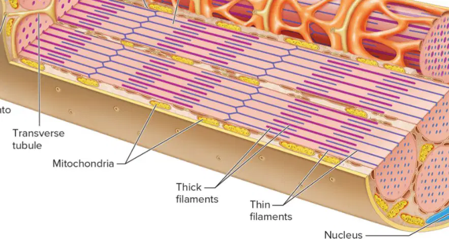
The thick filaments in muscle cells are composed of:
Actin
Myosin
Tropomyosin
Troponin
Which structure is responsible for the contraction of muscle fibers?
What stimulates skeletal muscle fibers to contract?
Synapse
Motor neuron
Neurotransmitter
Muscle fiber
Each skeletal muscle fiber is functionally connected to the axon of a motor neuron, creating a ______.
synapse.
junction.
gap.
connection.
What chemicals are released at the synapse to communicate with the muscle fiber?
Hormones
Neurotransmitters
Enzymes
Proteins
Define neuromuscular junction.
A synapse between a motor neuron and a muscle fiber that it regulates.
A connection between two neurons in the brain.
A junction between two bones in the body.
A type of blood vessel in the circulatory system.
The muscle fiber membrane in the area of the neuromuscular junction contains a specialized region called the ________.
motor end plate
neuromuscular junction
sarcomere
myofibril
What do synaptic vesicles store?
Hormones
Neurotransmitters
Enzymes
Proteins
What happens when an electrical impulse reaches the end of the axon of a motor neuron?
Synaptic vesicles release neurotransmitter into the synaptic cleft.
The muscle fiber contracts immediately.
The neuron stops functioning.
The synaptic cleft closes.
Fill in the blank: The neurotransmitters diffuse across the cleft, bind to the motor end plate, and stimulate the muscle fiber to _______.
contract
relax
expand
remain inactive
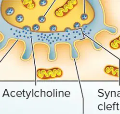
Acetylcholine released into the synaptic cleft reaches the muscle fiber membrane by:
diffusing across the synaptic cleft
being actively transported
binding to a carrier protein
undergoing endocytosis

