
 Worksheets
Chapter 5 Shoulder Girdle and Humerus
Total questions: 63
Worksheet time: 1hrs 3mins
Sonography is an effective diagnostic tool in studying the shoulder joint.
True
False
Where is the CR centered for the posterior oblique position for the glenoid cavity?
Acromion
2 inches (5 cm) medial and inferior to the superolateral border of shoulder
Coracoid process
1 inch (2.5 cm) superior to the coracoid process
What is the common term for idiopathic chronic adhesive capsulitis?
Bankart lesion
Tendinitis
Bursitis
Frozen shoulder
A patient enters the ED with a dislocated shoulder. The technologist attempts to position the patient into the transthoracic lateral projection, but the patient is unable to raise the unaffected arm over his head completely. What can the technologist do to compensate for the patient’s inability to raise his arm completely?
Perform the Grashey method instead.
Use a breathing technique.
Increase kV to penetrate through both shoulders.
Angle the CR 10° to 15° cephalad.
A patient is scheduled for an arthrogram. During the course of the study, the radiologist requests a projection to demonstrate the intertubercular groove. Which one of the following projections would best demonstrate this structure?
Fisk modification
Garth method
Grashey method
Pearson method
An inferosuperior axial projection (Clements modification) is performed on a patient with a nontraumatic shoulder injury. The patient cannot fully abduct the upper limb 90°. Which of the following modifications of the position should be performed for this patient?
Angle CR 30° toward the axilla.
Angle CR 5° to 15° toward the axilla.
Angle CR 45° toward the elbow AC joint separation.
Rotate shoulder slightly anterior to open joint space.
A radiograph of an AP axial projection of the clavicle demonstrates that the clavicle is within the midaspect of the lung apices. What should the technologist do to correct this error?
Do nothing; this is an acceptable AP axial clavicle projection.
Increase the caudal CR angle during repeat exposure.
Increase the cephalic CR angle during repeat exposure.
Make the exposure upon complete inspiration.
What is a possible radiographic sign for impingement syndrome of the shoulder?
Calcified tendons
Fluid-filled joint space
Fracture of the glenoid rim
Subacromial spurring
What type of CR angle is required for the superoinferior axial projection (Hobbs modification)?
No angle is required (CR is perpendicular to the axilla and humeral head).
CR is angled 15° cephalad.
CR is angled 30° caudad.
CR is angled 45° medially.
The Alexander method for AC joints requires a 15° cephalic CR angle.
True
False
For AC joint weight-bearing studies, patients should not be asked to hold on to the weights with their hands; rather, the weights should be attached to the wrists.
True
False
Which of the following AP shoulder projections demonstrates the greater tubercle in profile medially?
External rotation
Internal rotation
Neutral rotation
None of the above
Which of the following AP shoulder projections demonstrates the lesser tubercle in profile medially?
External rotation
Internal rotation
Neutral rotation
None of the above
A patient enters the ED with multiple injuries including a possible fracture of the left proximal humerus. Which positioning rotation should be performed to determine the extent of the humerus injury?
AP neutral shoulder rotation and carefully rotated internally proximal humerus
AP shoulder as is; show radiograph to the ED physician before attempting a rotational lateral projection
AP and horizontal beam transthoracic lateral shoulder projection
AP and apical oblique shoulder without any arm rotation
A patient enters the ED with a possible bony defect of the midwing area of the scapula. The patient is able to stand and move the upper limb freely. In addition to the routine AP scapula projection with the arm abducted, which of the following would best demonstrate the involved area?
Take an apical oblique projection of the shoulder.
Take a transthoracic lateral projection of the shoulder region.
Have the patient reach across the chest and grasp the opposite shoulder for a lateral scapula projection.
Have the patient drop the affected arm behind him or her and take a lateral scapula projection.
The recommended SID for AC joints is 72 inches (183 cm).
True
False
The Hill-Sachs defect is a fracture of the articular surface of the glenoid cavity.
True
False
The arm should be abducted about 45° for an AP scapula.
True
False
An orthostatic (breathing) technique can be performed for the AP projection of the scapula.
True
False
Which one of the following projections/positions should NOT be performed for a possible shoulder dislocation?
AP axial oblique (Garth method)
Transthoracic lateral (Lawrence method)
Inferosuperior axial (Clements modification)
Scapular Y
A posterior dislocation of the shoulder occurs more frequently than an anterior dislocation.
True
False
A patient enters the ED with a midshaft humeral fracture. The AP projection taken on the cart demonstrates another fracture near the surgical neck of the humerus. The patient is unable to stand or rotate the humerus because of the extent of the trauma. What other projection should be taken for this patient?
Scapular Y lateral-AP oblique projection
Apical oblique projection
Horizontal beam transthoracic lateral projection for humerus
Rotational lateral projection for humerus
A referring physician suspects that a subacromial spur may be the cause for a patient’s arm numbness. She asks the technologist for a projection that would best demonstrate any possible spurs. Which of the following projections would accomplish this objective?
PA scapular Y lateral with 10° to 15° caudal angle
PA scapular Y lateral with 10° to 15° cephalad angle
AP oblique shoulder with 45° caudal angle
AP shoulder with 10° to 15° caudal angle
A patient enters the ED with multiple injuries. The physician is concerned about a dislocation of the left proximal humerus. The patient is unable to stand. Which of the following routines is advisable to best demonstrate this condition?
AP shoulder and inferosuperior axiolateral projection
AP shoulder and 35° to 45° LPO position (Grashey method)
AP shoulder and Neer projection
AP shoulder and recumbent AP oblique scapular Y projection
A patient enters the ED with a possible AC joint separation. The patient is paraplegic; therefore, the study cannot be done erect. Which of the following routines would be performed to diagnose this condition?
Non–weight-bearing and weight-bearing type of projections performed with the patient recumbent by pulling down on the shoulders
Recumbent AP and AP axial projection of the clavicles to include AC joints
AP projections of shoulders with external and internal rotation performed recumbent
AP and AP apical oblique projection of the shoulder performed recumbent
A radiograph for an AP projection with external rotation of the proximal humerus reveals that the greater tubercle is profiled laterally. What should be changed to improve this image for a repeat exposure?
Rotate epicondyles so they are perpendicular to the image receptor.
Move the patient obliquely 10° to 15° toward the affected side.
Rotate the arm to place palm of the patient’s hand against the thigh.
Positioning is acceptable; do not repeat it.
A radiograph of an AP clavicle reveals that the sternal extremity is partially collimated off. What should the technologist do?
Repeat the AP projection and correct collimation.
Make sure the sternal extremity is included on the AP axial projection.
Only repeat it if the patient’s pain/symptoms involve the sternal extremity.
Ask the radiologist whether he or she wants the projection repeated.
A radiograph of a scapular Y lateral position reveals that the scapula is slightly rotated (the vertebral and axillary borders are not superimposed). The axillary border of the scapula is determined to be more lateral compared with the vertebral border. Which of the following modifications should be made for the repeat exposure?
Decrease rotation of thorax.
Decrease CR angle.
Increase rotation of thorax.
Abduct the arm more and flex it at the elbow.
A patient comes to radiology for treatment of an arthritic condition of the right shoulder. The radiologist orders AP internal/external rotation projections and an inferosuperior axiolateral projection of the scapulohumeral joint. However, the patient cannot abduct the arm for this projection. Which other projection will best demonstrate the scapulohumeral joint space?
Posterior oblique (Grashey method)
Scapular Y lateral
Transthoracic lateral
AP projection–neutral rotation
A patient comes to the emergency department (ED) with a possible right AC joint separation. Right clavicle and AC joint exams are ordered. The clavicle is taken first, and a small linear fracture of the midshaft of the clavicle is discovered. What should the technologist do in this situation?
Perform the weight-bearing phase as ordered.
Reduce the amount of weight that would normally be given to the patient and perform the weight-bearing study.
Consult with the ED physician before continuing with the AC joint study.
Slowly give the patient more weight to hold until he begins to complain and then
The inferosuperior axial projection (Clements modification) requires a CR angle of _____ toward axilla if a patient cannot fully abduct extremity 90°.
35°
none
40° to 45°
5° to 15°
How much CR angulation is required for an asthenic patient for an AP axial projection of the clavicle?
15°
30°
45°
No CR angulation should be used for this projection.
Where is the CR centered for the bilateral acromioclavicular (AC) joint projection on a single 14- × 17-inch (35 × 43 cm) image receptor?
At the affected AC joint
1 inch (2.5 cm) above jugular notch
At the level of the thyroid cartilage
At the sternal angle
A radiograph of an AP oblique projection for the glenoid cavity reveals that the anterior and posterior rims of the glenoid process are not superimposed. Which of the following modifications should produce a more acceptable image?
Angle CR 5° to 10° caudad.
Angle CR 5° to 10° cephalad.
Increase rotation of the body.
Abduct the arm slightly.
A radiograph of a transthoracic lateral projection reveals that it is difficult to visualize the proximal humerus due to the ribs and lung markings. The following analog exposure factors were used: 75 kV, 30 mAs, 40-inch (102 cm) SID, grid, and suspended respiration. Which of the following changes will improve the visibility of the proximal humerus?
Make the exposure on second inspiration.
Use a compression band to prevent patient movement.
Use a 72-inch (183 cm) SID.
Use an orthostatic (breathing) technique.
A radiograph of the inferosuperior axial projection (Lawrence method) demonstrates the acromion process of the shoulder to be located most superiorly (anteriorly).
True
False
For a Grashey method projection of the shoulder, the CR is centered to the acromion.
True
False
How much posterior CR angulation is required for the supine version of the tangential projection for the intertubercular (bicipital) groove?
20° to 30°
30° to 40°
10° to 15°
No angle is used for this projection.
Which ionization chamber(s) for the AEC should be used for a tangential projection for an intertubercular groove?
Center chamber
Both outside chambers
Left chamber
Cannot use AEC with this projection
Which of the following projections can be performed using a breathing technique?
Scapular Y lateral projection
Inferosuperior axiolateral projection
AP clavicle
AP scapula
How much CR angulation should be used for a scapular Y projection?
No CR angle should be used.
10° to 15°
20° to 30°
35° to 45°
Where is the CR centered for a transthoracic lateral projection for proximal humerus?
1 inch (2.5 cm) inferior to the acromion
Level of the greater tubercle
Level of surgical neck
Which AP projection of the shoulder and proximal humerus is created by placing the affected palm of the hand facing inward toward the thigh?
Internal rotation
Neutral rotation
External rotation
AP axial
The use of a grid during shoulder radiography will result in higher patient dose over nongrid procedures.
True
False
Which of the following shoulder positions is considered a trauma projection (can be performed safely for a possible fracture or dislocation)?
AP apical oblique axial (Garth method) projection
Inferosuperior axial (Clements modification) projection
AP Projection–internal rotation
None of the above
What medial central ray (CR) angle is required for the inferosuperior axial shoulder (Lawrence method)?
5° to 10°
40° to 45°
25° to 30°
10° to 15°
What additional maneuver must be added to the inferosuperior axial shoulder (Lawrence method) projection to best demonstrate a possible Hill-Sachs defect?
Increase medial CR angulation.
Angle the CR 10° to 15° downward or posteriorly in addition to the medial angle.
Perform exaggerated external rotation of the affected upper limb.
Increase abduction of the affected upper limb.
Which of the following shoulder projections best demonstrates the glenoid cavity in profile?
Grashey method
Clements modification
Garth method
AP shoulder, neutral rotation
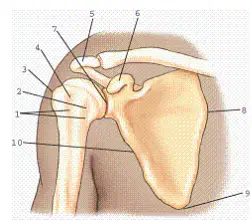
Refer to the diagram of the scapula. Which of the following is correct?
scapulohumeral joint.
glenohumeral joint.
glenoid cavity or fossa.
none of the above
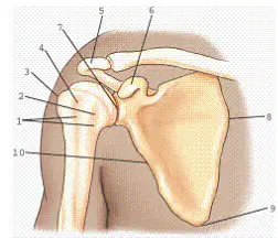
Part 10 refers to the vertebral border of the scapula.
True
False
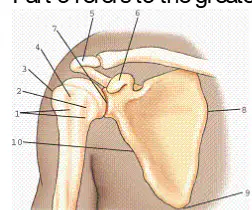
Refer to the diagram of the scapula. Part 3 refers to the greater tubercle.
True
False
Which rotation of the humerus will result in a lateral position of the proximal humerus?
Internal rotation (epicondyles perpendicular to image receptor)
Neutral rotation (epicondyles 45° to the image receptor)
External rotation (epicondyles parallel to the image receptor)
None of the above
All of the joints of the shoulder girdle are diarthrodial.
True
False
Which of the following joints is considered to have a spheroidal type of movement?
Acromioclavicular joints
Sternoclavicular joints
Bicipital joint
Scapulohumeral joint

Which view and projection of the proximal humerus is represented in the figure?
External rotation, anteroposterior (AP) projection
Neutral rotation, AP projection
Internal rotation, AP projection
External rotation, lateral projection

Part 5 refers to the:
coracoid process.
superior border of scapula.
lateral angle of scapula.
none of the above.
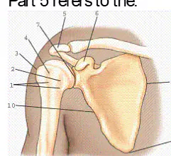
Part 3 refers to the:
Clavicle
Scapula
Humerus
Glenoid cavity
Which of the following structures is not part of the proximal humerus?
Lesser tubercle
Glenoid process
Intertubercular groove
Anatomic neck
Which term describes the medial end of the clavicle?
Acromial extremity
Acromion
Sternal extremity
Acromial tuberosity
The female clavicle is usually shorter and less curved than that of the male.
True
False
The anterior surface of the scapula is referred to as the:
scapular surface.
dorsal surface.
supraspinous and infraspinous fossa.
costal surface.
What is the name of the large fossa found within the anterior surface of the scapula?
Supraspinous fossa
Infraspinous fossa
Subscapular fossa
Glenoid fossa
What is the name of the insertion point for the deltoid muscle located on the anterolateral surface of the humerus?
Surgical neck
Deltoid protuberance
Deltoid tuberosity
Intertubercular groove

