
 Worksheets
MDH Mock Written Exam
Total questions: 155
Worksheet time: 2hrs 1mins
Which of the following laser–tissue interactions are matched correctly?
A. Photodisruption: selective absorption of light energy and conversion of energy to heat with thermally induced structural change in the target.
B. Photocoagulation: uses pulsed lasers to ionize the target and rupture the surrounding tissues.
C. Photoablation: high-powered ultraviolet light exceeds covalent bond strength of target protein precisely removing a submicron layer.
D. Photodisruption: primary type of laser tissue interaction used in LASIK surgery.
In a simple thin lens system, the object is upright with a height of 5 cm, the image is inverted with a height of 10 cm, and the object is 2 cm from the center of the lens. What is the distance of the image from the center of the lens?
A. 1 cm.
B. 2 cm.
C. 4 cm.
D. 8 cm.
Which of the following concerning linear magnification is true?
a. Magnification is equal to the ratio of object size to image size.
b. A magnification value < 0 implies inversion of the image relative to the object.
c. Magnification is equal to the image vergence divided by the object vergence.
d. A magnification value >–1, and <1 implies that the image is greater than the object.
You have gone fishing and see a fish in the water. You do not have a fishing rod. The only equipment that you have is a spear to catch the fish. Where do you throw the spear?
a. in front of the fish.
b. behind the fish.
c. directly at the fish.
d. It is not possible to hit the fish as it is a virtual image.
What is the power of a +20 D lens under water?
a. 6.2 D.
b. 6.5 D.
c. 7.3 D.
d. 8.0 D.
Which is a true statement regarding vergence?
a. If light rays converge to a point, the vergence is negative.
b. Vergence is directly proportional to distance from the object point to the image point.
c. As light travels away from an object point or toward an image point, its vergence stays the same.
d. Vergence is the reciprocal of distance.
What is the vergence of an object 50 cm away from a lens? What is the vergence of an object 25 cm away from the lens?
a. –4 D; –8 D.
b. –2 D; –4 D.
c. 2 D; 4 D.
d. 4 D; 8 D.
Consider an object 10 cm in front of a 5 D convex thin lens in air. What is the vergence after the light leaves the lens?
a. –5 D.
b. 5 D.
c. 10 D.
d. –10 D.
If an object is 50 cm in front of a plane mirror, where is the image in relation to the mirror? Is the image virtual or real?
a. 50 cm from the mirror; virtual.
b. 50 cm from the mirror; real.
c. 100 cm from the mirror; virtual.
d. 100 cm from the mirror; real.
Consider an image is 50 cm in front of a 5 D convex thin lens in air. What is the vergence after the light leaves the lens?
a. 1 D.
b. –1 D.
c. 3 D.
d. –3 D.
Which of the below answers is true regarding IOL calculations in eyes with previous retinal surgery and silicone-oil implantation?
a. A-scan velocity is faster in eyes with silicone oil versus vitreous.
b. A-scan velocity is slower in eyes with silicone oil versus vitreous.
c. Silicone oil acts as an intraocular positive lens when IOL’s are placed.
d. B-scan must be done to determine axial length.
Which change would allow a hard contact lens to fit more tightly to a cornea?
a. Decreasing the diameter from 8.80 mm to 8.40 mm.
b. Decreasing the base curve from 8.20 mm to 8.00 mm.
c. Increasing the optical zone from 8.20 mm to 8.40 mm.
d. Increasing the power from −3.00 D to −3.50 D.
Which one of the following single changes results in the selection of a lens more powerful than
intended?
a. Using an A-constant of 116.8 instead of 117.3.
b. Axial length measured as 20.5 mm rather than 20.2 mm.
c. Average keratometry of 44.50 D in place of 44.00 D.
d. Placing the lens in the ciliary sulcus instead of in the capsular bag.
Which of the following is a third-order refractive aberration?
a. Tetrafoil.
b. Spherical aberration.
c. Vertical coma.
d. Secondary astigmatism.
Image jump is most troublesome for bifocals of the
a. round-top type
b. flat-top type.
c. ribbon type.
d. progressive type.

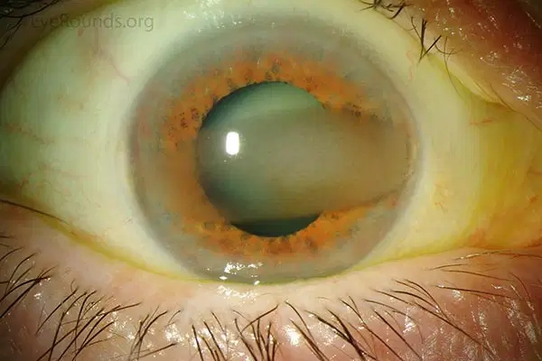
Which is a true statement about this patient?
a. A Vossius ring can be found on the anterior lens capsule.
b. The patient may have elevated intraocular pressure (IOP).
c. Patients may also have involvement of their liver and brain.
d. The cataract is usually visually significant.
Which of the following types of cataracts is classically associated with myotonic dystrophy?
a. Nuclear cataract
b. Polychromatic crystalline cataract
c. Posterior subcapsular cataract
d. Anterior polar cataract
A 5-year-old Caucasian boy presents with difficulty seeing the blackboard at school. Upon examination, he is found to be highly myopic. He is short of stature and has short stubby fingers with broad hands and tight joints. Which of the following is most likely?
a. Prone to angle-closure glaucoma aggravated by the administration of pilocarpine
b. Prone to angle-closure glaucoma aggravated by the administration of a cycloplegic
c. Normal-appearing parents
d. Higher risk of lens dislocation with minor trauma
Regarding age-related cataracts, which is correct concerning the following histopathologic changes?
a. Posterior subcapsular cataract—morgagnian globules
b. Cortical cataracts—Wedl (bladder) cells
c. Nuclear sclerotic cataracts—homogenous, loss of cellular laminations
d. Elschnig pearls—proliferation of lens capsule

Complications during or after cataract surgery in the patient shown will most likely be due to:
a. severe postoperative inflammation
b. zonular dehiscence
c. corneal decompensation
d. retention of viscoelastic
A dispersive ophthalmic viscosurgical device (OVD) used in phacoemulsification has which of the following properties?
a. Adheres to and protects endothelium from damage.
b. Easy to remove at the end of case.
c. Retained viscoelastic does not cause an increase in IOP postoperatively.
d. Prevents collapse of the anterior chamber during high vacuum
During a complicated cataract removal case, the lens and capsular bag were both removed and an anterior chamber lens was placed. The first day postoperatively, the IOP is 50 mm Hg and the iris is bowed forward around the intraocular lens (IOL). Which of the following measures would be most effective?
a. Topical glaucoma medications and close observation.
b. Laser peripheral iridectomy.
c. Increased frequency of the topical steroid.
d. Paracentesis to release aqueous fluid.
A patient has with-the-rule astigmatism. During uncomplicated cataract surgery, a one-piece toric PCIOL is placed into the capsular bag and aligned with the astigmatism. The day following surgery, the lens appears to have rotated 90°. Which of the following statements is most accurate?
a. Lens explantation is necessary because the lens is not stable.
b. The measured astigmatism has increased.
c. The lens should have been placed in the ciliary sulcus.
d. Rotation of the lens can be done anytime after the first 3 months.
What is the cause for posterior capsular opacification following phacoemulsification and implantation of an IOL?
a. Fibrovascular ingrowth stimulated by the lens.
b. Bacterial sequestration and colonization.
c. Proliferation of residual lens epithelium.
d. Toxicity of the IOL
Which of the following is true concerning the different designs of phacoemulsification machines?
a. The peristaltic pump requires a slow building of vacuum for aspiration.
b. The diaphragm pump allows instantaneous vacuum.
c. The Venturi pump has stepwise increases in vacuum.
d. The speed of rollers in the peristaltic pump allows linear control of vacuum.
Which of the following is correct concerning cataract surgery in patients with nanophthalmos?
a. The chances for a poor visual outcome are similar to those in other cataract surgeries.
b. Extracapsular cataract extraction without IOL insertion is the recommended procedure.
c. Trabeculectomy is not recommended at the time of cataract extraction.
d. Anterior sclerotomies may be indicated at the time of surgery.
A patient is sent to you for decreased visual acuity due to cataracts. On examination, you find significant nuclear sclerotic cataracts and a visual acuity of 20/80 in each eye. Dilated fundus examination shows proliferative diabetic retinopathy and clinically significant macular edema. Regarding cataract surgery, you should tell the patient that:
a. Cataract surgery should be performed as soon as possible to better visualize the fundus.
b. An attempt should be made to perform photocoagulation before cataract surgery.
c. Extracapsular cataract extraction without IOL implantation is the procedure of choice.
d. Extracapsular cataract extraction with intact posterior capsule is associated with a greater
chance of neovascular glaucoma postoperatively.
A 60-year-old man successfully undergoes phacoemulsification of the nuclear sclerotic cataract in his right eye. The surgeon accidentally leaves a moderate amount of viscoelastic in his eye. How long after completion of the case might the patient experience a significant spike in IOP?
a. 30 minutes
b. 4 hours
c. 10 hours
d. 24 hours
The risk of complications during cataract extraction in an eye that has previously undergone pars plana vitrectomy is primarily due to:
a. Collapse of the globe during surgery.
b. Positive pressure from the vitreous cavity.
c. The excessive mobility of the posterior capsule.
d. Iridodonesis.
When the ciliary muscle contracts, the diameter of the muscle ring is:
a. reduced, relaxing tension on the zonules, allowing the lens to become more spherical.
b. increased, relaxing tension on the zonules, allowing the lens to become more spherical.
c. reduced, increasing tension on the zonules, allowing the lens to become less spherical.
d. increased, increasing tension on the zonules, allowing the lens to become less spherical.
A 77-year-old female presents 12 hours after cataract surgery with pain, redness, and photophobia in the operated eye. On exam she is found to have 20/400 vision with marked anterior chamber reaction and trace hypopyon. Which detail in the history above favors a diagnosis of Toxic Anterior Segment Syndrome (TASS) over infectious endophthalmitis?
a. the age of the patient.
b. the timing of the findings.
c. the level of vision.
d. the amount of anterior chamber reaction.
During cataract surgery, the surgeon notices that the patient’s pupil dilates poorly and undulates in response to intraocular irrigation. What medication is this patient likely on?
a. Metformins.
b. Tamsulosin.
c. Aspirin.
d. Beta-blockers.
Which of the following lenses has the LOWEST likelihood of posterior capsular opacification (PCO)?
a. Silicone lens with round edge.
b. Silicone lens with square edge.
c. Acrylic lens with round edge.
d. Acrylic lens with square edge.
The lens capsule is made of a transparent basement membrane composed of what type of collagen?
a. Type I.
b. Type II.
c. Type III.
d. Type IV.
Anterior lenticonus can be found in association with what syndrome?
a. Alport’s syndrome.
b. Down’s syndrome.
c. Edwards’ syndrome.
d. Patau’s syndrome.
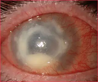
A 29-year-old contact lens wearer presents with pain, photophobia, decreased vision, and clinical findings as indicated in the photograph. Which clinical feature is nearly universal in this disease process?
a. tearing.
b. hypopyon.
c. severe pain.
d. ring infiltrate.
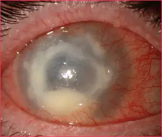
Which of the following is TRUE regarding the condition shown in the picture.
a. Epithelial dendrites may be found.
b. Stromal infiltration should be primarily managed by debridement.
c. Primary infection usually occurs on skin and mucosal tissues innervated by the trigeminal nerve.
d. Gram-negative rods are seen on smears obtained by corneal scraping.
A conjunctival inflammatory response characterized by multiple polygonal nodules with central fibrovascular cores is consistent with a
a. follicular response.
b. papillary response.
c. phlyctenular response.
d. ligneous response.
Clumps of calcific degeneration and eosinophils at the limbus are termed
a. Herbert’s pits.
b. von Arlt’s line.
c. Fuchs’ spots.
d. Horner-Trantas dots.
A patient presents approximately 6 days after daily use of antibiotic ointment following cataract surgery. The patient describes a gradual onset of scaling and itching of the skin and increasing redness of the eye. The hypersensitivity pattern most likely at play is
a. Type I.
b. Type II.
c. Type III.
d. Type IV.
An obese 35-year-old man presents to an ophthalmologist complaining of increasing redness and irritation of his left eye (pictured below), progressive over the previous 4 to 6 months. Examination discloses mildly edematous and erythematous left eyelids with mild conjunctival injection and scant mucus discharge. The conjunctival findings are much more prominent superiorly. The right eye appears normal. With this patient, the critical historical feature to inquire about is
a. Use of over-the-counter eye medications.
b. Any history of previous sexually transmitted diseases.
c. Which side of his body he generally chooses to sleep on.
d. Any previous history of allergic disorders.
A circular, superficial distribution of neutrophils around an area of corneal edema or inflammation is called
a. Wessely ring.
b. Disciform keratitis.
c. Superficial punctate keratitis
d. Ring ulcer.
Which one of the following associations between a microorganism and a useful medium for growth is correct?
a. Moraxella—blood agar in 5% to 10% carbon dioxide
b. Fungi—Sabouraud dextrose agar with cycloheximide
c. Mycobacterium tuberculosis—Loeffler medium
d. Haemophilus—blood agar
Which one of the following medications is commonly used to treat cases of filamentous fungal keratitis caused by Fusarium spp.?
a. Flucytosine
b. Natamycin
c. Amphotericin B
d. Miconazole

Which one of the following statements is CORRECT for the condition shown in the photograph?
a. The steepest meridian of the cornea is adjacent to this lesion.
b. These are benign with no malignant potential.
c. They can grow rapidly.
d. Patients may also have congenital cardiac defects.
Which of the following conditions is thought to have a similar pathophysiology to staphylococcal marginal keratitis?
a. Phlectenulosis
b. Pterygium
c. Pseudogerontoxon
d. Pseudomembranes
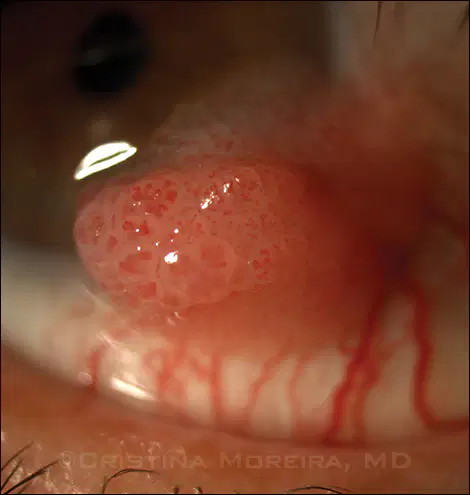
A 24-year-old student presents after noticing a localized area of redness in his right eye near his caruncle as shown in the photograph. He has had similar previous lesions that have resolved spontaneously. What would the lesion likely show histopathologically?
a. Spindle-shaped atypical cells with dark nuclei
b. Acanthotic epithelium over fibrovascular cores
c. Small caliber vascular channels in collagenase stroma
d. Lined nonkeratinizing stratified squamous epithelium

Which therapy might be most appropriate at this time?
a. Observation
b. Simple excision
c. Photocoagulation
d. Excision with frozen section controls
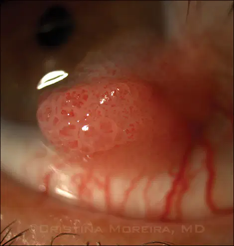
Which virus has been implicated in causing this lesion?
a. Epstein–Barr virus
b. Human papilloma virus
c. HSV
d. Molluscum contagiosum virus
A 68-year-old woman complains to her ophthalmologist that her stye just will not go away, despite 3 months of warm compresses and two surgical drainages. She undergoes full-thickness biopsy of her lower lid; a light microscopic section is shown below. Which of the following is true regarding her situation?
a. The disorder typically affects middle-aged or elderly people.
b. Prompt drainage of the initial chalazion would have been curative.
c. The lesion is likely derived from Moll’s glands.
d. Mohs’ micrographic techniques can be curative at this stage.
The key structure preventing local invasion of squamous cell carcinoma of the cornea is
a. Corneal stroma.
b. Bowman’s layer.
c. Endothelium.
d. Descemet’s membrane.
A patient presents to the ophthalmologist unhappy with his latest refraction. Examination discloses vision of 20/50 with each eye through his new pair of spectacles. This improves to 20/25+ with a pinhole over either lens. Keratometry reveals 45.5 D at 180° and 53.5 D at 90° in each eye. The next logical intervention should be
a. Photorefractive keratectomy.
b. Toric soft contact lens fitting.
c. Rigid gas permeable contact lens fitting.
d. Penetrating keratoplasty.
A 62-year-old woman presents complaining of slow loss of vision in each eye. She denies any previous ocular history. Visual acuities are 20/400 in the right eye and 20/100 in the left eye. Slit-lamp examination reveals corneal guttata in both eyes with central stromal thickening more prominent on the right. The corneal epithelium is normal bilaterally. There is dense nuclear sclerosis on the right and moderate nuclear sclerosis on the left. The view of the fundus is consistent with the patient’s vision. The next appropriate step for visual rehabilitation in this patient might be
a. Cataract extraction only in the right eye.
b. Cataract extraction with posterior chamber intraocular lens (PCIOL) in the right eye.
c. Penetrating keratoplasty (PK) alone in the right eye.
d. PK, cataract extraction, and PCIOL in the right eye.
Which is true of pellucid marginal degeneration?
a. Decreased vision results from lipid deposition.
b. Protrusion of the cornea is at the point of maximal thinning.
c. This is a bilateral condition.
d. Best surgically treated with PRK.
Which one of the following is true regarding congenital hereditary endothelial dystrophy?
a. Nystagmus is absent in the recessive form of the disease.
b. There are usually associated systemic abnormalities.
c. The recessive form is nonprogressive, whereas the dominantly inherited form is slowly
progressive.
d. Corneal clouding is present at birth in both forms of the disease.
A 57-year-old woman presents with a history of chronic ocular surface disease since a bilateral upper lid blepharoplasty 3 years ago. Clinical findings include a fine papillary reaction on the superior tarsal conjunctiva associated with hypertrophy of the superior limbus and fine punctate epithelial erosions in the superior one-third of the cornea. What would be the most likely diagnosis?
a. Floppy eyelid syndrome
b. Exposure keratopathy
c. Foreign body under superior eyelid
d. Superior limbic keratoconjunctivitis

A 5-day-old female with no systemic abnormalities presents with left eye corneal opacities as shown in the given figure. What would the most likely cause of the findings?
a. Forceps trauma
b. Congenital glaucoma
c. Cortical cataract
d. Corneal ulcer
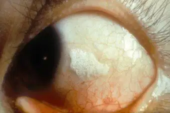
A 45-year-old female with a history of diabetes mellitus type 2 and end-stage renal disease presents with recurrent episodes of eye pain and photophobia in the left eye. On examination, she is noted to have a horizontal white plaque at 3 and 9 o’clock on the right eye and a large one across the inferior visual axis in the left eye (see the figure provided). Which of the following is not advised as primary treatment of this condition?
a. Ethylenediaminetetraacetic acid (EDTA) chelation
b. Phototherapeutic keratectomy (PTK)
c. Artificial tears
d. Bandage contact lens
A 15-year-old male with hyperextensibility of his joints presents with worsening visual acuity and high irregular astigmatism. Corneal topography reveals an asymmetric bowtie pattern. Which of the following patterns is most likely to be present?
a. Protrusion below the area of maximal thinning
b. Protrusion above the area of thinning
c. Steepening 90 degrees away from the area of thinning
d. Thinning nasal and temporally
A corneal scraping from a 23-year-old male who presented to the emergency room was obtained. The patient presented with right eye pain, photophobia, and tearing. On initial examination, visual acuity was 20/70 in the affected eye with a central 3 x 3 mm epithelial infiltrate with feathery edges. Cultures revealed a microorganism that can invade an intact epithelium. Which of the following is the most likely pathogen?
a. Fusarium species
b. Candida albicans
c. Acanthamoeba
d. Pseudomonas aeruginosa
A 65-year-old female visits your clinic for a third opinion. The patient had been experiencing left eye redness, tearing, and photophobia for 3 weeks. She was diagnosed with iritis after YAG capsulotomy at an outside hospital and managed with topical steroids with recurrent bouts after tapering attempts. At examination, visual acuity is 20/40 in the affected eye, intraocular pressure is 21, and keratic precipitates are observed. There is no retained lens material on gonioscopy. Which of the following pathogens are you most concerned for?
a. Gram-positive cocci in clusters
b. Gram-positive cocci in pairs
c. Gram-positive rods
d. Gram-negative diplococci
An 88-year-old female with a history of Crohn’s disease presents due to progressive visual loss over the past 10 years. Her past ocular history includes an uneventful bilateral cataract surgery. On examination, her initial visual acuity is 20/40 OU, with intra- ocular pressure of 12 mm on the right and 10 mmHg on the left. Ishihara color plates are within normal limits. Slit lamp exam reveals the finding in the photograph below (see the figure provided). Automated perimetry shows central constriction bilaterally. Electroretinography reveals reduced scotopic and photopic responses. Which of the following pathogens have been associated with this condition?
a. Gram-positive rods
b. Gram-negativecocci
c. Gram-positivecocci
d. Gram-positive filaments
A 25-year-old male patient with history of uneventful LASIK surgery 2 months ago presents with right eye pain, photophobia, and blurry vision for the past 2 weeks. On examination, his visual acuity is 20/50 on the right and 20/20 on the left. Slit lamp examination revealed a corneal epithelial defect 2 x 2 mm at 3 to 5 o'clock, originating from the flap and intrastromal infiltration. Corneal scraping revealed gram-positive aerobic filaments. Which of the following growth media was most likely to be used?
a. Blood agar
b. MacConkey agar
c. Lowenstein-Jensen medium
d. Thayer Martin agar
A 25-year-old presents with 4 days of left eye redness and mucopurulent discharge associated with tender preauricular lymph node. On examination, visual acuity is 20/20 on the right and 20/40 on the left. A follicular reaction is noted in the inferior tarsal conjunctiva, and there are superficial punctate erosions in the inferior cornea. Giemsa stain reveals basophilic intracytoplasmic bodies. Which of the following culture media is indicated?
a. McCoy cell culture
b. Sabouraud agar
c. Non-nutrient agar with Escherichia Coli
d. Lowenstein–Jensen medium
Which of the following statements is correct regarding penetrating keratoplasty?
a. Large diameter grafts cause higher astigmatism.
b. Donorbuttonisusuallyabout1mmlargerthan
the trephination site.
c. Small-diameter grafts are associated with lower tendency to cause peripheral anterior synechiae.
d. Host tissue cornea excision precedes preparation of donor cornea.
Which of the following is true in regard to aqueous humor dynamics?
a. Aqueous humor is produced in the anterior chamber and flows through the pupil.
b. Aqueous humor is produced by the inner nonpigmented cells of the ciliary processes.
-
c. Aqueous humor exits the eye by passing through the trabecular meshwork and into Schlemm’s canal before draining into the suprachoroidal spaces.
d. Aqueous humor exits the eye by passing through the uveoscleral pathway that ultimately drains into the venous system through a plexus of collector channels.
Which of the following gonioscopy lenses would most likely narrow the angle on indentation gonioscopy?
a. Goldmann.
b. Posner.
c. Sussman.
d. Zeiss.
Which method of gonioscopy is considered best for evaluating a patient with potential traumatic (angle-recession) glaucoma?
a. Goldmann.
b. Koeppe.
c. Zeiss.
d. Sussman.
Which of the following was a finding in the Early Manifest Glaucoma Trial (EMGT)?
a. Treatment reduced IOP by 30%.
b. In multivariate analyses, progression risk was halved by treatment.
c. Progression risk decreased by 20% with each millimeter of mercury of IOP reduction from baseline to the first follow-up visit.
d. At 6 years, 62% of untreated patients showed progression, whereas 30% of treated patients progressed.
Which of the following is true in regard to treating glaucoma secondary to elevated episcleral venous pressure.
a. Medications that increase trabecular outflow are more effective than drugs that reduce aqueous humor formation.
b. Glaucoma filtering surgery may be complicated by a ciliochoroidal effusion or a suprachoroidal hemorrhage.
c. Prostaglandin analogs are not effective in any patients.
d. Laser trabeculoplasty is effective unless there are secondary changes in the outflow channels.
In a patient who presents with traumatic hyphema, elevated IOP, and a history of sickle cell disease, which of the following is safest to use for IOP reduction?
a. Apraclonidine.
b. Brimonidine.
c. Epinephrine.
d. Dipivefrin.
A 24-year-old myope presents with acute angle closure glaucoma. Examination reveals that the central anterior chamber appears to be of normal depth and the iris plane appears to be rather flat for an eye with angle closure. What entity is most likely to be found on gonioscopy?
a. Plateau iris.
b. Blood in Schlemm’s canal.
c. Heavily pigmented trabecular mesh work.
d. Neovascularization of the angle.
Which of the following is true in regard to microspherophakia?
a. It is often familial and may occur as part of either Weill-Marchesani or Down’s syndrome.
b. It can lead to ectopia lentis.
c. Treatment with miotics may tighten zonules, flatten the lens, and pull it posteriorly, breaking pupillary block.
Cycloplegics may make pupillary block worse by rotating the ciliary body forward, loosening the zonule, and allowing the lens to become more globular.
A mother brings in her 7-month-old son for evaluation of excessive tearing from both of his eyes. On examination, the patient is noted to be photophobic and have bilateral corneal enlargement and corneal clouding. What is the best course of action to take?
a. Send the patient home and instruct the mother on how to perform nasolacrimal sac massage.
b. Perform corneal scrapings and treat as a corneal ulcer.
c. Examine the patient under general anesthesia.
d. Perform B-scan ultrasonography.
A 60-year-old man from India presents with a painful red eye. He has had poor vision in this eye for years, but he feels it has worsened over the past 2 weeks. He denies any ocular trauma. On examination, he is noted to have epithelial edema, heavy flare and cell (large cells), and a dense, brunescent cataract. The IOP is 56 mm Hg. What is the definitive management of this condition?
a. Trabeculectomy
b. Anterior chamber washout
c. Extracapsular cataract extraction
d. Posterior pole vitrectomy
A 67-year-old Asian woman calls complaining of 3 hours of acute right eye pain, blurred vision, and redness of her eye. She has a cloudy cornea with epithelial edema, shallow anterior chamber, 4-mm nonreactive pupil, and IOPs of 62 mm Hg OD and 17 mm Hg OS. Gonioscopy of the left eye reveals a narrow potentially occludable angle with virtually no angle structures visible. She has never had any previous similar episodes. Other findings expected on examination include:
a. keratic precipitates
b. glaukomflecken
c. optic nerve pallor and cupping
d. optic nerve hyperemia and swelling
A 75-year-old white man underwent intracapsular cataract extraction 25 years ago. Approximately 15 years ago, he underwent secondary anterior chamber lens placement. Two years ago, he had a
penetrating keratoplasty on this eye. Over the past year, he has had recurrent uveitis in this eye, and on referral evaluation in your office, his IOP is 25 mm Hg. Gonioscopy reveals an open angle and a small hyphema. The most appropriate management of his eye would be:
a. laser iridotomy
b. topical corticosteroids
c. AC washout
d. surgical removal of his anterior chamber lens
A young man is seen in your office over a 5-year period with several episodes of unilateral elevation of IOP to the 40 to 50 mm Hg range. During these episodes, fine keratic precipitates and faint flare are noted. A mild ciliary flush is noted. No iris changes are noted. Each episode seems to respond well to topical corticosteroids and topical and systemic aqueous suppression. What is the most likely cause of his episodic glaucoma?
a. Fuchs heterochromic iridocyclitis
b. Juvenile rheumatoid arthritis
c. Posner–Schlossman syndrome
d. Uveitis–glaucoma–hyphema syndrome
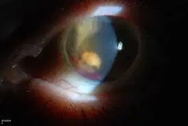
A 46-year-old woman with a long history of insulin-dependent diabetes mellitus presented with a non-clearing vitreous hemorrhage in her left eye. She underwent a vitrectomy to clear the hemorrhage. One week after surgery, she presented with left eye pain, an IOP of 58 mm Hg, and the slit-lamp appearance shown in the photograph. The most likely diagnosis is:
a. Hemolytic glaucoma
b. Phacolytic glaucoma
c. Ghost cell glaucoma
d. Hyphema
A 34-year-old lawyer is struck in the eye by a golf ball launched by an ophthalmologist. The ophthalmologist rushes the lawyer to his office and examines his eye. A 20% hyphema is present in the anterior chamber. No rupture of the globe is present. The hyphema clears within a week; however, the eye remains hypotonus for several months while retaining good vision. Suddenly, while on vacation in a remote region of the country, the lawyer experiences extreme pain and blurred vision in the previously traumatized eye. Hours later, he is examined in an emergency room of a local rural hospital; his eye has an IOP of 62 mm Hg by Schiotz tonometry. Treatment with timolol drops and acetazolamide tablets is instituted, and the lawyer rushes home to the care of his ophthalmologist. What is the most likely cause for this sudden elevation in IOP?
a. Angle-recession glaucoma
b. Ghost cell glaucoma
c. Recurrent hyphema
d. Spontaneous closure of a cyclodialysis cleft
A patient presents complaining of decreased vision in the left eye. His acuities are 20/20 in the right eye and 20/100 in the left eye. The examination is entirely unremarkable, and the diagnosis of factitious visual loss is considered. The test that would be most useful in establishing this diagnosis is
a. Optokinetic nystagmus testing.
b. Gently rocking a large mirror in front of the patient with the good eye occluded.
c. Introducing a prism base up in front of the left eye while the patient is reading binocularly.
d. Performing a fogging refraction.
Which of the following concerning the optic chiasm is true?
a. As many fibers cross as do not cross.
b. The posterior portion of the chiasm has a high density of macular fibers.
c. The chiasm typically lies 1 mm above the anterior pituitary gland.
d. The superior fibers are the first to cross.
Which of the following features is consistent with a temporal lobe lesion?
a. unformed visual hallucinations.
b. optokinetic nystagmus (OKN) abnormalities.
c. formed visual hallucinations.
d. high congruity of visual field deficits.
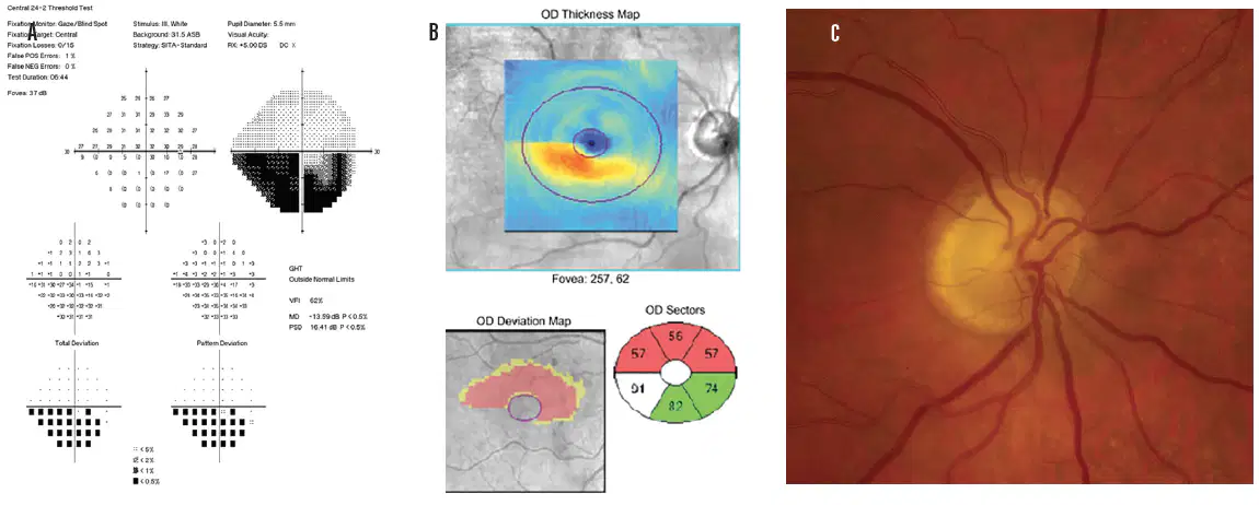
A 63-year-old male walks in with complaints of sudden onset of blurring of vision in his right eye. His visual acuity when checked is 20/30 in that eye. The patient’s history includes hypertension and diabetes. The patient currently has no other systemic symptoms. A photo of his right optic nerve and his visual field is shown. His left optic nerve is normal with a cup/disc ratio of 0.1. What is his most likely diagnosis?
a. Arteritic anterior ischemic optic neuropathy (AAION).
b. Nonarteritic anterior ischemic optic neuropathy (NAION).
c. Myelinated nerve fiber layer.
d. Optic disc drusen.
Which of the following concerning optic nerve glioma is true?
a. Optic nerve glioma is more frequently seen in children than adults.
b. The vast majority of patients will have associated neurofibromatosis.
c. Optociliary shunt vessels of the disc occur more commonly with optic nerve gliomas than with meningiomas of the optic nerve.
d. These tumors are more likely to be aggressively malignant in children than in adults.
Which of the following concerning optic nerve meningiomas is true?
a. It primarily affects children.
b. The majority of patients will have associated neurofibromatosis.
c. On computed tomography (CT) scanning, affected optic nerves have a “kinked” appearance.
d. These tumors are more likely to be aggressively malignant in children than in adults.

A patient comes in for a routine eye exam. He does not have any visual complaints and his vision is 20/20 OU. The photo of the right optic nerve is seen. What is the diagnosis?
a. Papilledema.
b. Myelinated nerve fiber layer.
c. Optic disc drusen.
d. Optic nerve hypoplasia.
Which of the following is true regarding morning glory disc anomaly?
a. Occult transsphenoidal basal encephaloceles may be present.
b. It is more common in males than females.
c. Visual acuity is normal in these patients.
d. Retinal vessels emanate from the center of the optic disc.
In a patient with a right fourth nerve palsy, you would expect a
a. Left hyperdeviation in primary position that worsens on right gaze and left head tilt.
b. Right hyperdeviation in primary position that worsens on left gaze and right head tilt.
c. Right hyperdeviation in primary position that worsens on right gaze and right head tilt.
d. Right hyperdeviation in primary position that worsens on left gaze and left head tilt.
A 69-year-old female was found to have an intra-cranial neoplasm on MRI after complaining of severe headaches. The patient has miosis of his left eye and is “unable to abduct that eye. The neoplasm is most likely found in the
a. Cerebellopontine angle.
b. Left cavernous sinus.
c. Superior orbital fissure.
d. Petrous bone.
A 72-year-old female presents with normal visual acuity, diminished corneal sensation, and global impairment of ocular motility of the right eye. The patient also has a severe boring headache. The MRI shows an enhancing mass within the right cavernous sinus. The pain and ophthalmoplegia respond dramatically to corticosteroid therapy. The most likely diagnosis is
a. Carotid cavernous sinus fistula.
b. Meningioma of the cavernous sinus.
c. Internal carotid aneurysm.
d. Tolosa-Hunt syndrome.
The saccadic movement that is often affected in progressive supranuclear palsy (PSP) is
a. upward.
b. downward.
c. leftward.
d. rightward.
Hypertropia associated with ipsilateral adduction weakness is most suggestive of
a. Decompensated congenital fourth nerve palsy.
b. Dorsal midbrain syndrome.
c. “One-and-a-half ” syndrome.
d. skew deviation with internuclear ophthalmoplegia (INO).
Which one of the following autoantibodies are most commonly found in patients with generalized myasthenia gravis (MG)?
a. binding antibodies to acetylcholine receptors.
b. blocking antibodies to acetylcholinesterase.
c. blocking antibodies to acetylcholine receptors.
d. blocking antibodies to acetylcholine.
A patient presents with irritation and conjunctival injection of the right eye. You find right orbicularis weakness, decreased ability to wrinkle the right forehead, slight right corneal anesthesia, and a small angle esodeviation develops on right gaze. What test would be most helpful?
a. Tensilon test
b. CT to rule out a left parietal lesion
c. Thyroid function tests
d. MRI to evaluate right cerebellopontine angle
The factor primarily responsible for the shallow anterior chamber in a normal infant eye is:
a. The infant cornea is flatter than the adult cornea.
b. The infant iris is relatively thicker than the adult iris.
c. The infant lens is relatively thicker than the adult lens.
d. There is more positive vitreous pressure in the infant eye than in the adult eye.
Which of the following about extraocular muscles is true?
a. Both oblique muscles are characterized by a physical distinction between the anatomic origin and the mechanical origin.
b. The superior oblique tendon passes between the superior rectus muscle and the globe on the way to its insertion.
c. The inferior oblique muscle passes between the inferior rectus muscle and the globe on the way to its insertion.
d. The superior oblique muscle becomes tendinous after turning through the trochlea.
Which of the following changes in lid position are consistent with the muscle surgery described?
a. Narrowing of palpebral fissure with superior rectus recession.
b. Narrowing of palpebral fissure with inferior rectus recession.
c. Narrowing of the palpebral fissure with inferior rectus resection.
d. Narrowing of the palpebral fissure with superior oblique tenotomy.
Which of the following about amblyopia is true?
a. The incidence in the general population is approximately 0.2%.
b. The presence of an afferent pupillary defect clearly establishes an organic etiology for visual loss, rather than amblyopia.
c. Patients with amblyopia will frequently perform better with single-symbol acuity test targets than with line targets (crowded stimuli).
d. A neutral density filter placed over an amblyopic eye will generally cause a greater decrement in visual acuity than the same filter placed over an eye with maculopathy.
A 42-year-old patient presents to the emergency room with a manifest right esotropia. A red glass is placed over the left eye and the patient is asked to fixate at a distant point-light target. In the absence of suppression, and with normal retinal correspondence, the patient should perceive the red light
a. above the white light.
b. below the white light.
c. to the right of the white light.
d. to the left of the white light.
A patient with strabismus is asked to fixate a penlight held by the examiner. The examiner notes that the corneal reflex in the right eye is central, whereas that in the left eye is displaced approximately 3 mm temporal to the center of the pupil. Using Hirschberg’s method for estimating the angle of strabismus, the examiner concludes that the patient has a
a. 45-degree esotropia.
b. 45-prism-diopter esotropia.
c. 45-degree exotropia.
d. 45-prism-diopter exotropia.
When tested with a Maddox rod held over the affected eye with its cylinders running horizontally, a patient with new-onset excyclotropia will perceive
a. a horizontal line.
b. a vertical line.
c. an oblique line running superotemporal to inferonasal.
d. an oblique line running superonasal to inferotemporal.
A 31-year-old man with moderate hyperopia presents for routine examination. There is a 10-prism-diopter alternating esotropia at distance. While reading through his distance correction at 20 cm, there is a 35-prism-diopter esotropia. Eye movements are full, and he denies any history of prior surgery. Yo u conclude that
a. He must have amblyopia in one eye.
b. He probably has restrictive strabismus.
c. He probably will note double vision if questioned appropriately.
d. He has a high AC/A ratio.
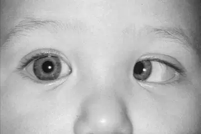
Which of the following concerning the patient represented in the photograph is most likely true?
a. Although it is typically seen in isolation, these findings may be associated with neurologic abnormalities.
b. Amblyopia will likely develop as the child cross-fixates.
c. Characteristically, the esotropia is small (<25 prism diopters).
d. There is never an accommodative component found in this clinical scenario.
Which of the following regarding the treatment of esotropia is true?
a. Accommodative esotropia is more likely to require surgical intervention than infantile esotropia.
b. Bifocals are most helpful in the management of patients with refractive accommodative esotropia.
c. If refractive correction fails to solve the problem, the only solution is surgical.
d. Accommodative esotropia may progress over the first 5 to 7 years of life and should be monitored carefully.
Which of the following regarding the treatment of accommodative esotropia is true?
a. No improvement of esotropia with miotic therapy rules out the possibility of an accommodative component.
b. Delay in refractive correction of an accommodative esotropia increases the probability of a permanent residual esotropia after full correction is given.
c. It is important to attempt surgical realignment before prolonged occlusion therapy.
d. Surgical realignment resulting in a residual esotropia of <20 prism diopters may permit the development of peripheral fusion.
A patient presents for evaluation of “wandering eyes.” On alternate-cover testing, with the left eye covered, the right eye fixes a distance target. As the cover is shifted to the right eye, the left eye moves down to pick up fixation. As the cover is shifted back over the left eye, the right does not move in order to reassume fixation. This set of findings is most consistent with
a. Right hyperdeviation.
b. Left hyperdeviation.
c. Overaction of the inferior obliques.
d. Dissociated vertical deviation (DVD).
Which of the following about A and V patterns of horizontal strabismus is true?
a. A patterns must measure at least 15 prism diopters difference between upgaze and downgaze to be considered significant.
b. V patterns must measure at least 10 prism diopters between upgaze and downgaze to be considered significant.
c. These forms of noncomitance are seen in fewer than 5% of horizontal strabismus.
d. All the extraocular muscles (in varying combinations) have been implicated as responsible for these patterns.
A 3-year-old child presents to the ophthalmologist with parents complaining of “cross-eyes” for approximately 1 year. Examination discloses visual acuity of 20/30 in the right eye and 20/100 in the left eye with Allen cards. There is a 35-prism-diopter esotropia at distance increasing to 45 prism diopters at near. Refraction reveals +3.50 D in both eyes. Initial steps in managing this patient should include
a. Bimedial recessions.
b. Bilateral resections.
c. Prescription of +3.50 in both eyes with add +3.50 in both eyes (bifocals).
D. prescription of +3.50D in both eyes and patching of the right eye.
A 7-year-old boy presents with an exotropia. His deviation measures 30 prism diopters in primary position, 20 prism diopters in right gaze, and 40 prism diopters in left gaze. Near deviation is 15 prism diopters in all directions. Fixation appears to alternate, and visual acuity is 20/20 in both eyes. The patient’s parents strongly desire some form of correction. You recommend
a. addition of -2.00 D to his current distance refraction.
b. bilateral rectus recessions, equal on each side.
c. bilateral rectus recessions with a greater distance of recession on the left.
d. bilateral rectus recessions with a greater distance of recession on the right.
Which statement concerning the medial canthal area is true?
a. All of the attachments anchoring the tarsi to the medial orbital wall lie anterior to the lacrimal sac and attach to the maxillary portion of the frontal bone.
b. The lacrimal sac lies posterior to the orbital septum.
c. The muscle pump of the lacrimal pump mechanism is innervated by the fifth cranial nerve.
d. Lockwood ligament attaches posterior to the lacrimal sac.
Features of the orbicularis muscle include:
a. Closure of the eyelid, depression of the eyebrow, and facilitation of tear drainage.
b. Pretarsal orbicularis inserts temporally to become the lateral canthal tendon, contraction
narrows the palpebral fissure, and the orbital portion of the muscle inserts medially on the
posterior lacrimal crest.
c. The deep head of the medial pretarsal muscle is called Horner tensor tarsi and innervation of
the orbicularis muscle by cranial nerve III is divided into three segments (pretarsal, preseptal,
and orbital).
d. The zygomaticofacial nerve innervates the upper lid orbicularis, the frontal branch of cranial
nerve VII sends motor fibers to the upper lid orbicularis, and the preseptal orbicularis divides to encompass the lacrimal gland.
Which of the following glands are matched with their correct types of secretions?
a. Moll—apocrine, main lacrimal gland—eccrine, meibomian glands—apocrine
b. Glands of Krause—holocrine, gland of Zeis—apocrine, goblet cells—holocrine
c. Glands of Wolfring—eccrine, gland of Moll—apocrine, goblet cells—holocrine
d. Main lacrimal gland—eccrine, meibomian glands—holocrine, gland of Zeis—apocrine
In patients with ptosis, the 2.5% phenylephrine hydrochloride test:
a. May activate the sympathetic receptors in Müller muscle, resulting in elevation of the lid
b. Can be used to assess the approximate elevation of the lid with external levator advancement
c. Dilates the pupil so that the contralateral eyelid may drop
d. Does not affect blood pressure through systemic absorption of the phenylephrine
Adult patients presenting with epiphora with a complete obstruction at the sac–duct junction would be expected to have:
a. negative dye disappearance test/positive Jones III
b. positive dye disappearance test/positive Jones I
c. positive dye disappearance test/negative Jones I
d. negative dye disappearance test/negative Jones II
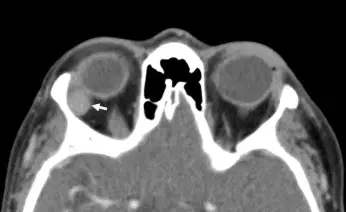
The CT scan is from a 60-year-old woman who presented with painless swelling of the lacrimal gland and anterior orbit for approximately 2 months. There was no significant past or current ophthalmic history. The patient’s medical history was noncontributory. What is the most likely diagnosis?
a. Primary lacrimal gland lymphoma
b. Pleomorphic adenoma
c. Adenoid cystic carcinoma
d. Malignant pleomorphic adenoma
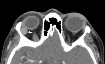
What is the most accurate description of the pathology specimen taken from this lesion?
a. Spindle cells with both ductal epithelium and a mixed stromal pattern
b. “Swiss cheese” pattern—hyperchromatic small cells proliferating around nerves
c. Ductal epithelium in a tubular formation with malignant degeneration
d. Mixture of both B and T cells, with predominance of B cells
A patient with suspected orbital cellulitis suddenly worsens with a virtually frozen globe, without a measurable increase in proptosis. Corneal sensation is likewise diminished, but visual acuity remains grossly intact. The most likely explanation for these events is
a. Cavernous sinus thrombosis.
b. Orbital compartment syndrome.
c. Meningitis secondary to orbital cellulitis.
D. orbital apex syndrome.
Which one of the following signs is considered classic for CT scanning in Graves’ ophthalmopathy?
a. kinking of extraocular muscles.
b. nodular muscle enlargement.
c. fusiform muscle enlargement with sparing of tendons.
d. solitary muscle enlargement.
After eyelid retraction, what is the expected order of eyelid findings in patients with Graves’ ophthalmopathy (in order from most frequent to least frequent)?
a. von Graefe’s sign > lid lag > lagophthalmos.
b. lagophthalmos > von Graefe’s sign > lid lag.
c. lid lag > von Graefe’s sign > lagophthalmos.
d. von Graefe’s sign > lagophthalmos > lid lag.
A patient with known neurofibromatosis presents with pulsating proptosis of long duration. CT scan of the orbit will most likely reveal
a. Orbital neurofibroma.
b. Abnormality of the sphenoid bone.
c. Optic nerve glioma.
d. Carotid–cavernous fistula.
The procedure of choice in a patient with ptosis following cataract surgery (good levator function and a high or effaced upper eyelid crease) would be
a. levator muscle resection.
b. unilateral frontalis suspension.
c. Müller’s muscle resection.
d. reinsertion of levator aponeurosis.
The most likely outcome following inadvertent suturing of the orbital septum into subcutaneous tissues while repairing a partial-thickness eyelid laceration is
a. Ectropion.
b. Lid retraction in downgaze.
c. Entropion.
d. Blepharoptosis.
The best choice below for surgical repair of a ptotic upper eyelid exhibiting a high eyelid crease, a margin to reflex distance (MRD) of 0 mm, and excellent levator function would be:
a. Resection of the superior tarsal muscle
b. Unilateral frontalis suspension using autogenous fascia lata
c. Reattachment of the dehisced levator aponeurosis
d. Plication of the levator muscle (16 mm)
Which one of the following is found in the blepharophimosis syndrome?
a. Euryblepharon
b. Ankyloblepharon
c. Epiblepharon
d. Telecanthus
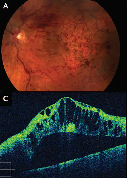
A 45-year-old male complains of slowly progressive decreased vision in his left eye for 2 months. Visual acuity is 20/200. His fundus examination and OCT are shownA 45-year-old male complains of slowly progressive decreased vision in his left eye for 2 months. Visual acuity is 20/200. His fundus examination and OCT are shown. Which of the following best represents the pathophysiology of this patient’s condition?
a. Embolic phenomenon
b. Thrombosis at the level of the lamina cribrosa
c. Carotid stenosis
d. Compression of the central retinal vein due to an atherosclerotic arteriole
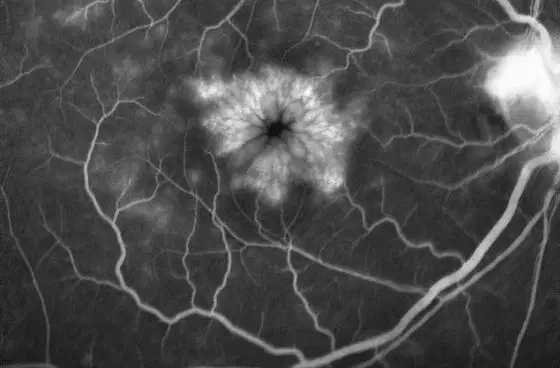
A 73-year-old female with a history of well-controlled diabetes recently underwent uncomplicated phacoemulsification with a posterior chamber intraocular lens and complains of blurry vision. Best-corrected visual acuity (BCVA) is 20/60. Her fluorescein angiogram is provided. Which retinal layer accounts for the appearance of this patient’s vision loss?
a. Nerve fiber layer
b. Outer plexiform layer
c. Inner plexiform layer
d. Outer nuclear layer
Which of the following statements regarding the neurosensory retinal anatomy is correct?
a. The internal limiting membrane is formed by the footplates of Müller’s cells.
b. The retinal vascular system generally supplies half of the oxygen used by the fundus.
c. Retinal photoreceptors generally synapse with bipolar cells, which then generally synapse with Müller’s cells.
d. The macula, as defined histologically, contains photoreceptors with two or more layers.
Which of the following patients should generally be considered for focal laser treatment of central serous chorioretinopathy (CSR)?
a. A patient with pigment epithelial detachment and surrounding sensory retinal detachment lasting for 3 weeks.
b. A retinal surgeon with a subfoveal lesion.
c. A patient whose prior episodes of CSR have been associated with permanently decreased acuity.
d. A patient with a history of bilateral CSR.
Fundus autofluorescence helps evaluate the function of which of the following structures within the eye?
a. Retinal pigment epithelium.
b. Bipolar cells.
c. Ganglion cells.
d. Retinal photoreceptors.
Which factor is most strongly correlated with the development of choroidal effusion following panretinal photocoagulation?
a. Systemic hypertension.
b. Increasing age.
c. Total retinal surface area treated.
d. Short axial length (<23 mm)
Which of the following statements regarding diabetic retinopathy is true?
a. The ETDRS showed that focal laser treatment of clinically significant diabetic macular edema leads to an improvement in vision in twice as many treated patients as untreated patients.
b. The Diabetic Retinopathy Study (DRS) showed that panretinal photocoagulation (PRP) could reduce the incidence of severe visual loss in certain patients by 50%.
c. One definition of high-risk proliferative diabetic retinopathy mandating immediate PRP neovascularization is optic disc neovascularization covering greater than half of its area (greater than standard photograph 10A), only if associated with vitreous hemorrhage.
d. One definition of high-risk proliferative diabetic retinopathy is moderate to severe neovas-cularization elsewhere, only if associated with vitreous hemorrhage.
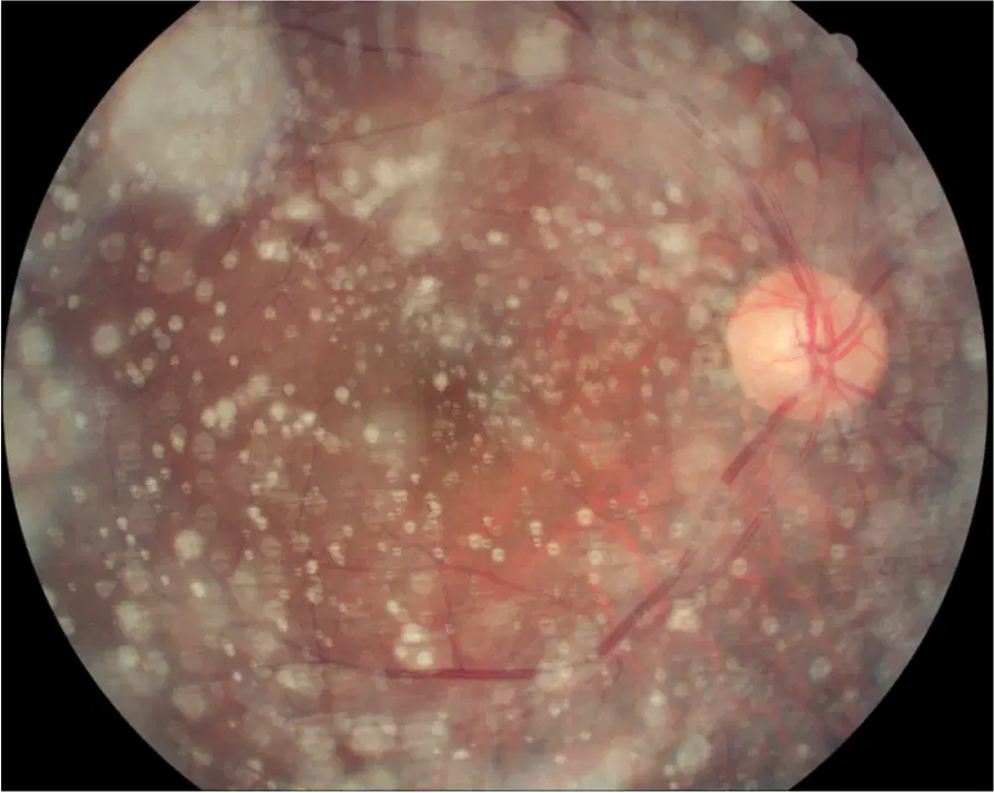
Which of the following statements regarding the condition shown is true?
a. Most cases of this condition are bilateral.
b. Visual acuity in patients with this condition is generally <20/200.
c. The prevalence of this condition is increased in patients with diabetes.
d. The condition is common in young Asian males.
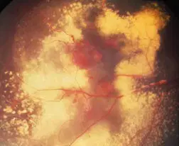
An 8-year-old boy is having difficulty seeing in his right eye. His ophthalmic (including funduscopy) examination is normal except for his right fundus. A peripheral photograph of his right fundus is shown in the photograph. Which of the following statements is true regarding the patient’s condition?
a. Although not so in this specific case, most cases of this condition are bilateral.
b. The condition is typically inherited in an X-linked recessive pattern and has a high male preponderance.
c. The condition can generally be managed with a single session of laser photocoagulation or cryotherapy.
d. Although it can occur, retinal neovascularization is not common.
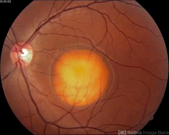
A 10-year-old boy is found to have the fundus appearance as shown. Which of the following statements is TRUE regarding the patient’s condition?
a. The condition is typically unilateral and idiopathic.
b. The ERG is generally normal, and the EOG is abnormal in eyes with this condition.
c. Visual acuity is generally 20/200 or worse at the stage shown.
d. To date, the gene responsible for this condition has not been identified.
Which of the following statements regarding retinoschisis is true?
a. Typical degenerative retinoschisis is associated with an increased risk of retinal detachment.
b. The majority of patients with retinoschisis are hyperopic and have bilateral retinoschisis.
c. The absolute scotoma caused by posterior extension of retinoschisis is highly symptomatic.
d. Retinoschisis is almost always associated with retinal dialysis.
What is, in order of frequency, the likelihood of traumatic retinal tears after blunt ocular injury?
1. Tears around lattice
2. Giant retinal tears
3. Inferotemporal dialysis
4. Superonasal dialysis
5. Flap tears
a. 3 > 2 > 4 > 5 > 1
b. 4 > 3 > 2 > 1 > 5
c. 3 > 4 > 2 > 5 > 1
d. 4 > 2 > 3 > 5 > 1
Commotio retinae represents:
a. retinal edema from contusion injury
b. traumatic disruption of choroidal circulation resulting in retinal edema
c. disruption of photoreceptor elements and damage to photoreceptor cells
d. retinal edema from damage to retinal vasculature
A 27-year-old had a ruptured right globe with uveal prolapse repaired 6 weeks before presenting with photophobia, blurry vision, and pain in the left eye. Which one of the following statements regarding this patient is true?
a. Granulomatous keratic precipitates are found in both eyes.
b. Enucleation of the right eye will be beneficial in this condition.
c. This is endogenous endophthalmitis and will benefit from IV antibiotics.
d. This condition occurs in 5% of cases of penetrating ocular trauma.
What is the most common cause of recurrent retinal detachment or failure of scleral buckling procedure?
a. Scleral buckle too large causing retinal folds and fish-mouthed tears
b. Scleral buckle too small resulting in inadequate support of tears
c. No circumferential band to support vitreous base
d. Proliferative vitreoretinopathy
A 10-year-old girl with a history of arthralgia and a chronic anterior chamber inflammation of both eyes presents to your office. She has no cells, but mild flare is present in the anterior chamber. Given the natural history of this disease, which one of the following statements is most likely true?
a. This patient is antinuclear antibody (ANA)–negative.
b. This patient has pauciarticular disease involving the hands and wrists.
c. This patient has pauciarticular disease involving the lower extremities without involvement
of the wrist joints.
d. This patient is rheumatoid factor–positive.
A 40-year-old white man is referred for ophthalmologic examination because the vision in one eye could not be refracted better than 20/40. On examination, you note a mild unilateral anterior chamber inflammation and stellate keratic precipitates diffusely dispersed over the posterior surface of the cornea. The patient has hazel irides with a mild difference of iris pigmentation between the eyes. Which one of the following statements is most likely true?
a. The involved eye is darker brown than the uninvolved eye.
b. Less than 2% of patients with this condition will have bilateral involvement with no obvious
heterochromia.
c. Pathologic specimens of this condition have revealed the presence of plasma cells within the
ciliary body.
d. Posterior synechiae may be prominent.
Which one of the following characteristics regarding serpiginous choroidopathy is true?
a. Recurrent, indolent course
b. Primarily affects children
c. Multifocal lesions
d. Responds promptly to corticosteroids
Which one of the following statements about the drugs used to treat ocular toxoplasmosis is true?
Pyrimethamine blocks the production of dihydrofolate from para-aminobenzoic acid.
Sulfadiazine inhibits the dihydrofolate reductase enzyme.
Clindamycin can effectively kill Toxoplasma organisms.
Systemic and periocular corticosteroids are contraindicated because they may cause
proliferation of Toxoplasma organisms.

This patient has a granulomatous uveitis with multiple serous retinal detachments as shown. Fluorescein angiography would reveal:
a. single “smokestack” of fluorescein leakage into the subretinal space
b. multiple pinpoint areas of fluorescein leakage into the subretinal space
c. diffuse retinal venous staining and leakage
d. well-defined lacy hyperfluorescence with late leakage
You examine a 78-year-old white man with complaints of decreased vision in his right eye. Three weeks earlier, he had undergone coronary artery bypass surgery with a complicated postoperative course requiring prolonged ventilatory support. He has been receiving hyperalimentation and IV antibiotics since the time of his surgery. On dilated fundus examination of his right eye, you note a fluffy white choroidal lesion under the macula. There is minimal overlying vitritis present. Which one of the following statements concerning the organism most likely responsible for this lesion is true?
a. The responsible organism grows on blood agar and Sabouraud glucose within 24 to 48 hours.
b. This organism exists exclusively as an oval budding cell known as a blastoconidia.
c. Ocular infection caused by this organism is generally accompanied by positive blood
cultures.
d. This organism often colonizes the respiratory tract and is a frequent cause of pneumonia.
Posner–Schlossman syndrome:
a. requires systemic or periocular corticosteroid therapy
b. is not associated with late-onset glaucoma
c. is painless
d. is often self-limited
A 70-year-old man infected with virus A. He has never been exposed to this virus in the past. What subtype of immunoglobulin will be made in response to the initial exposure to the virus?
a. IgA.
b. IgD.
c. IgM.
d. IgG.
A 34-year-old man presents with a 1-week history of right eye pain and light sensitivity. The left eye is without symptoms. There are 2+ cells and 2+ flare in the anterior chamber. There is no posterior inflammation present. Upon further questioning, he has had lower back stiffness and pain upon awakening for several years. The back pain improves during the day. He has no other general medical symptoms. His social history is negative for promiscuous sexual history. What radiographic test would be appropriate and most cost effective in this scenario?
a. CT scan of head.
b. MRI of spinal column.
c. sacroiliac x-ray films.
d. CT scan of thoracic spine.
The most common cause of decreased vision in intermediate uveitis is
a. disc edema.
b. posterior subcapsular cataract.
c. vitritis.
d. cystoid macular edema.
Which of the following is true regarding Behçet’s disease?
a. The course of the ocular disease generally is self-limited.
b. There is no cardiovascular association in Behçet’s disease.
c. A shifting hypopyon may be present in the anterior chamber.
d. Steroids are generally the only long-term therapy
What are the characteristic fluorescein angiography findings in APMPPE?
a. early hypofluorescence, late hyperfluorescence.
b. early hypofluorescence, late hypofluorescence.
c. early hyperfluorescence, late hypofluorescence.
d. early hyperfluorescence, late hyperfluorescence.
Periocular steroid injection should be AVOIDED in which of the following conditions?
a. Pars planitis associated with sarcoidosis and cystoid macular edema.
b. Necrotizing scleritis associated with rheumatoid arthritis.
c. Cystoid macular edema following cataract surgery.
d. Punctate inner choroidopathy.
Which one of the following concerning congenital syphilis is true?
a. The interstitial keratitis of congenital syphilis is generally bilateral and asymptomatic.
b. Syphilitic interstitial keratitis is a direct manifestation of active corneal infection.
c. The chorioretinitis of congenital syphilis is generally bilateral and nonprogressive.
d. A diagnosis of syphilitic interstitial keratitis mandates a full course of treatment for neurosyphilis.
The organism with the poorest prognosis in post-traumatic endophthalmitis is
a. Staphylococcus aureus.
b. Streptococcus pneumoniae.
c. Aspergillus.
d. Bacillus cereus.

