
 Worksheets
Gallbladder and Bile Duct HW
Total questions: 113
Worksheet time: 2hrs 45mins
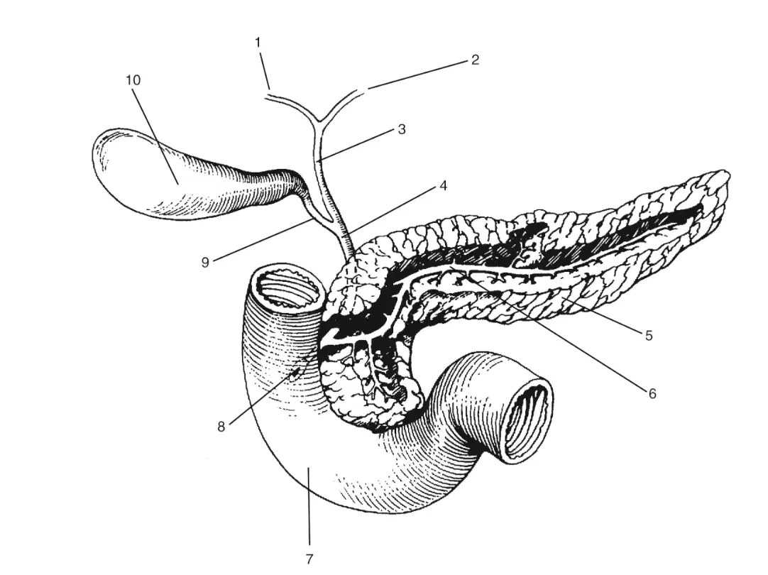
In this image, what is labeled 1
(a)
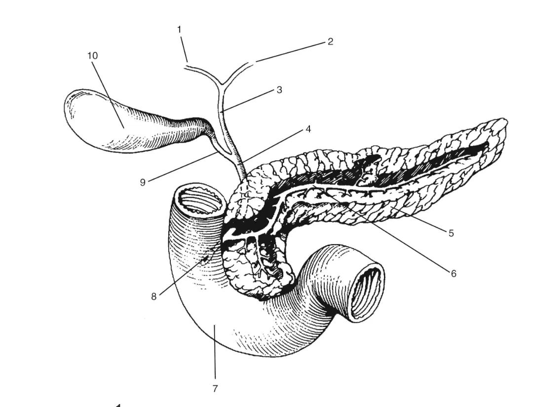
In this image, what is labeled 2
(a)
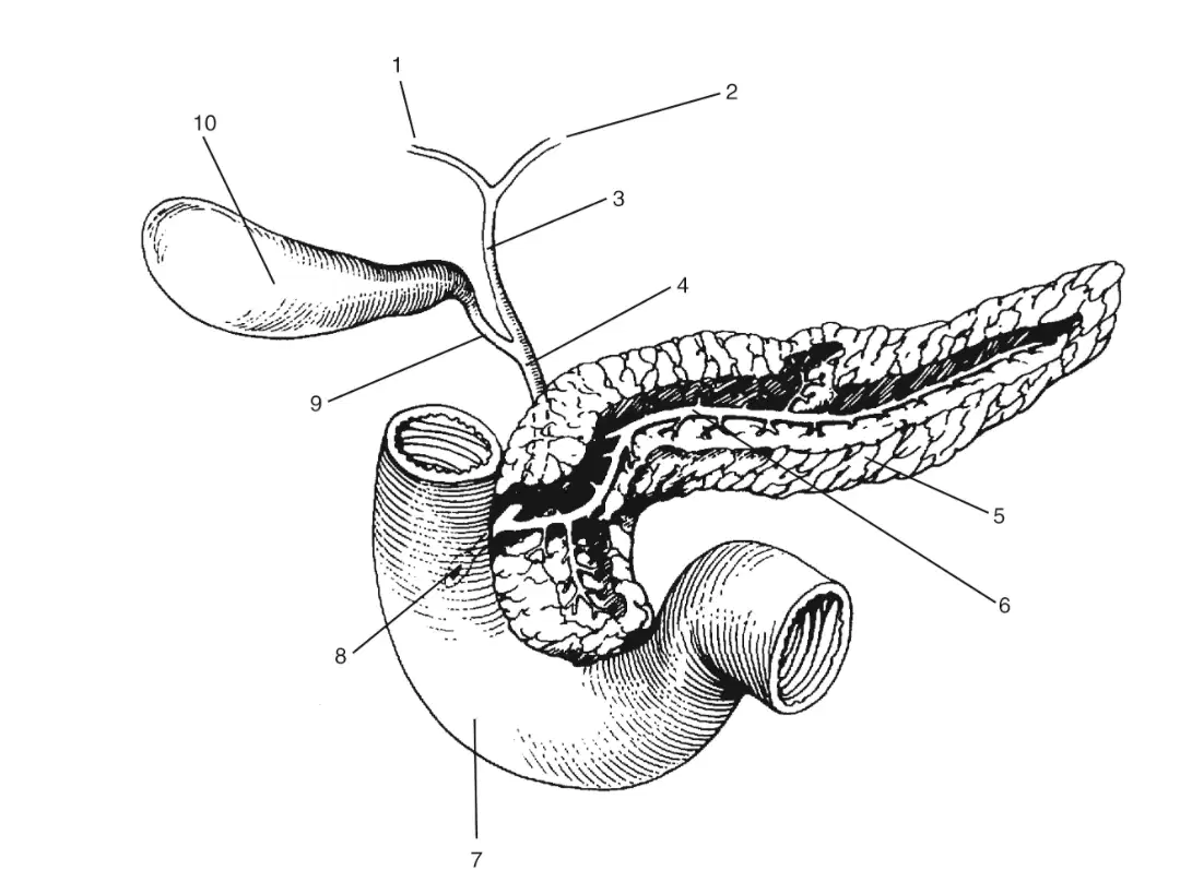
In this image, what is labeled 3
(a)
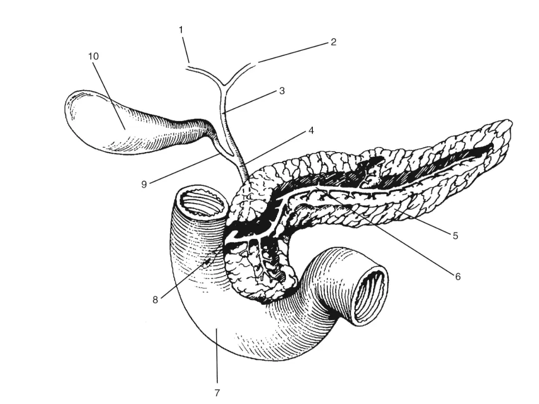
In this image, what is labeled 4
(a)
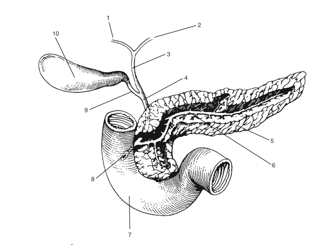
In this image, what is labeled 5
(a)

In this image, what is labeled 6
(a)
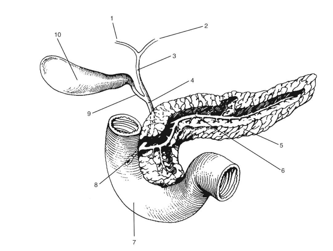
In this image, what is labeled 7?
(a)
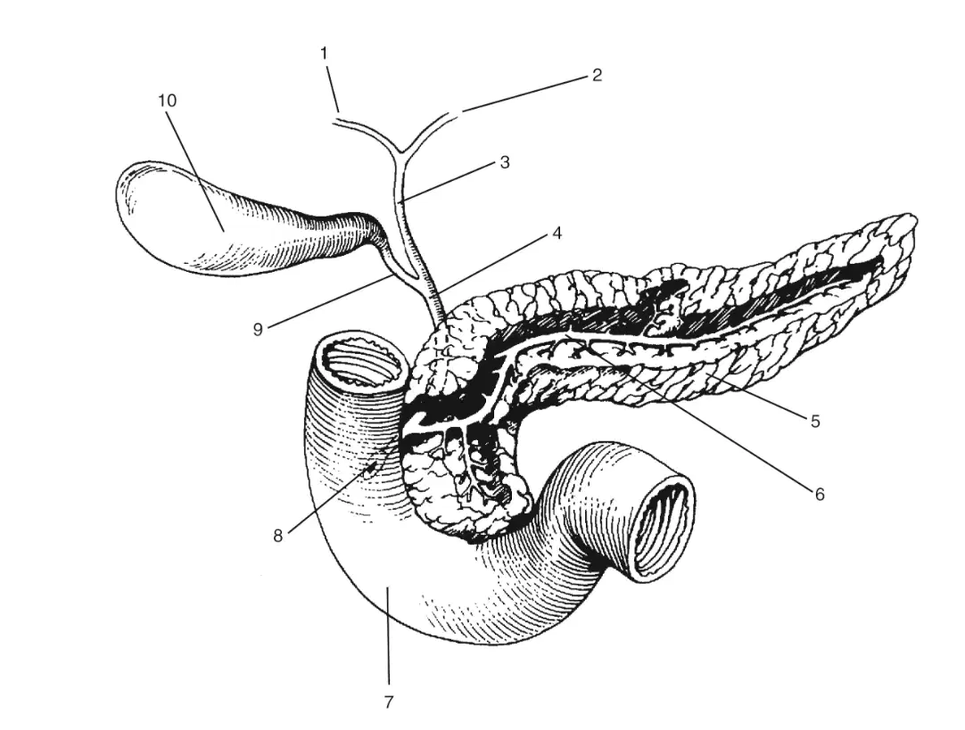
In this image, what is labeled 8?
Duodenum
Area of ampulla of vater with sphincter of odd
Pancreatic duct
Cystic duct
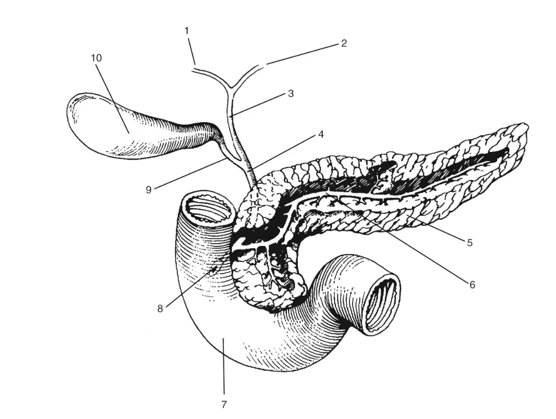
In this image, what is labeled 9?
(a)
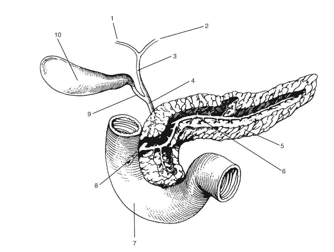
In this image, what is labeled 10
(a)
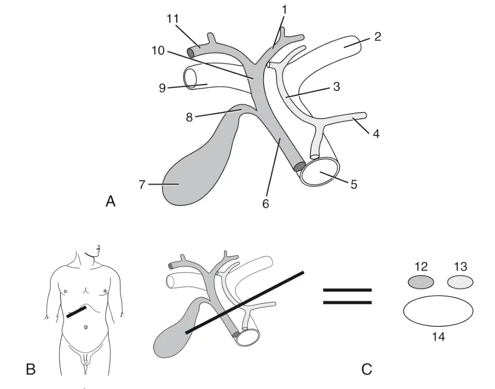
In this image, what is labeled 1
(a)
In this image, what is labeled 2
(a)
In this image, what is labeled 3
(a)
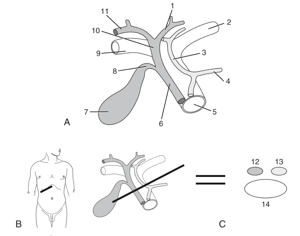
In this image, what is labeled 4
(a)
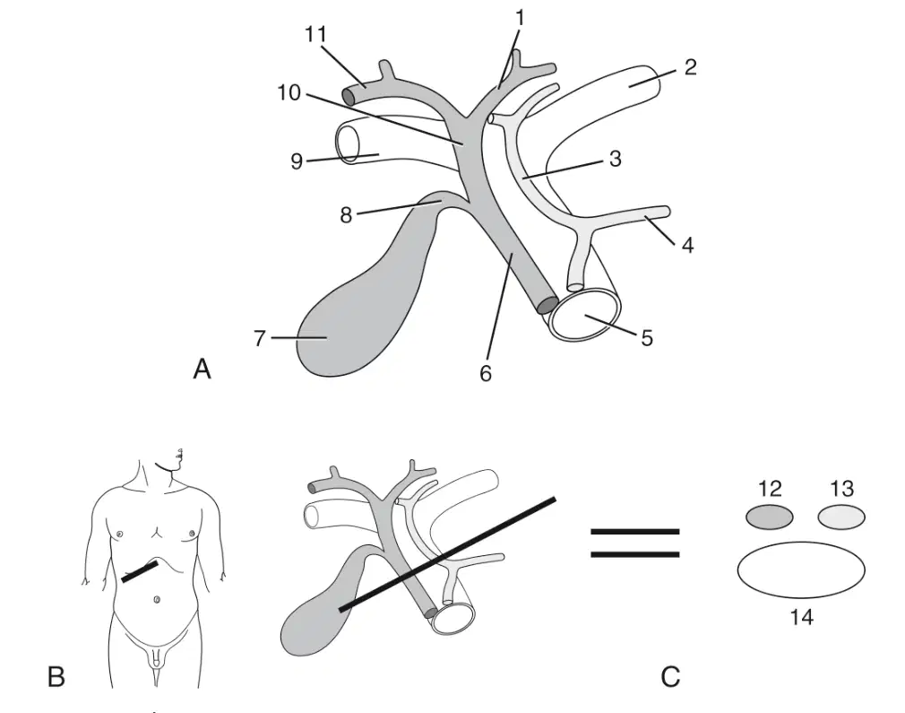
In this image, what is labeled 5
(a)

In this image, what is labeled 6
(a)

In this image, what is labeled 7
(a)
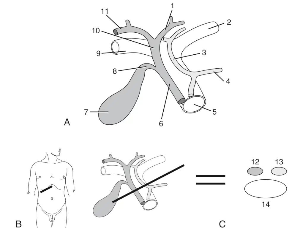
In this image, what is labeled 8
(a)

In this image, what is labeled 9
(a)
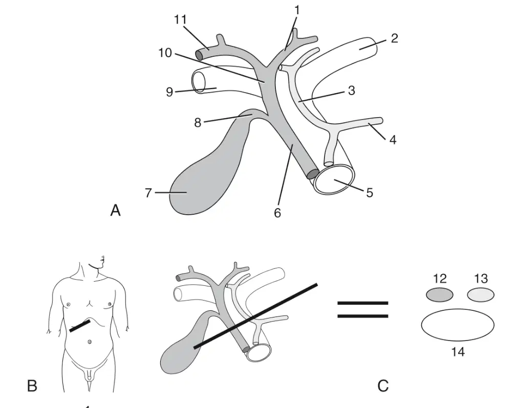
In this image, what is labeled 10
(a)
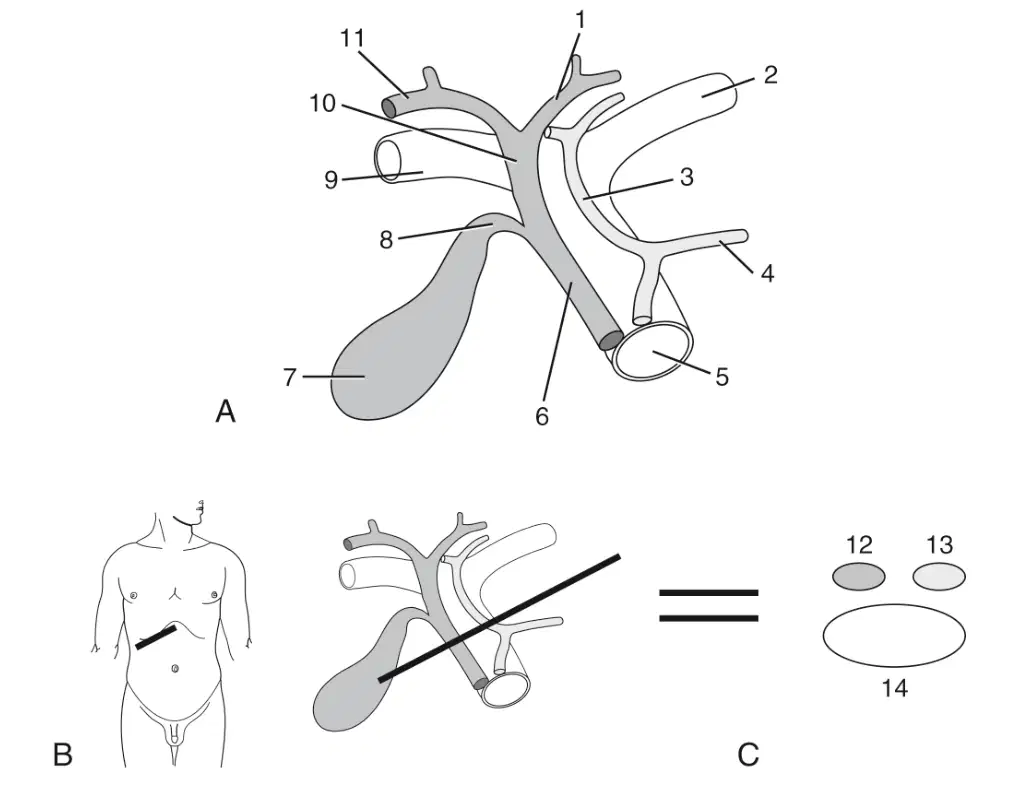
In this image, what is labeled 11
(a)
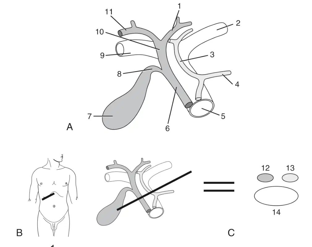
In this image, what is labeled 12
(a)

In this image, what is labeled 13
(a)
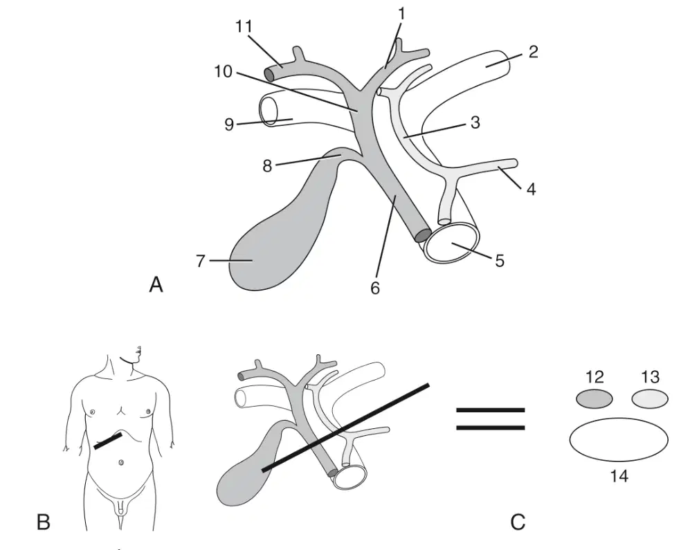
In this image, what is labeled 14
(a)
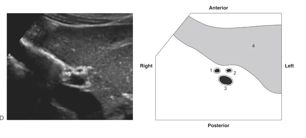
In this image, what is labeled 1
(a)
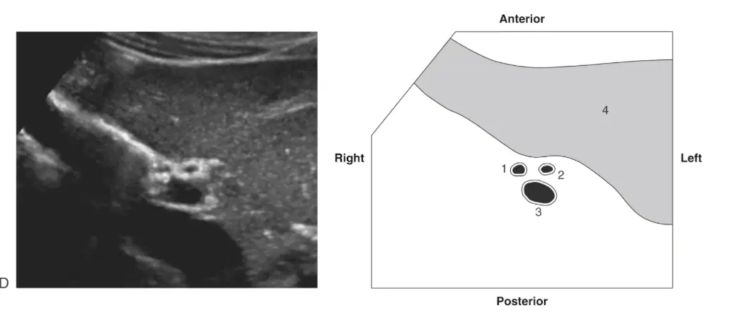
In this image, what is labeled 2
(a)
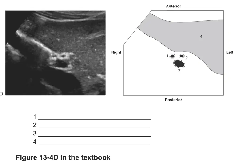
In this image, what is labeled 3
(a)

In this image, what is labeled 4
(a)
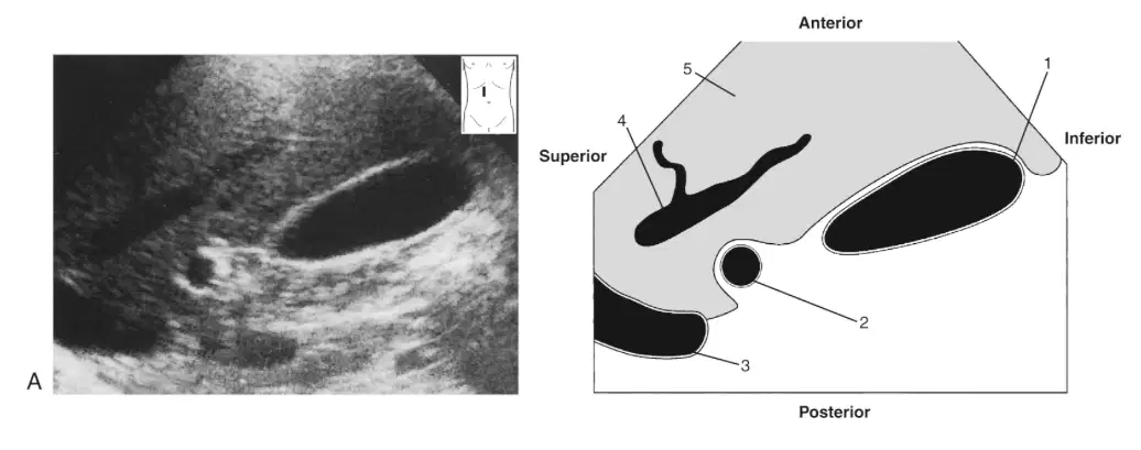
In this image, what is labeled 1
(a)
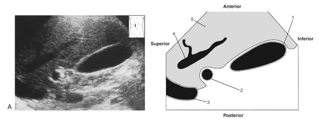
In this image, what is labeled 2
(a)
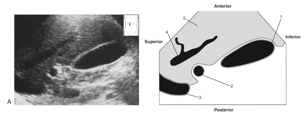
In this image, what is labeled 3
(a)
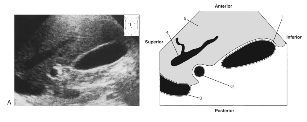
In this image, what is labeled 4
(a)
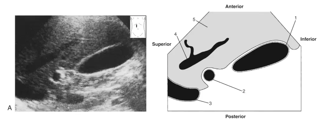
In this image, what is labeled 5
(a)
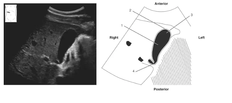
In this image, what is labeled 1
(a)
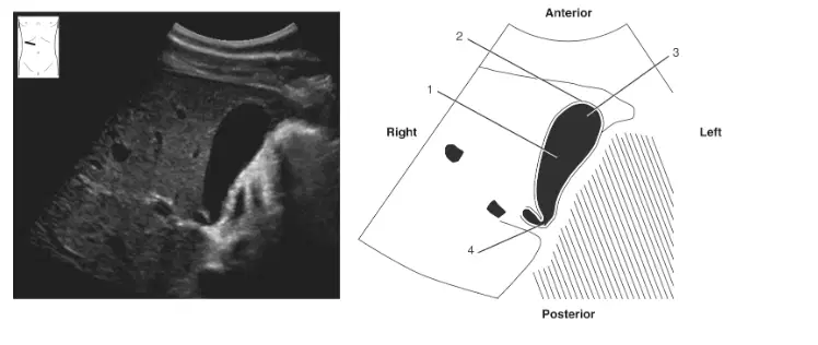
In this image, what is labeled 2
(a)

In this image, what is labeled 3
(a)

In this image, what is labeled 4
(a)
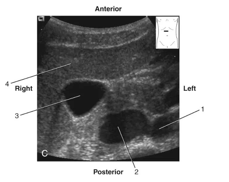
In this image, what is labeled 1
(a)
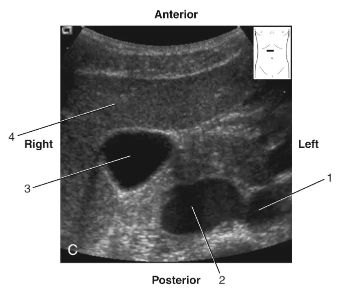
In this image, what is labeled 2
(a)
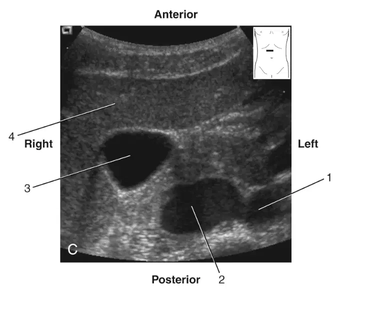
In this image, what is labeled 3
(a)
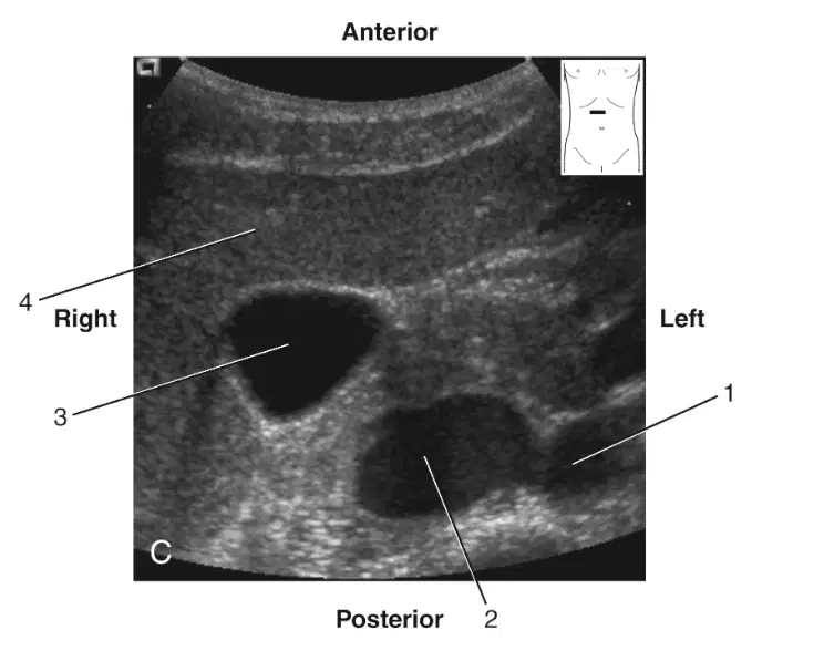
In this image, what is labeled 4
(a)
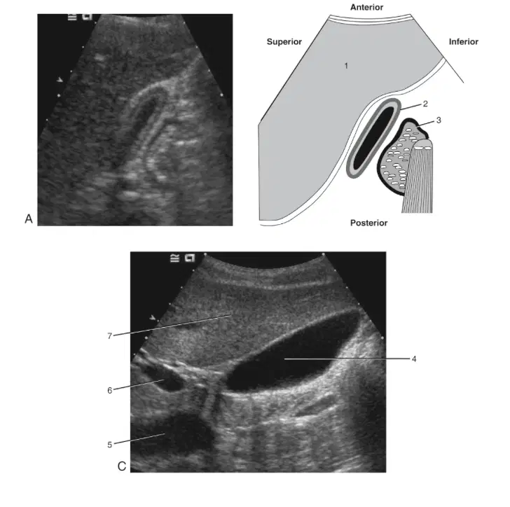
In this image, what is labeled 1
(a)
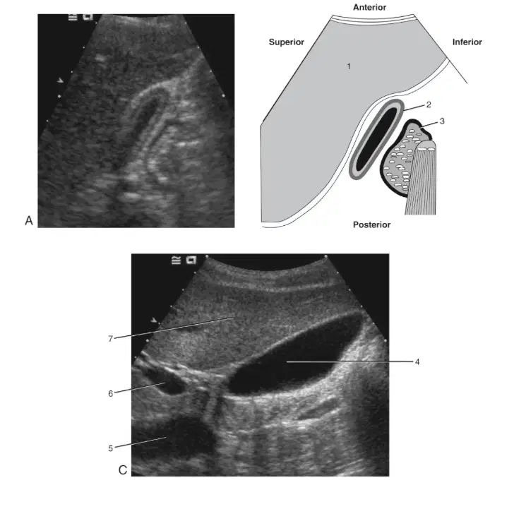
In this image, what is labeled 2
(a)
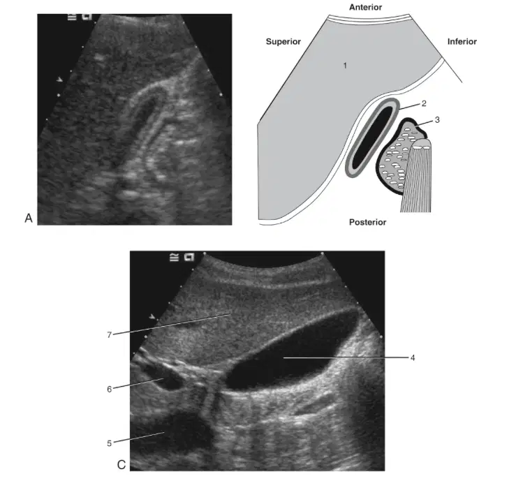
In this image, what is labeled 3
(a)
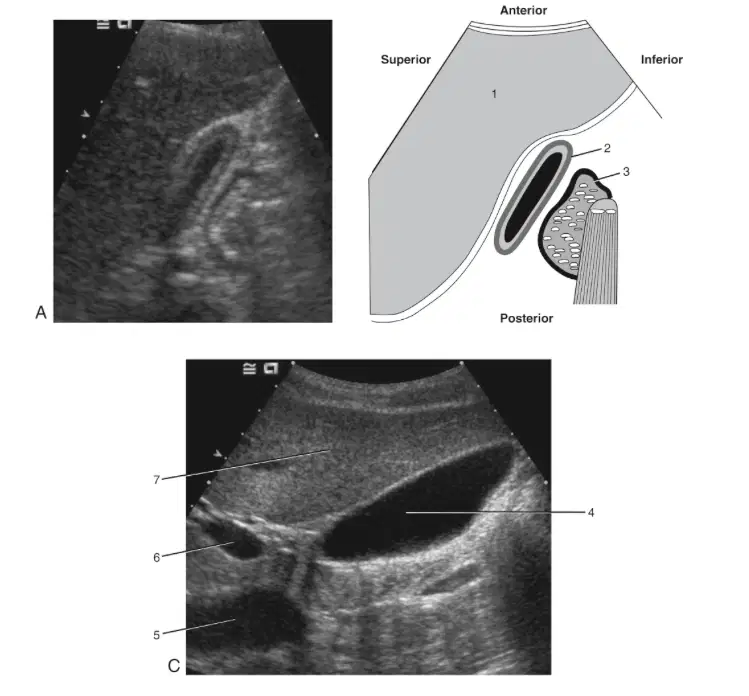
In this image, what is labeled 4
(a)
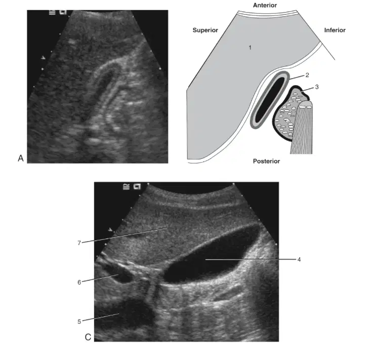
In this image, what is labeled 5
(a)
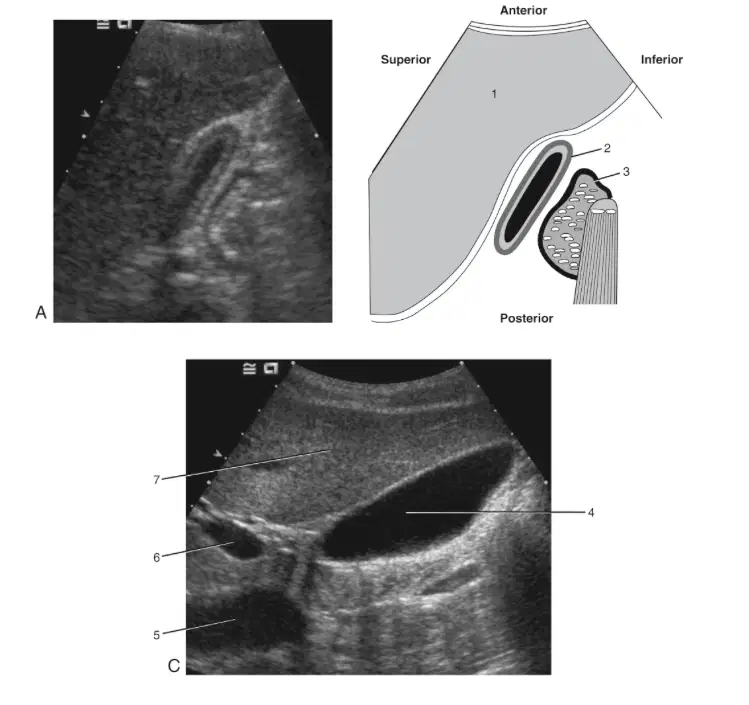
In this image, what is labeled 6
(a)

In this image, what is labeled 7
(a)
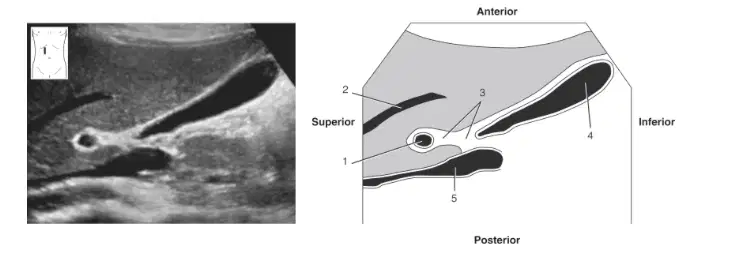
In this image, what is labeled 1
(a)
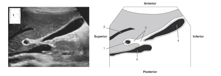
In this image, what is labeled 2
(a)

In this image, what is labeled 3
(a)
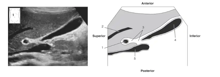
In this image, what is labeled 4
(a)
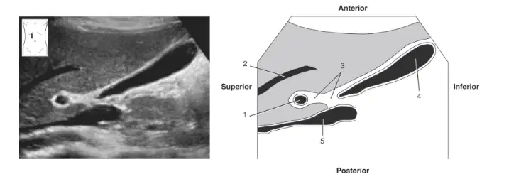
In this image, what is labeled 5
(a)
Which maximum measurement is noted for a bile-filled gallbladder
12cm
5cm
9cm
15cm
Which aspect of the common duct is considered the common bile duct?
Proximal
Superior
Distal
Anterior
Which congenital sonographic finding can appear similar to an hour glass?
Duplicated gallbladder
Phrygian cap
Junctional fold
Bilobed
Which sonographic finding is associated with the presence of stones in the ductal system?
Cholelithiasis
Choledocholithiasis
Nephrocalcinosis
Sludge
Which clinical indication presents as yellowish skin
Jaundice
Cyanosis
Pallor
Erythema
Which imaging test is used to assess the function of the gallbladder?
HIDA scan
CT scan
OCG
ERCP
Which term best characterizes the widest portion of the gallbladder?
Neck
Body
Tail
Fundus
Which group of terms is associated with an extrahepatic portal triad?
Right portal vein, common bile duct, and cystic artery
Main portal vein, proper hepatic artery, and common bile duct
Left portal vein, left hepatic artery, and common bile duct
Main portal vein, common hepatic duct, and main hepatic artery
Which sonographic feature best characterizes a choledochal cyst?
Outpouching of the gallbladder neck
Dilatation of the CBD
Pedunculated areas around the gallbladder fundus
Dilation of the cystic duct
Which muscle sheath regulates the flow of bile into the duodenum?
Ampulla of Vater
Rectus abdominis muslce
Sphincter of oddi
Pylorus
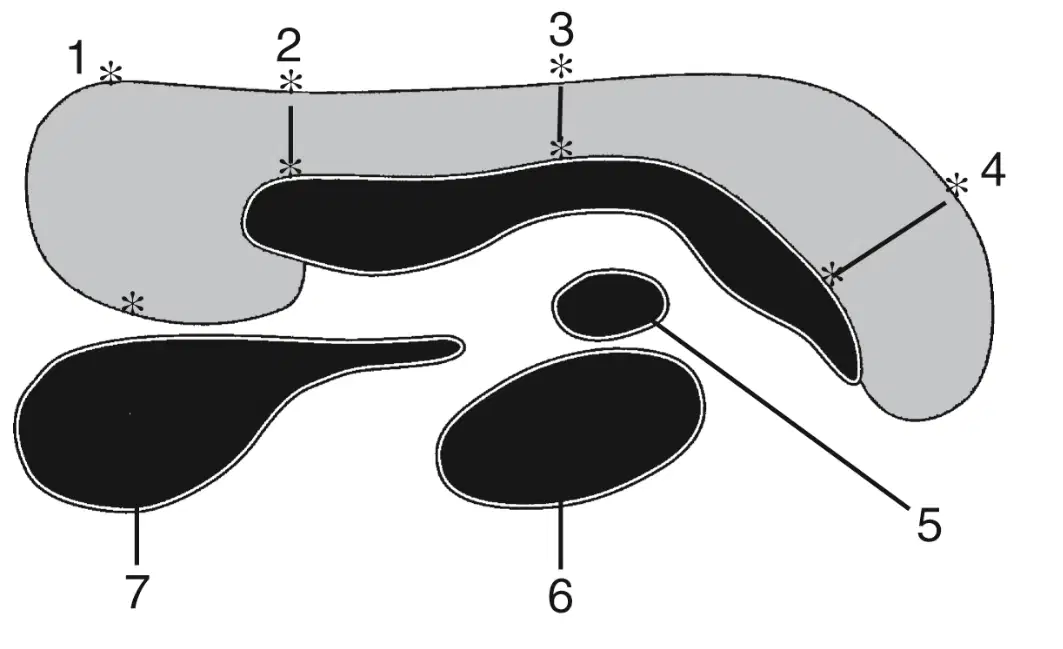
In this image what is labeled 1
(a)
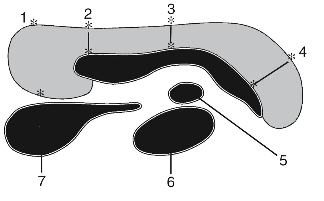
In this image what is labeled 2
(a)
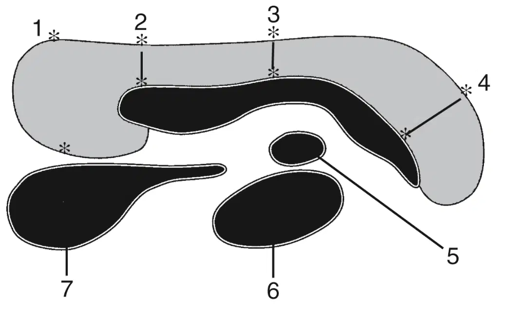
In this image what is labeled 3
(a)

In this image what is labeled 4?
(a)
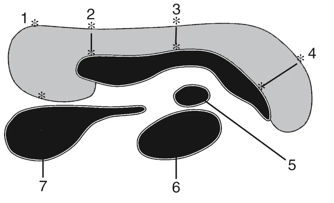
In this image what is labeled 5?
(a)

In this image what is labeled 6
(a)
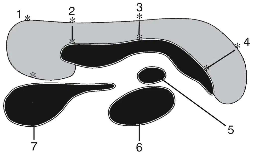
In this image what is labeled 7
(a)
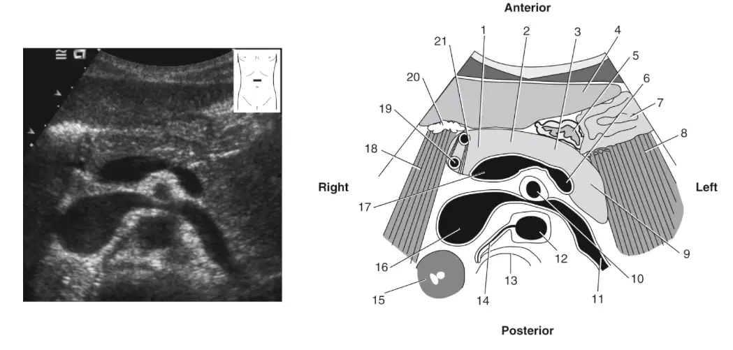
In this image what is labeled 1
(a)
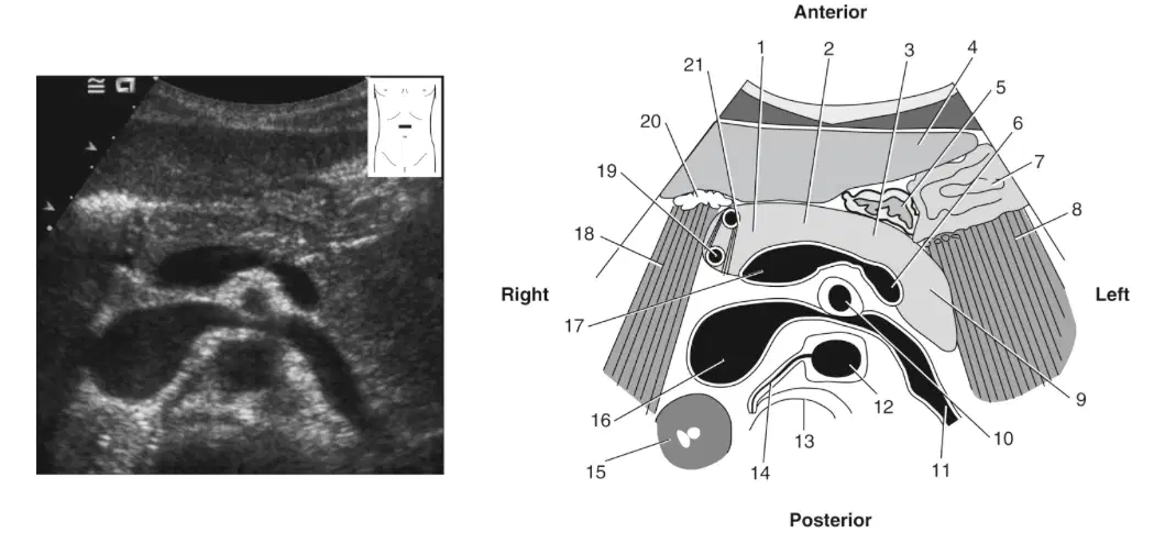
In this image what is labeled 2?
(a)
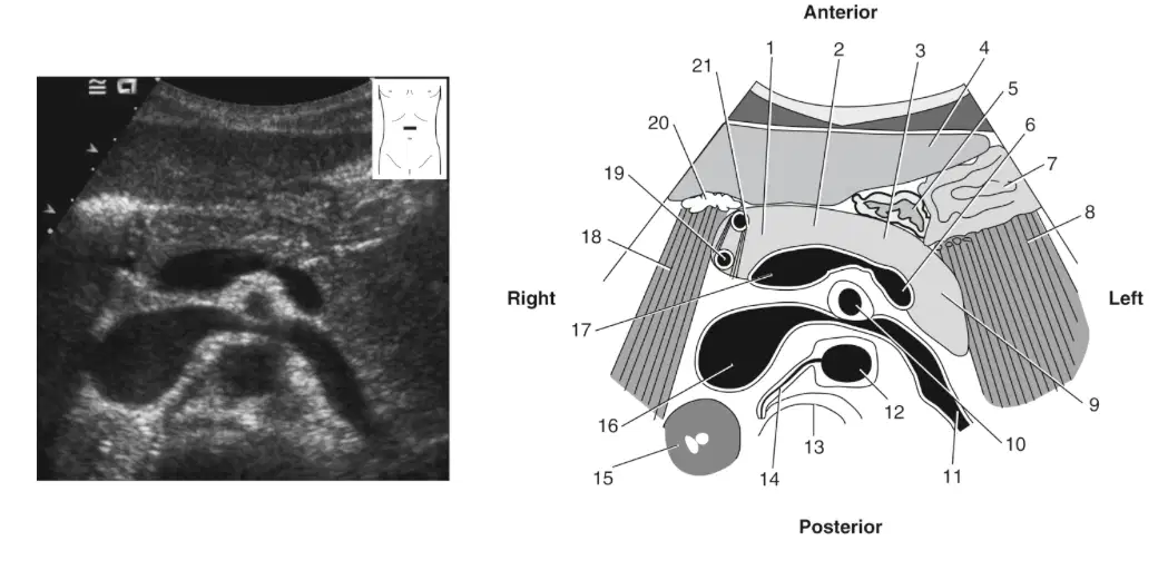
In this image what is labeled 3
(a)
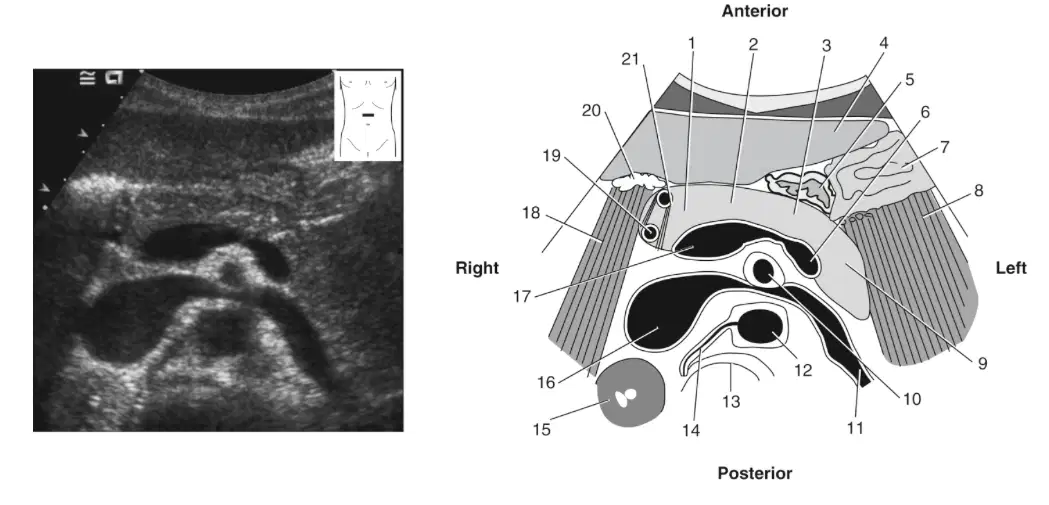
In this image what is labeled 4
(a)
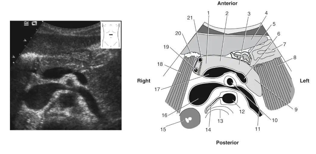
In this image what is labeled 5
(a)
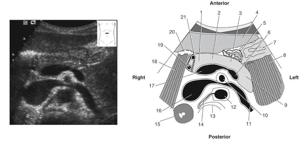
In this image what is labeled 6
(a)
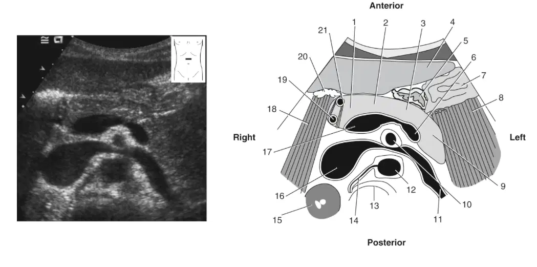
In this image what is labeled 7
(a)
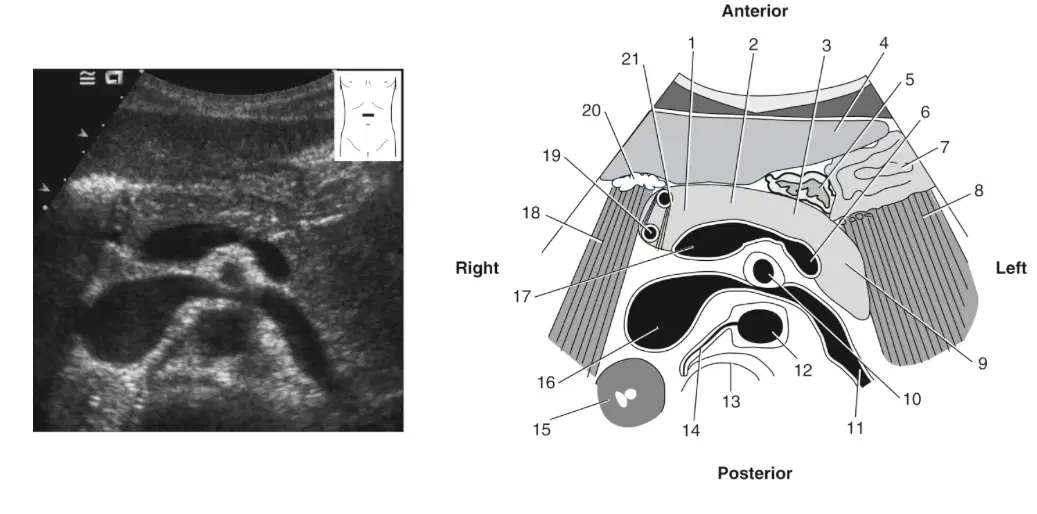
In this image what is labeled 8 & 18
(a)

In this image what is labeled 9
(a)
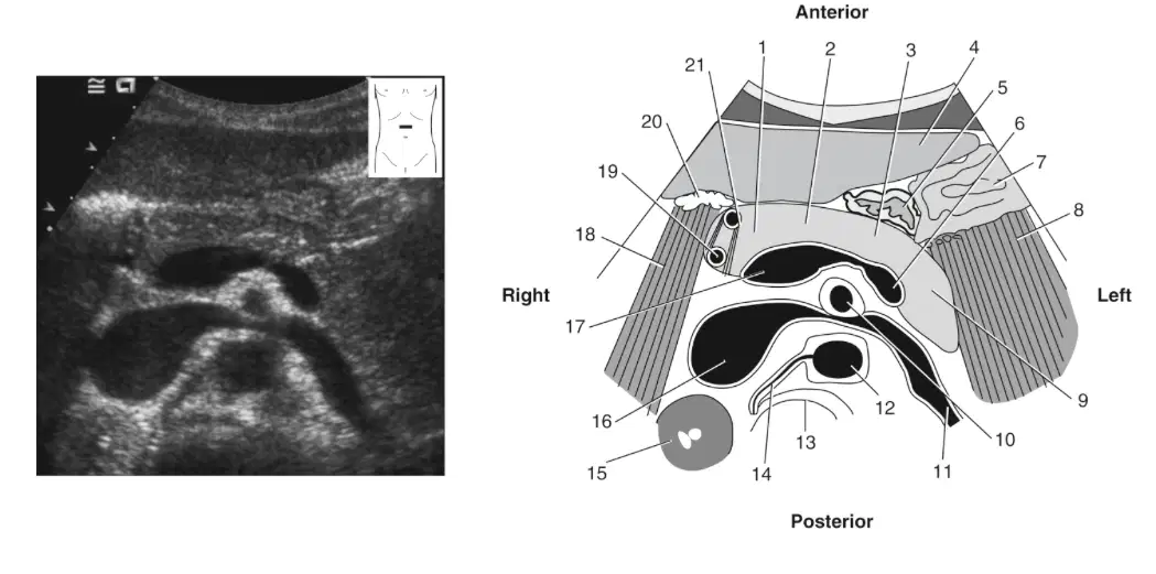
In this image what is labeled 10
(a)
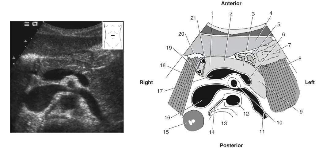
In this image what is labeled 11
(a)
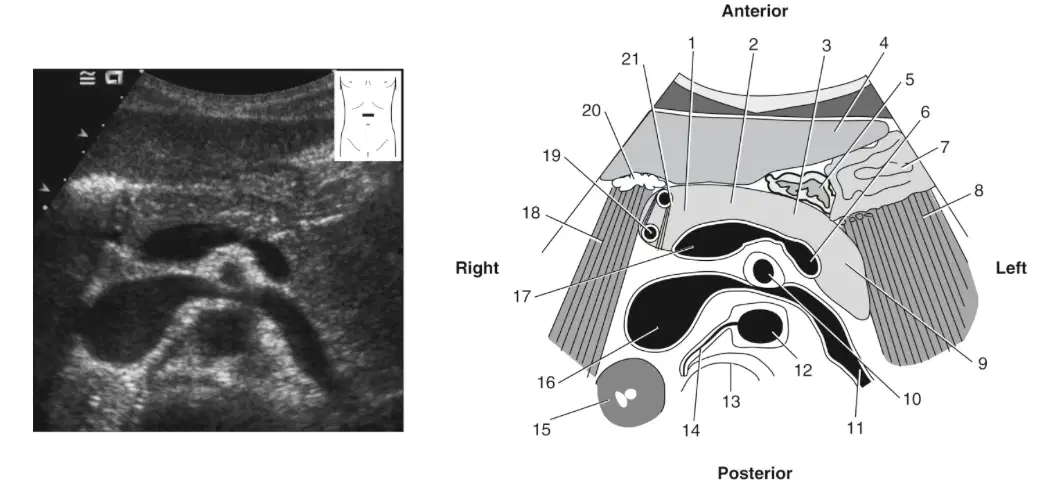
In this image what is labeled 12
(a)
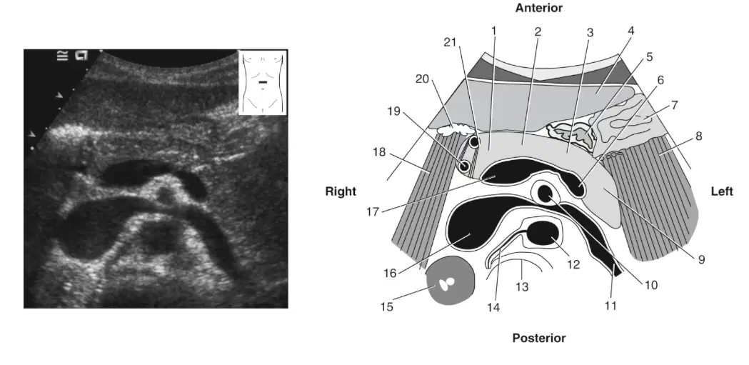
In this image what is labeled 13
(a)
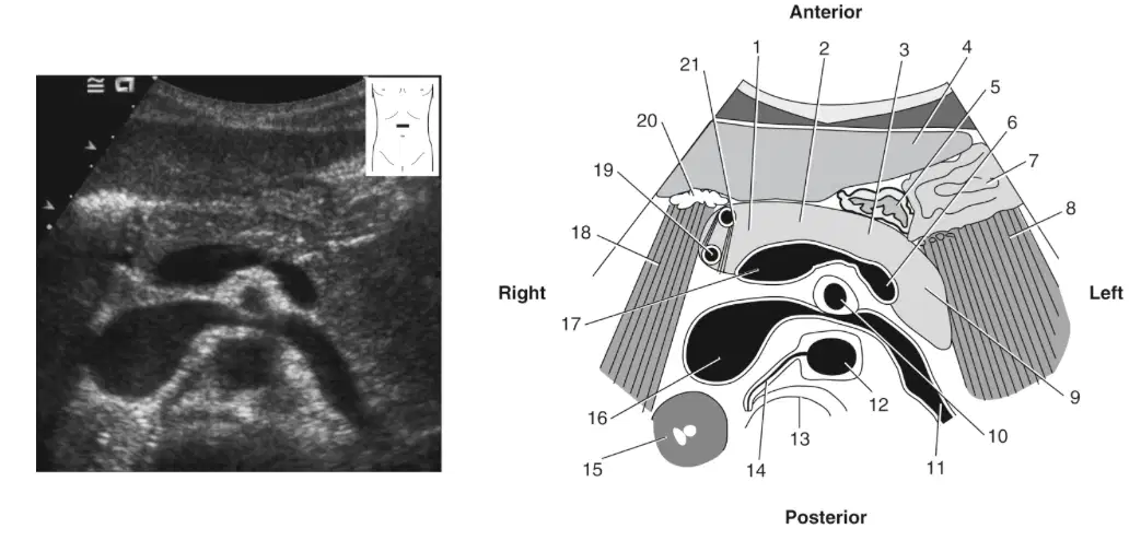
In this image what is labeled 14
(a)
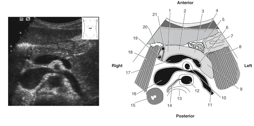
In this image what is labeled 15
(a)
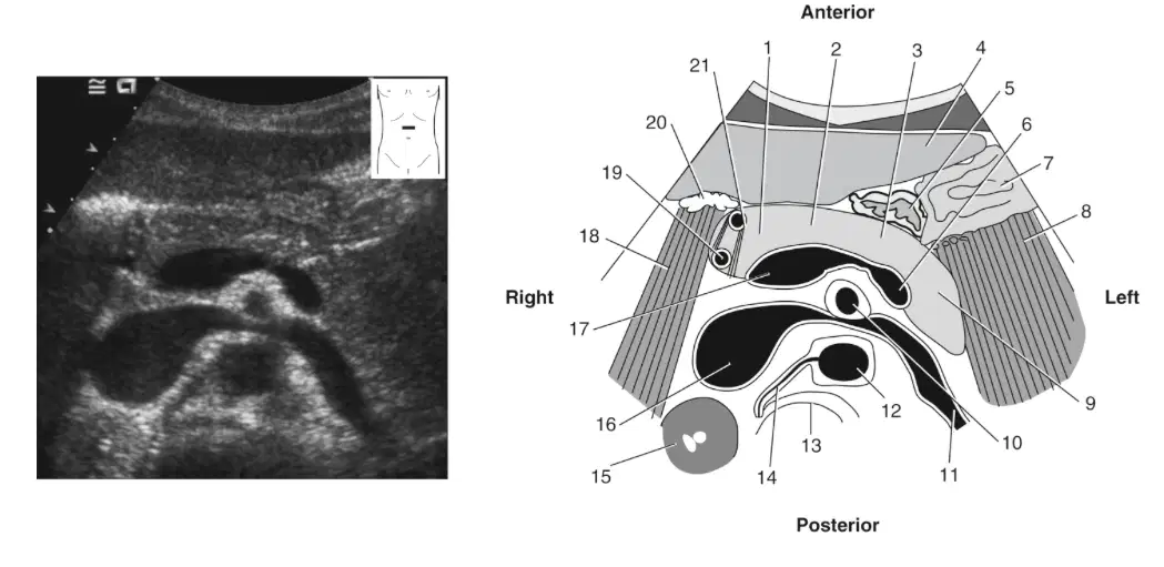
In this image what is labeled 16
(a)
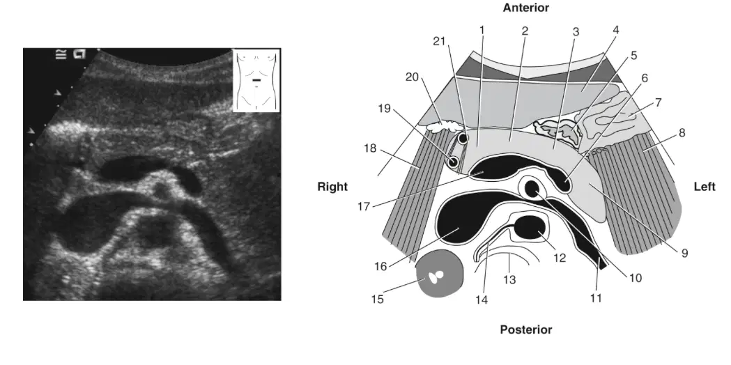
In this image what is labeled 19
(a)
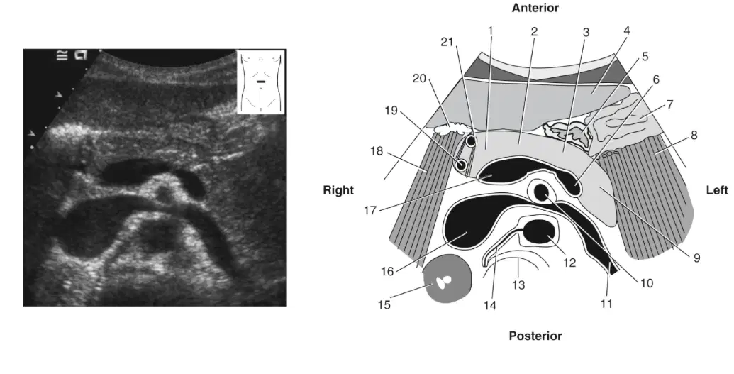
In this image what is labeled 20
(a)
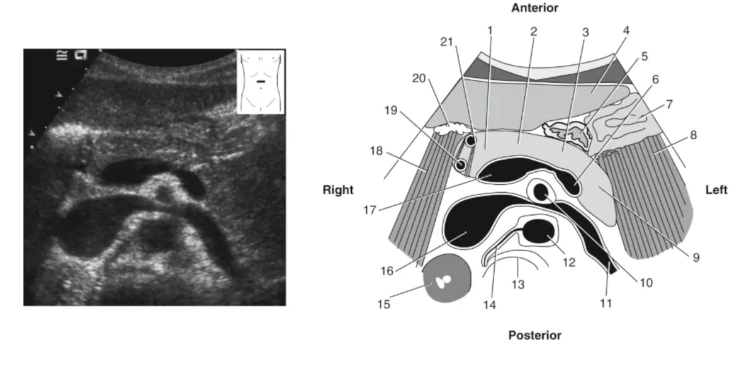
In this image what is labeled 21
(a)
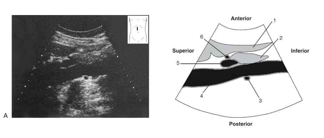
In this image what is labeled 1
(a)
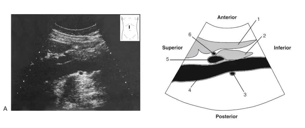
In this image what is labeled 2
(a)
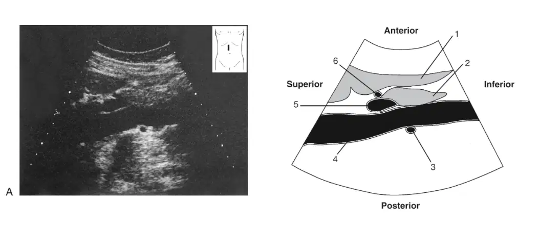
In this image what is labeled 3
(a)
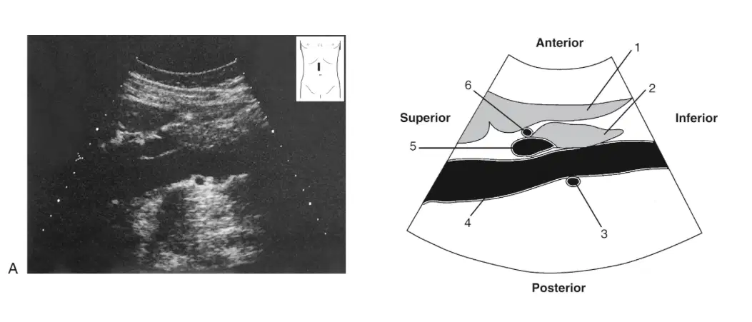
In this image what is labeled 4
(a)
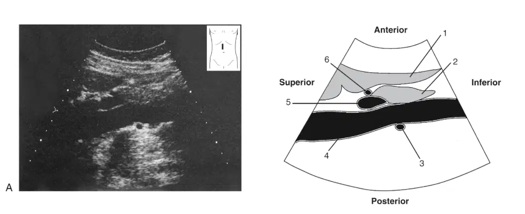
In this image what is labeled 5
(a)
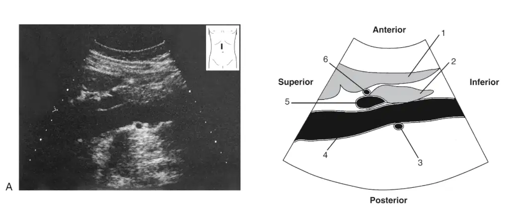
In this image what is labeled 6
(a)
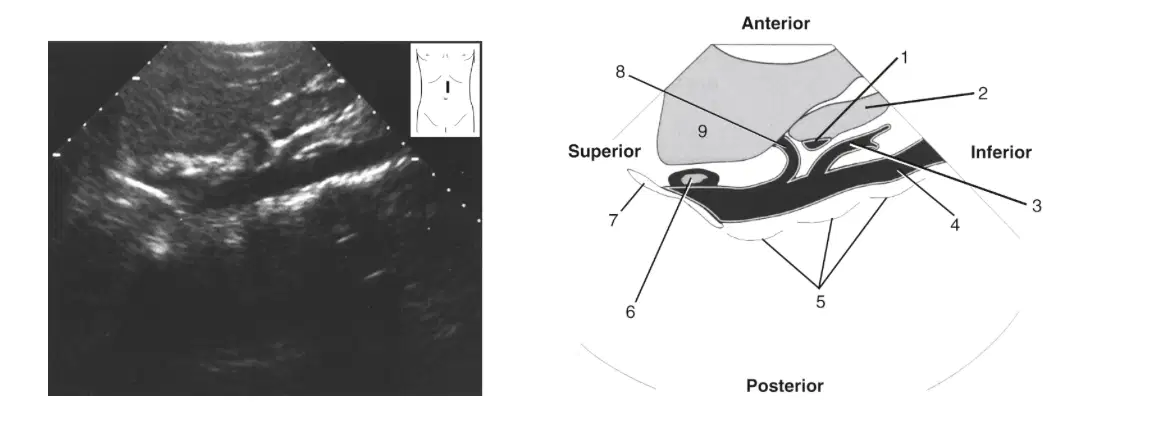
In this image what is labeled 1
(a)
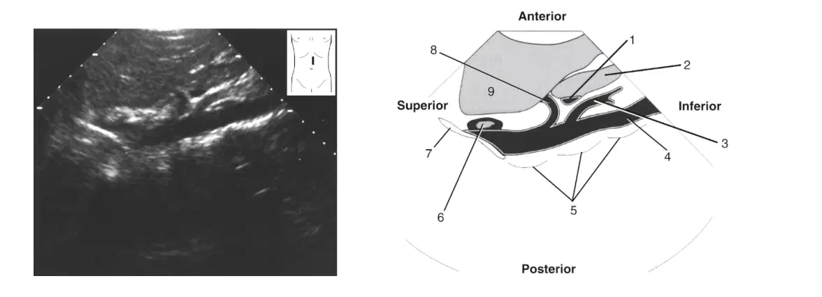
In this image what is labeled 3
(a)
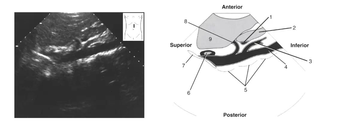
In this image what is labeled 4
(a)

In this image what is labeled 5
(a)
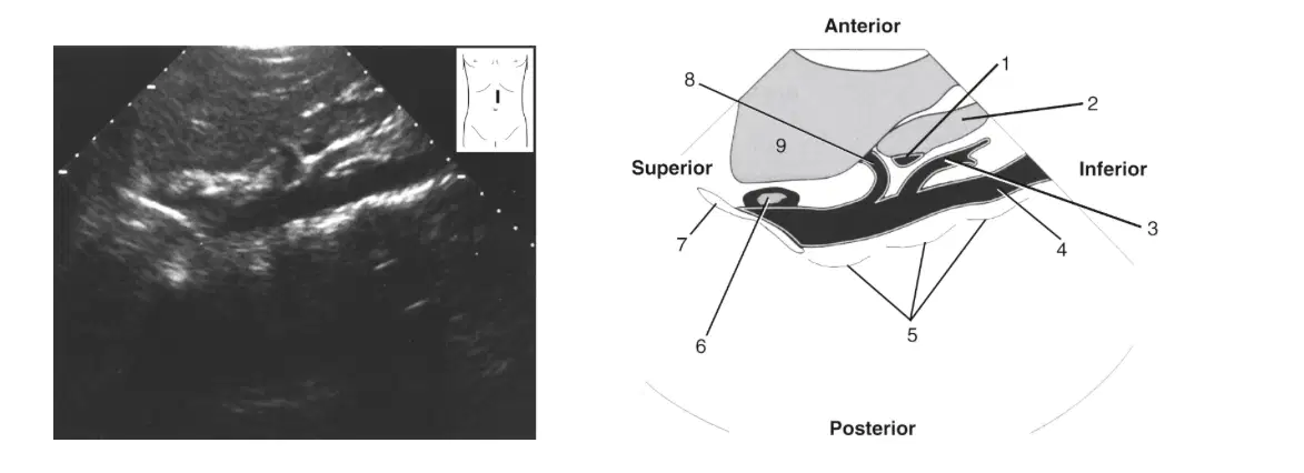
In this image what is labeled 6
(a)
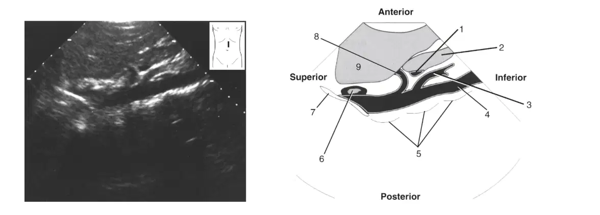
In this image what is labeled 7
(a)
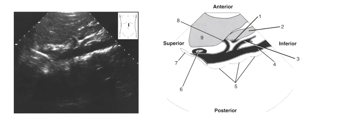
In this image what is labeled 8
(a)
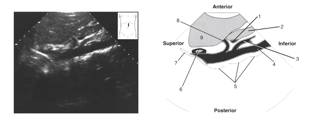
In this image what is labeled 9
(a)
The pancreas is shaped like the upside down letter
H
N
U
C
Which structures are posterior to the pancreas?
Inferior vena cava (IVC), stomach, transverse colon
IVC, diaphragm, aorta (AO)
AO, stomach, duodenum
Duodenum, connective prevertebral tissue, diaphragm
Which of the following structures enter the duodenum?
Common bile duct, accessory pancreatic duct, duct of Wirsung
Common bile duct, common hepatic duct, accessory pancreatic duct
Common hepatic duct, cystic duct, main pancreatic duct
Celiac duct, common hepatic duct, common bile duct
The body of the pancreas can best be described as lying anterior to
superior mesenteric artery (SMA), splenic vein (SV), AO.
SMA, SV, IVC
IMA, common bile duct (CBD), AO.
inferior mesenteric artery (IMA), SV, IVC
The tail of the pancreas can best be described as medial to the
Stomach
Left kidney
Splenic Artery
Splenic hilum
Anteroposterior measurement of pancreatic head (a)
Anteroposterior measurement of pancreatic neck (a)
Anteroposterior measurement of pancreatic body (a)
Anteroposterior measurement of pancreatic tail (a)
Total length of the pancreas (a)

