
 Worksheets
Intro wk 6
Total questions: 121
Worksheet time: 2hrs 59mins
Assessment of ultrasound system components
(a)
Provide repairs
Perform preventative maintenance
(a)
Keep records of assessments
(a)
Maintain high function of equipment
(a)
Detect gradual degradation of images
(a)
Minimize equipment downtime
(a)
Protect patient from overexposure
(a)
Sonography departments have ___ ___ who perform analysis of equipment using specific instruments
(a)
Any cracks in the transducer housing
(a)
Tears in equipment wiring
Patient safety hazards
(a)
Degradation of imaging
(a)
Minimal exposure to the patient should always be practiced
(a)
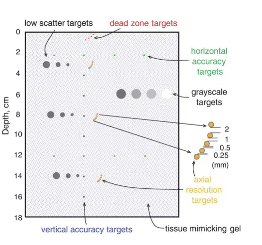
General-purpose ultrasound quality assurance (a) are typically composed of several scanning modules.
AIUM 100 mm test object
(a)
Tissue equivalent or phantom
(a)
Doppler phantom
(a)
Beam profile/slice thickness phantom
(a)
When are sonography examinations most useful?
When performed for no reason
When performed for the purpose of answering a specific, well informed question
When performed for a broad question
None of the above
Studies performed as surveys tend to be (a) informative
Is there a mass in the liver?
Is there an aneurysm?
Does the patient have ascites?
These are all examples of: (a)
Are there gallstones?
Is the mass solid, cystic or complex?
Is there a blood clot or stenosis?
These are all examples of (a)
Sonographer should adopt a (a) attitude toward examination technique
Don’t hesitate to go beyond what is request on the examination
(a)
Look at other structures for origin of pain
(a)
Demonstrate incidental findings and comment on them
(a)
Change patient’s position to produce adequate study
(a)
It is useful to move the patient while scanning, especially to determine if a fluid collection is free, contained in bowel, or loculated
(a)
Preparation depends on (a) of examination
Preparation (a) always needed
__ __ __ may not be able to prepare
(a)
For an ___ ___ patients are kept NPO (fasting) overnight or 6-8 hours
(a)
Insures visualization of normal, distended gallbladder
(a)
May reduce bowel air content
(a)
Previous barium studies may impair sound transmission or mimic an abnormal mass
(a)
US should be done before upper GI or other barium studies
(a)
For kidneys, patient should be well (a)
Evaluate in longitudinal and transverse scanning planes
Compare to adjacent organs
Evaluate all lobes
(a)
Evaluate in longitudinal and transverse scanning planes in supine position
(a)
Assess for painful gallbladder using probe pressure
Image entire length of biliary tree
(a)
Turn patient into different positions
Take appropriate measurements
(a)
Focus, tgc, gain repairs
(a)
Image via transverse and longitudinal planes
Identify all portions
Assess for abnormalities, ductal dilatations, and echo texture alterations
(a)
Assess peripancreatic region (relational anatomy) for fluid and adenopathy (enlarged lymph nodes)
Measure as needed
(a)
Image in the longitudinal and transverse planes
Compare echogenicity to the left kidney
(a)
Evaluate for left pleural effusion (fluid around the lung)
Measure spleen size
(a)
This is an example of a (a)
- Evaluate for wall thickness, dilatation, muscular hypertrophy, masses, hyperemia, and other abnormalities
- Evaluate at least two perpendicular scanning planes
(a)
- Provide location and extent of fluid (small, moderate, or large)
- Evaluate in longitudinal and transverse plane images through specific organs according to quadrant
(a)
- Demonstrate relationship of an identified mass with the peritoneum
- Evaluate surrounding muscles
(a)
Kidneys, spleen, pancreas, GI tract, kidney, bladder = (a)
- Evaluate in the longitudinal and transverse scanning planes, usually with the patient in a supine position
- Assess the cortices, renal pelvis, and echogenicity of each kidney
(a)
-Record maximum renal length (required measurements)
-Evaluate the perirenal area and the adrenal glands
(a)
- Evaluate the bladder for thickening and masses
- Exame the intraluminal contents for signs of debris
(a)
Use Doppler imagery to make various assessmentsR
(a)
- Obtain the greatest dimension of the different segments of the aorta and the proximal common iliac vessels
(a)
Record details about any aneurysms
(a)
-Maximum size and location
-Relationship to the renal arteries and to the aortic bifurcation
-Utilize color flow and Doppler waveforms
(a)
There is no patient prep for which exam?
OB
Abdomen
Thyroid, cervical lymph, parathyroid, salivary glands
Vacular
Which abdominal sonogram does not need to be NPO?
(a)
Position the patient supine with the neck hyperextended.
Image the right and left lobes of the thyroid gland in the longitudinal and transverse planes.
(a)
- Images of the superior, mid, and inferior portions of both lobes should be done in the transverse plane.
- Measure each lobe in three dimensions: length, width, and height.
(a)
A general assessment of the neck may be required when there are palpable lesions outside of the ___ ___.
(a)
Patient positioning for a (a) scan is the same as that for the thyroid.
Search the common locations of adenoma, as it may not be palpable
(a)
Patient positioning for a (a) scan is the same as for the thyroid
Typically, no patient preparation.
Position the patient so both testicles can be imaged simultaneously.
(a)
Typically, no patient preparation.
Position the patient so both testicles can be imaged simultaneously.
(a)
Compare the size of each testicle and epididymis.
Examine scrotal wall thickness and any focal palpable masses.
(a)
Color and spectral Doppler should be employed to evaluate the vascularity of both (a)
Measure gallbladder wall in (a)
- There is typically no patient preparation.
- Correlate the sonogram with clinical signs and/or symptoms and with other imaging studies (mammography)
(a)
- View any lesions or areas being studied in two perpendicular projections.
- Determine the size of any lesion by recording its maximal dimensions in at least two orthogonal planes.
(a)
Obtain at least one set of images of a lesion without calipers.
Annotate the image appropriately and completely.
(a)
- The BI-RADS sonographic categories help characterize (a) masses.
-Elastography may be used by some institutions.
- Mass characterization with sonography is highly dependent on technical factors.
(a)
A rotating chair may be needed to perform the sonogram.
(a)
Image the shoulder according to the structure examined
(a)
Perform dynamic imaging with the patient moving the arm into different positions throughout.
(a)
- A comparison of the contralateral side may be helpful.
- The bones of the shoulder may be examined for signs of fracture.
(a)
Patient preparation requires that the patient's urinary bladder can be distended with urine
(a)
Transabdominal pelvic: In emergency situations it can be retrofitted with (a) through a Foley catheter
Is a Foley catheter commonly used anymore?
Yes
No
The uterus is imaged in the __ and __ planes
(a)
Evaluate the size, shape, orientations, myometrium, endometrium, and cervix
(a)
Evaluate the depth, length, and width of ovaries
(a)
Evaluate the adnexa for fluid, dilated tubes, and masses
(a)
Color, duplex doppler can be used to evaluate
(a)
There is typically no patient preparation required.
The uterus is imaged in longitudinal and transverse planes
(a)
Evaluate the size, shape, orientations, myometrium, endometrium, and cervix
Evaluate the depth, length, and width, of ovaries
(a)
Evaluate the adnexa for fluid, dilated tubes, and masses
(a)
Color, and duplex doppler can be used to evaluate blood flow
(a)
For a (a) sonography, the patient should have a distended bladder.
For (a) scanning, the bladder should be emptied
Evaluate the maternal uterus as if performing a pelvic sonogram
(a)
Analyze for evidence of a gestational sac to confirm an intrauterine pregnancy (IUP)
If an IUP is identified, the crown-rump length is a good indicator of gestational age.
(a)
- Document amnionicity and chorionicity for all multiple gestations.
- Assess fetal anatomy appropriate for the first trimester
(a)
NT measurements in conjunction with serum biochemistry can determine risk of anatomic abnormalities.
(a)
There is typically no patient preparation needed.
Document fetal number, fetal cardiac activity, and presentation.
(a)
Assess amniotic fluid volume.
Assess location and appearance of the placenta
(a)
Evaluate the umbilical cord for presence of two arteries and one vein as well as its insertion into the placenta.
(a)
Biometric parameters can be used to estimate gestational age and fetal weight.
Evaluate the maternal adnexa and cul-de-sacs when technically feasible.
(a)
No patient preparation
Assess the common and internal and external carotid arteries
(a)
- Scan arteries using appropriate grayscale and Doppler techniques
- Use color/duplex Doppler to detect areas of narrowing and abnormal flow
(a)
No patient preparation
Examining all vessels in the longitudinal and transverse scan planes
(a)
Apply venous compression to (a) the normal vein lumen
-Record right and left common femoral or right and left external iliac venous spectral Doppler waveforms
- Determine location and duration of reversed blood flow when evaluating for venous insufficiency
(a)
- No patient preparation
- Evaluate via grayscale and Doppler all portions of the subclavian, innominate, internal jugular veins, and axillary veins
(a)
Evaluate the brachial, basilica, and cephalic veins using compression sonography
(a)
No patient preparation
Conduct grayscale/color Doppler imaging and spectra Doppler waveforms in the appropriate segments
(a)
Use color/duplex Doppler imaging to improve detection of arterial lesions
(a)
There is typically no patient preparation, though institutional requirements may vary
(a)
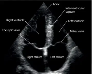
(a)
Minimum documentation should include
Patient name and or identifying information (birthday, gender, etc.)
Date of examination
Identifying information of the facility where the exam was performed
Unrelevant clinical information
First trimester up to (a) weeks
Minimum documentation should include
No indications
Relevant clinical information
Image orientation when appropriate
Standard presentation and annotation (labeling) of images to include anatomic location laterality (right, left, midline when appropriate)
Minimum documentation should include:
No name
Signs and symptoms, pertinent lab information
Indication for exam
Name of healthcare provider
Completed oral or written summary of findings for the supervising interpretating provider or physician
(a)
Description of sonographic findings only without offering a conclusion or diagnosis
(a)
Shadowing
Acoustic enhancement
Measurements
(a)
Always adhere to code of medical ethics/and or professional conduct available from professional associations
(a)
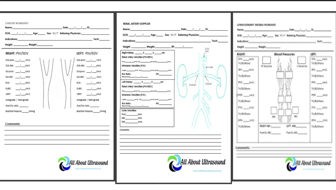
These are examples of (a)

