
 Worksheets
Martha
Total questions: 55
Worksheet time: 28mins
Bones in the body
206
200
204
222
Axial Skeleton:
80 bones in the core of the body, including the sku l, ears, neck, back, and ribcage
126 bones. Limbs or appendages
70 bones in the core of the body, including the skull, ears, neck, back, and ribcage
Osteoblast:
Build/repair bone
destoy bone
bone mass
bone damage
Osteoclas
destroy bone
repair bone
heals bone
What is the longest bone?
Femur
ulna
tibula
hemur
The patela/kneecap
a sesamoid bone
flat bones
legs ankle
sesamoid bone
a bone embedded within a tendon or a muscle
in the ankle
near the chest cavity
types of bones
long bones
short bones
flat bones
irregular
Skeletal system
provides support and protects organs. It’s the bodies foundation. It makes blood for the body, and stores necessary minerals.
It links the nervous and the skeletal systems together. A lows for movement. There are three types of muscle: skeletal, smooth, and cardiac.
only axial bones
only appendicular bones
Muscular system:
It links the nervous and the skeletal systems together. A lows for movement. There are three types of muscle: skeletal, smooth, and cardiac.
provides support and protects organs. It’s the bodies foundation. It makes blood for the body, and stores necessary minerals.
The brain and the spinal cord. Responsible for receiving, processing, and responding to sensory information.
Synovial Joints (most common in body)
pivot, hinge, condyloid, saddle, plane, and ba l-and socket-joints
only pivot cant hinge
only hinge cant pivot
can only hinge and pivot
Central nervous system:
The brain and the spinal cord. Responsible for receiving, processing, and responding to sensory information
Nerves that extend from the spine to a l other parts of the body. Sends info back to the CNS.
It links the nervous and the skeletal systems together. A lows for movement. There are three types of muscle: skeletal, smooth, and cardiac.
Peripheral nervous system:
Nerves that extend from the spine to a l other parts of the body. Sends info back to the CNS
The brain and the spinal cord. Responsible for receiving, processing, and responding to sensory information.
Basic unit of the nervous system. Axon, ce l body, dendrites. A lows you to do everything (breathe, run, etc.)
Sarcomere:
The basic contractile unit of muscle fiber.
Made up of a motor neuron and the muscle fibers it innervates
Made up of a motor neuron and the muscle fibers it innervates
Motor unit:
Made up of a motor neuron and the muscle fibers it innervates
Sympathetic Nervous System
Part of the autonomic nervous system. "Fight-or-flight"
The basic contractile unit of muscle fiber.
Sympathetic Nervous System:
Part of the autonomic nervous system. "Fight-or-flight
The basic contractile unit of muscle fiber
Part of the autonomic nervous system. “Rest and Digest”
Parasympathetic Nervous System
Somatic Nervous system:
A lows you to control your body's movements and receive external stimuli.
Sliding Filament Theory
When actin and myosin (myosin larger), slide past each other to generate movement. This occurs in the sarcomere (basic muscle unit)..
Type 1 muscle fibers
Slow twitch. More important for endurance events. Lighter and less powerful. Mostly aerobic (fat/food fuel).
Type 2 muscle fibers:
T Fast twitch. Larger and paler than type 1. Anaerobic, short, explosive periods of physical activity (carbs, glucose fuel)
Arteries:
Carry oxygen-rich blood away from the heart to the rest of the body.
Veins
Carry deoxygenated blood from the body to the heart, where goes to the lungs to be re-oxygenated. .
Capillaries:
The smallest blood vessels, and the site of exchange for nutrients (oxygen,CO2,etc.) to tissues
Atria:
Top reservoir chambers (smaller)
Ventricles
Bottom chambers (bigger)
Right Atria
Receives deoxygenated blood from body
Left ventricle:
Pumps oxygen-rich blood to the body through the aorta
SA Node
Pacemaker of heart
Steps of oxygen exchange in the heart
1. Oxygen-poor blood enters the right atrium 2. Blood flows through the right ventricle 3. Blood is pumped to the lungs from right ventricle 4. Oxygenated blood returns to the heart 5. Oxygenated blood goes from Left atrium to Left Ventricle, and the LV pumps O2 blood to the entire body.
Kinetic Chain Checkpoints
Feet and ankles- Knees- Lumbo-pelvic-hip-complex- Shoulders- Head and neck
cheeks, knees, belly
neck, titties. hips
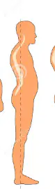
lordotic
Lordosis describes an exaggerated inward curve of the spine, often in the neck (cervical lordosis ) or lower back (lumbar lordosis).
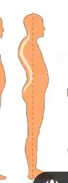
Kyphotic
offten referred as upper crossed syndrom. A condition of excessive spinal curvature. The anterior chest, shoulder muscles, lats and neck extensors are too tight and shortened. The rhomboids, lower and mid traps, and neck flexors are lengthened and weak.
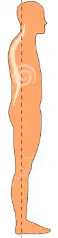
swayback
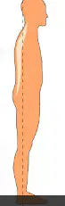
flat back
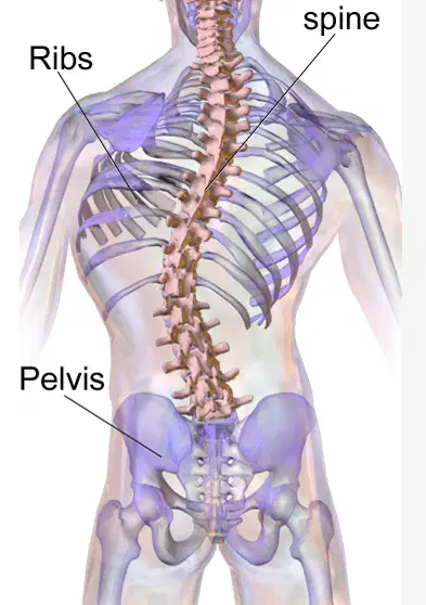
scoliosis
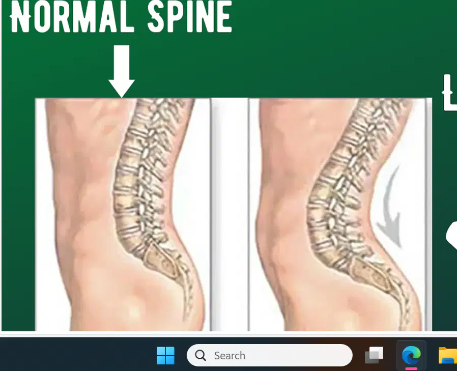
Lower crossed syndrome
overly lordotic posture, the one that looks like youre sticking your butt out for instagram,
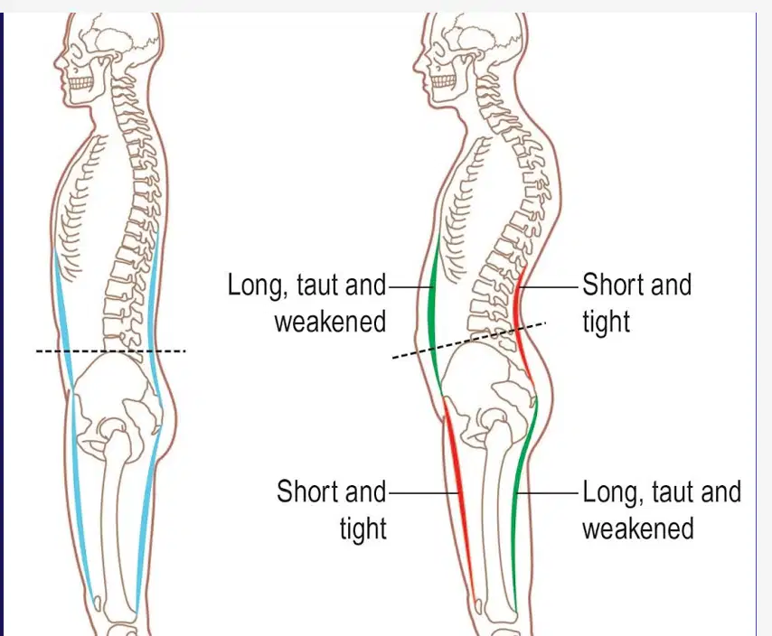
Anterior Pelvic Tilt
It usually occurs alongside a lordotic posture, as well as kyphosis.
Forward Head Posture & Upper Crossed
The overactive muscles here are the cervical spine extensors, the upper traps, pecs, SCM, and the levator scapula. Forward Head Posture and Upper Crossed Syndrome are related conditions that involve muscle imbalances in the upper body.
Underactive Muscles upper crossed and forward head
Cervical flexors: Muscles in the front of the neck Rhomboids: Muscles in the lower shoulders Lower and middle traps: Muscles in the lower back of the shoulder blades Deep neck flexors: Muscles in the neck
Rhomboids: Muscles in the lower shoulders Lower and middle traps: Muscles in the lower back of the shoulder blades Deep neck flexors: Muscles in the neck
Overhead Squat assessment
Typically the first movement assessment done- Assess dynamic posture, core stability, neuromuscular control- A good way to spot muscle imbalances (knee valgus, limited ankle mob.)
Foot and ankles: The feet should stay straight ahead Knees: They should track straight forward or over the second and third toes.,
overhead squat assessment steps
Your client should attempt squatting to parallel (femur parallel with ground).- The squat depth can be reduced if the client can't achieve that depth or has discomfort/pain.- The client will perform roughly 5 reps while the trainer views from the front and side.
Overhead Squat Anterior
Foot and ankles: The feet should stay straight ahead Knees: They should track straight forward or over the second and third toes.
Overhead Squat Lateral assessment
-View the LPHC (hips) and shoulders from the side.
assessment overhead Squat (feet turn out) Overactive
- Gastrocnemius/soleus (calves)- Hamstrings complex
assessment Overhead Squat (feet turn out) Underactive
Anterior and posterior tibialis (shin muscles)- Gluteus maximus and medius
Overhead Squat (knees cave in) underactive
Glutes (med and max)- Anterior and posterior tibialis
overhead Squat (low back arches) Overactive
Hip Flexors- Lumbar Extensors- Lats
Overhead Squat (low back arches) underactive
Glute max- Hamstrings- Abdominals
overhead Squat (forward lean) Overactive
Hip flexors and gastrocnemius and soleus - rectus abdominus and external obliques
Overhead Squat (forward lean) Underactive
Glute max- Hamstrings- Lumbar extensors
Overhead Squat (arms fall forward) Overactive
Lats- Pecs- Teres Major
Overhead Squat (arms fall forward) Underactive
Middle and Lower Traps- Rhomboids- Posterior Delts & some R.cuff.
____-Leg Squat- Strong inter- and intrarater reliability- Uses an anterior view- Up to 5 reps will be completed for both sides-The client squats as deep as possible (while maintaining balance) and returns to the starting position.
Single-Leg Squat
The client stands on a flat, stable surface, with hands on hips and their eyes focused forward - The client lifts one foot approximately 6 in. off the floor. The foot, ankle and knee should be in a neutral, straight ahead position.
Single-Leg Squat form

