
 Worksheets
Male Reproductive System
Total questions: 87
Worksheet time: 1hrs 26mins
Penis
Prostate
Seminal Vesciles
Testicles
(a)
located just below the bladder and in front of the rectum
(a)
about the size of a walnut and surrounds the urethra (the tube that empties urine from the bladder)
(a)
Prostatic and seminal secretions are conveyed through numerous ducts to __ __
(a)
Fluids are then carried outside the body through penis via the __ __ and finally exit through the external urethral orifice
(a)
Shaped like a cone with a central core: the prostatic urethra
(a)
How many zones is the prostate gland divided into?
2
3
4
5
(Largest) zone of the prostate, the area lateral and posterior to the distal prostatic urethra
(a)
This zone of the prostate is located at the superior edge bordering the bladder and seminal vesicles. The ejaculatory ducts course through this zone
(a)
The (a) zone of the prostate borders the central zone posteriorly and laterally and the fibromuscular tissue anteriorly
The 4th zone of the prostate is the (a)
Size and echo texture
❑ Prostatitis (infection)
❑ Detection of masses ❑Evaluation of benign prostatic
hypertrophy (BPH)
(a)
Sonographic correlation
evaluated serum prostatic specific antigen (PSA)
❑Evaluation of postoperative transurethral resection (TURP)
❑Ultrasound-guided biopsies of
prostatic lesions
(a)
Prostate Gland is (a) , with medium-level echoes Symmetric in shape and size
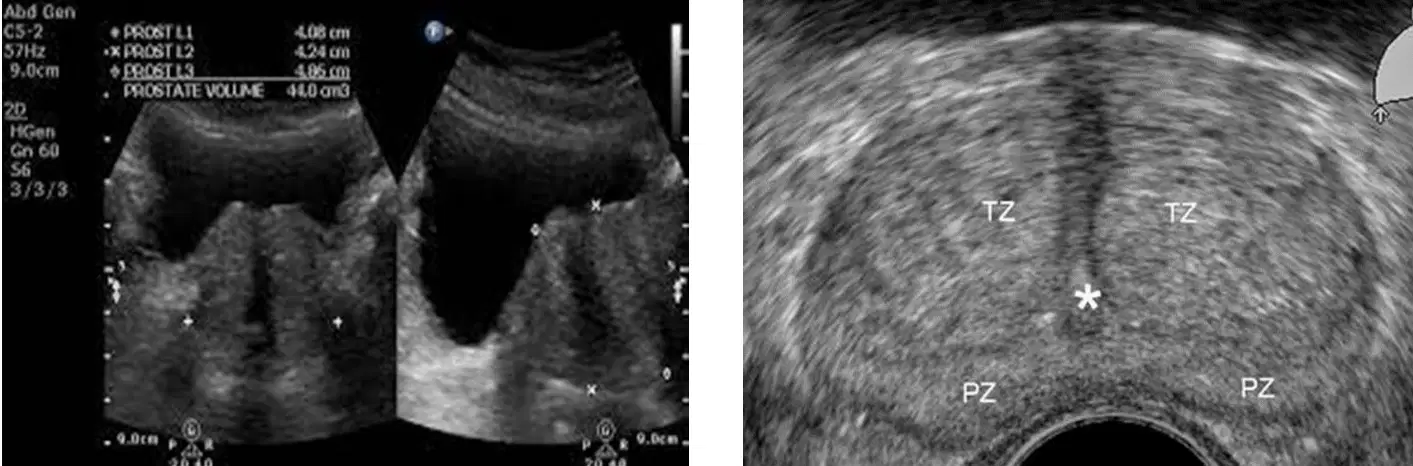
What structure is shown in this image?
(a)
Evaluation of size, symmetry, and echo texture
❑Ruling out presence of cysts or calculi
(a)
Paired glands, each encapsulated by connective tissue
(a)
Inflammatory processes
❑Congenital anomalies
(a)
__ ___ are convoluted, pouchlike structures emptying into distal portion of ductus deferens to form ejaculatory ducts
(a)
Ovoid structures with low-level echoes superior to Prostate gland
(a)
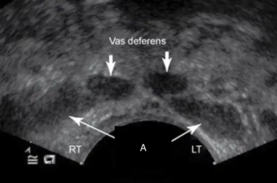
What is labeled A?
(a)
3.8 cm in length
3.0 cm anteroposterior
4.0 cm in width
(a)
5.0 cm in length
<1.0 cm in diameter
(a)
<4.0
(a)
Is continuous with the skin of the lower abdomen
(a)
Located directly behind the penis and in front of the anus
(a)
Sac of cutaneous tissue that supports testicles
(a)
The scrotum is (a) divided into lateral portions by median ridge called the Median Raphe
The scrotum is (a) divided into sacs by a septum called the Dartos or Tunica Dartos
(a) surrounds each testicle and extends into abdomen over spermatic cord
Contraction of Cremaster muscle performs the important function of regulating (a) of testicles
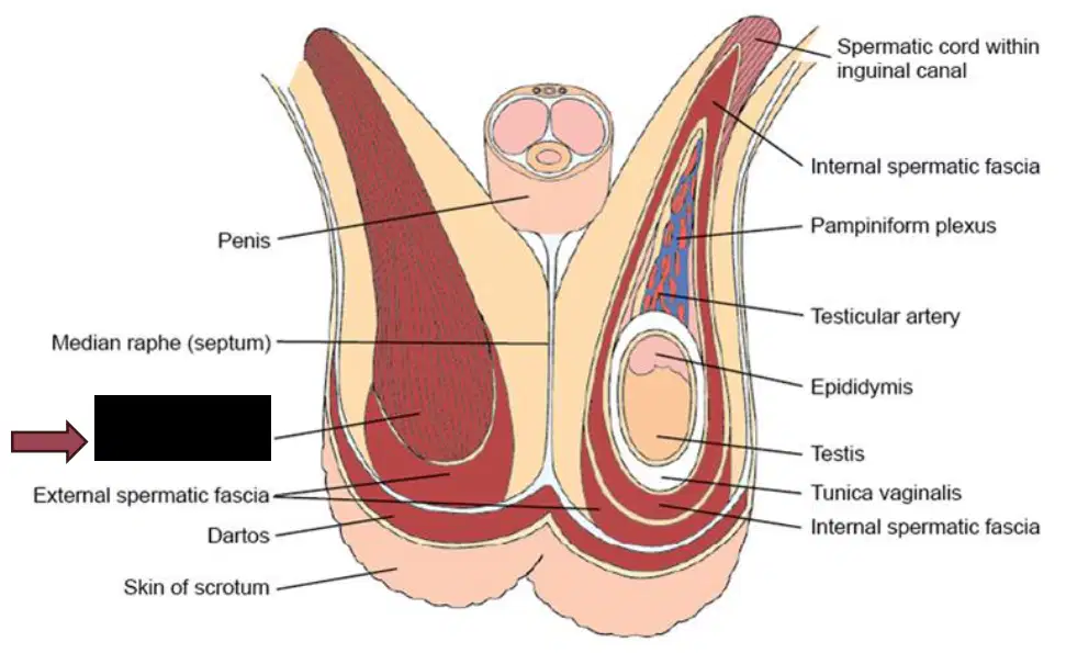
The arrow is pointing to
(a)
(a) lines inner walls of scrotum, covering each testis and epididymis.
Consists of two layers: parietal and visceral
(a)
(a) is inner lining of scrotal wall
(a) surrounds testis and epididymis
Tunica vaginalis has a Small bare area, which is (a)
Prevents torsion
(a)
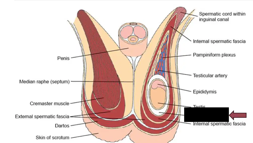
The red arrow is pointing to the (a)

A =
(a)
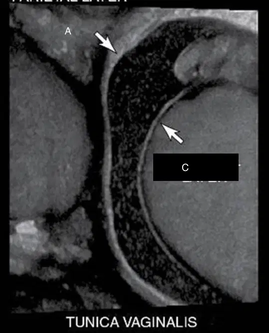
C =
(a)
Each testis is covered by dense, white fibrous tissue called
(a)
Tunica Albuginea extends into posterior wall of testicle and forms __ testis and __ septa
(a)
Mediastinum Septa radiate into testicle and separate into __ to __ lobules
200 to 400
200
200 to 300
200 to 500
Each lobule contains __ to __ convoluted Seminiferous tubules
(a)
Seminiferous tubules are connected to (a) , which lead to the Rete testis
(a) is located within Mediastinum testis
Is a narrow, tightly-coiled tube connecting rear
of the testicles to the Deferent duct (Ductus deferens or Vas deferens)
(a)
Divided into:
• Head (located on superior pole of testis)
• Body
• Tail
(a)
Empties into Ductus deferens
(a)
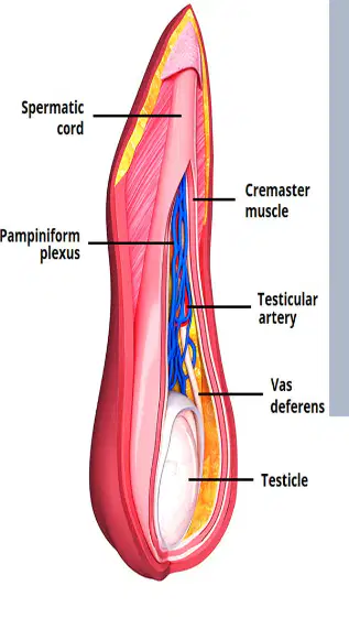
Two (a) extend from Inguinal canals and internal inguinal rings into pelvis
Ductus deferens
Testicular arteries
Venous Pampiniform plexus
Lymphatics
Autonomic nerves
Fibers of Cremaster muscle
(a)
AKA the Vas Deferens
(a)
A long, muscular tube that carries mature sperm from
the Epididymis to the ejaculatory ducts, ultimately leading to the Urethra
(a)
The sperm's highway from storage to the point of
ejaculation
(a)
Originates at the tail of the Epididymis, ascends through
the spermatic cord, passes through the inguinal canal, and then loops over the bladder before joining the ejaculatory duct.
(a)
❑ Testicular size
❑ Inflammatory processes
(epididymitis, orchitis)
❑ Presence and composition of
masses
❑Detection of peritesticular fluid
collections (hydrocele)
(a)
❑Evaluation of scrotal trauma ❑Doppler evaluation to rule out
testicular torsion
❑Evaluation of scrotal pain
(a)
❑Location of undescended testicles
(Cryptorchidism) (Inguinal canal, external inguinal ring, abdomen).
(a)
❑ Homogeneous
❑ Medium-level echoes
(a)
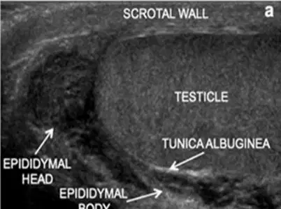
Hyperechoic linear structure is the (a)
Low-resistance arterial blood flow
(a)
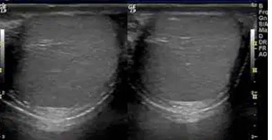
This image shows the (a)
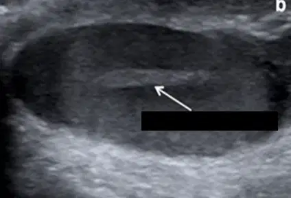
The arrow in this image is showing the (a)
Iso - to hypoechoic compared with normal testicle
(a)
Coarse in appearance compared to normal testicle
(a)
This image is taken from a (a) scanning plane
3 to 5 cm in length
2 to 3 cm height
2 to 3 cm in width
(a)
Head: 10-12 mm AP / 5-12 mm length
Body: 2 – 4 mm AP
Tail: 2-5 mm AP
(a)
The testicle is supplied by the (a) artery (aortic branch)
The vas deferent is supplied by the (a) artery (internal iliac branch)
Pampiniform plexus draining the testis and epididymis.
• Right testicular vein→IVC
• Left testicular vein→left renal vein
(a)
Composed of Two Corpora Cavernosa situated dorsolateral
(a)
Single Corpus Spongiosum in midventral region, which contains the spongy urethra
(a)
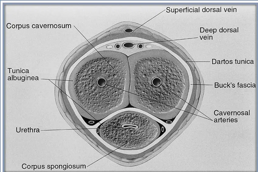
(a) , a thick, fibrous, loosely applied covering of skin that envelops penis
Blood supply to the penis and urethra is from the paired (a) arteries (branches of the Internal Iliac arteries
Corpora Cavernosa is supplied by the (a) artery (AKA the Cavernosal artery)
Corpus Spongiosum is supplied by branches of the __ artery and __ artery
(a)
Main veins of the penis:__ Dorsal vein and __ dorsal vein
(a)
❑Detection of fibrosis (Peyronie’s disease)
❑Detection of scar tissue and plaques
❑Evaluation of tumors
(a)
❑Penile hematoma
❑Evaluation of periurethral disease
❑Doppler evaluation of vasculogenic impotence
(a)
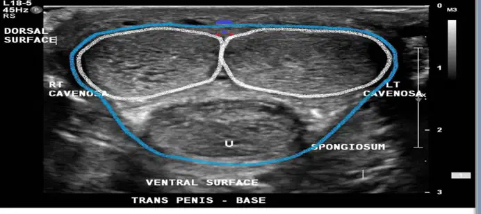
Lie anterior or above the
Corpus Spongiosum
(a)
Appear symmetrical, round or oval, with a medium-level homogeneous echo texture
(a)
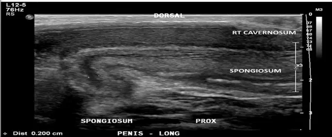
Lies posterior or below to the paired Corpus Cavernosum
(a)
Homogeneous texture composed of medium-level echoes
(a)
Highly echogenic covering of the Corpora cavernosa
(a)
Echogenic plane dividing the two Corpora Cavernosa
Extension of the Tunica Albuginea
(a)

