
 Worksheets
OBGYN WK 2 (Transvaginal/Muscles)
Total questions: 100
Worksheet time: 2hrs 39mins
An internal ultrasound exam similar to a pelvic examination using a transvaginal transducer , covered with a sterile transducer cover
(a)
This approach requires an empty bladder, and it is performed after TA examination is completed
(a)
It provides closer view of the pelvic structures
(a)
Which exam requires an empty bladder?
transabdominal
transvaginal
When examining in the transvaginal plane, the patient should be __
Supine
prone
LLD
RLD
Position – supine, knees are bent, feet are placed flat on the table, pelvis may need to be elevated so that the probe can be tilted downward.
(a)
TV ultrasound requires (a)
The orientation of the transvaginal probe is controlled by probe __ and __
(a)
Movement of the transducer is centered around the (a)
Rotation of the probe along its long axis provides __-degree longitudinal visualization of the __
(a)
Pushing or pulling the probe can bring the tip close to a region of interest and provide a method of (a) , allowing evaluation of focal tenderness or fixation
The notch of the transducer is along the top surface of the handle so the
beam is projected in the midline (a) aspect of the body
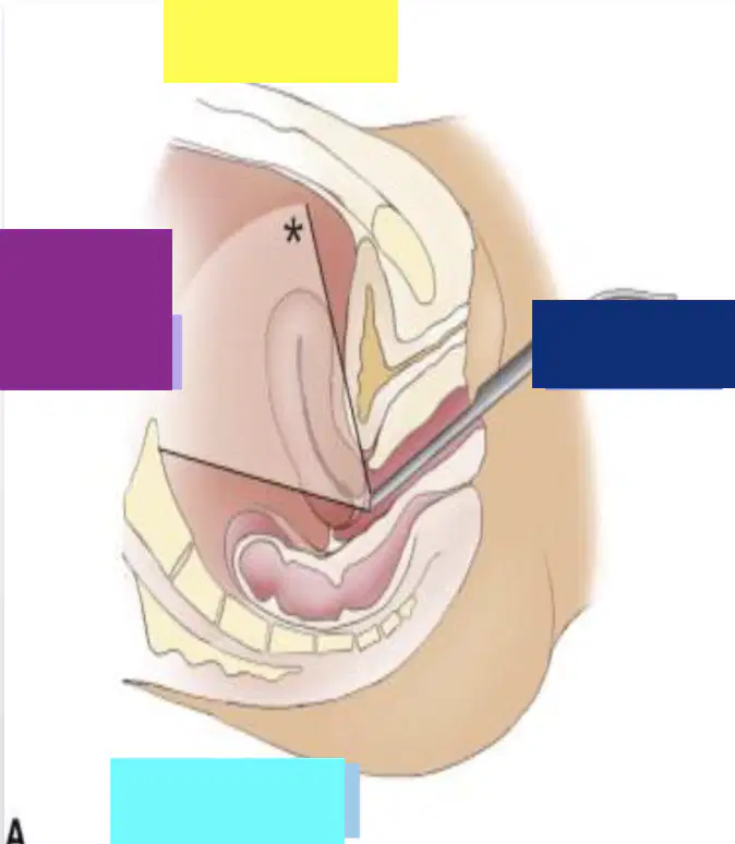
Yellow
(a)

Dark blue
(a)
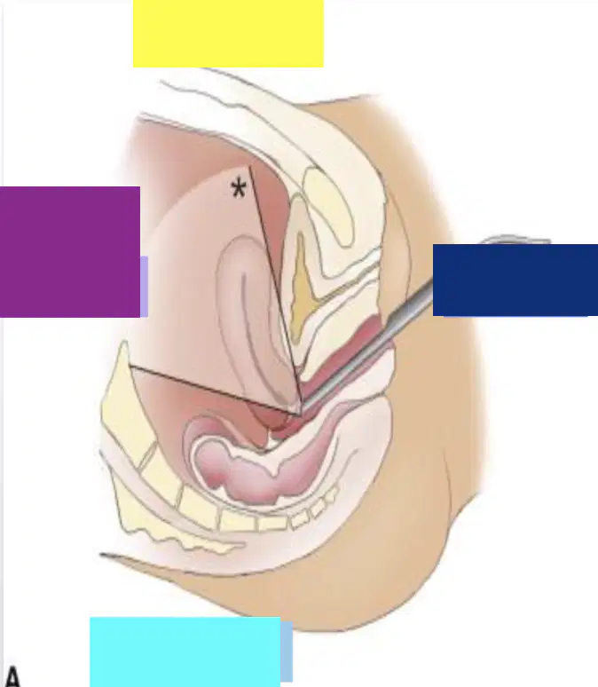
Light blue
(a)

Purple
(a)

Dark blue
(a)
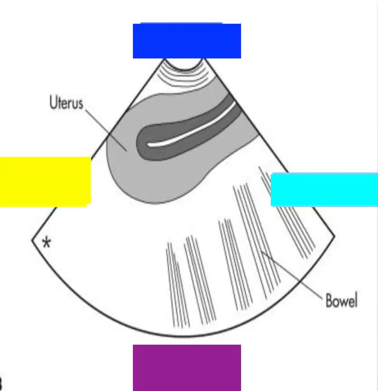
Light blue
(a)
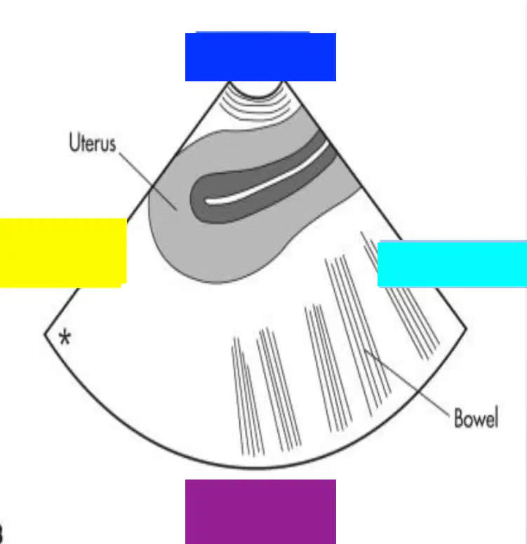
Purple
(a)

Yellow
(a)
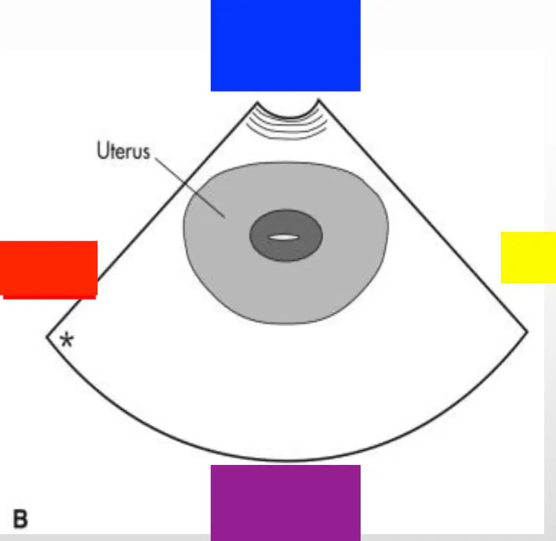
Yellow
(a)
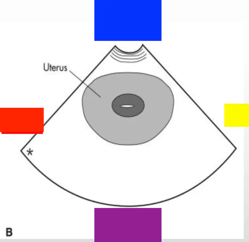
Purple
(a)
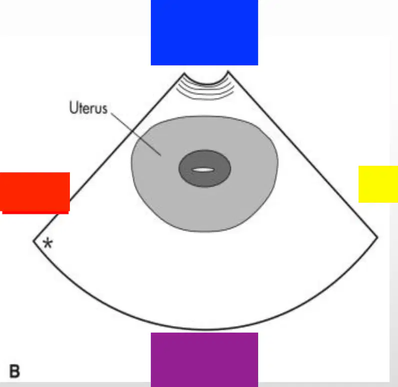
Red
(a)

Blue
(a)
The notch of the transducer is rotated (a) the sonographer so the beam may image the uterus in a coronal view
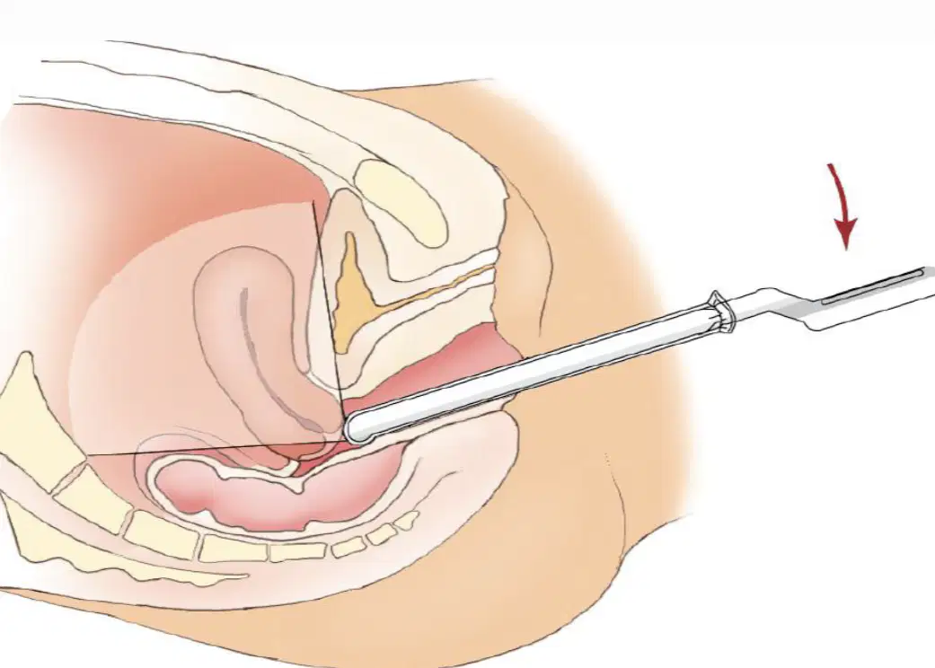
This image shows (a) angulation
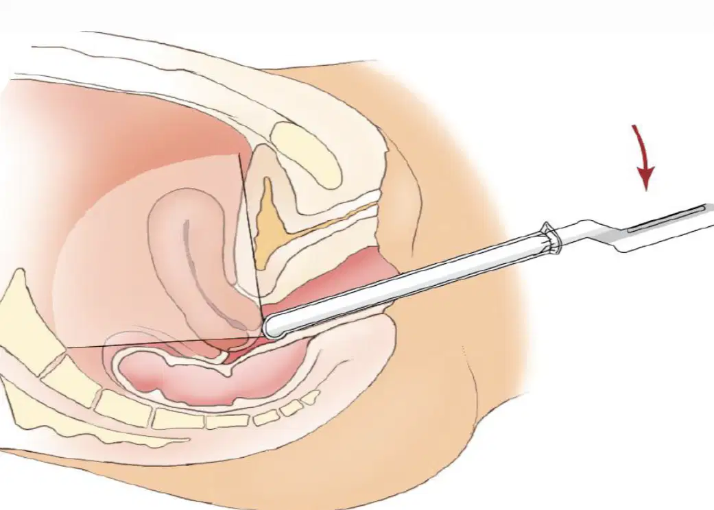
Transvaginal sagittal scanning with anterior angulation to better visualize the (a)
of a normal anteflexed uterus

This image shows (a) angulation
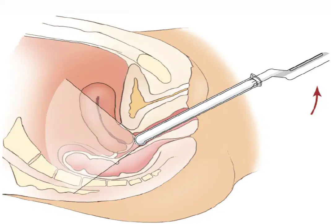
Transvaginal sagittal scanning with posterior angulation to better visualize the __
and __
(a)

This image shows (a)

This image shows (a)

This image shows (a) transvaginally
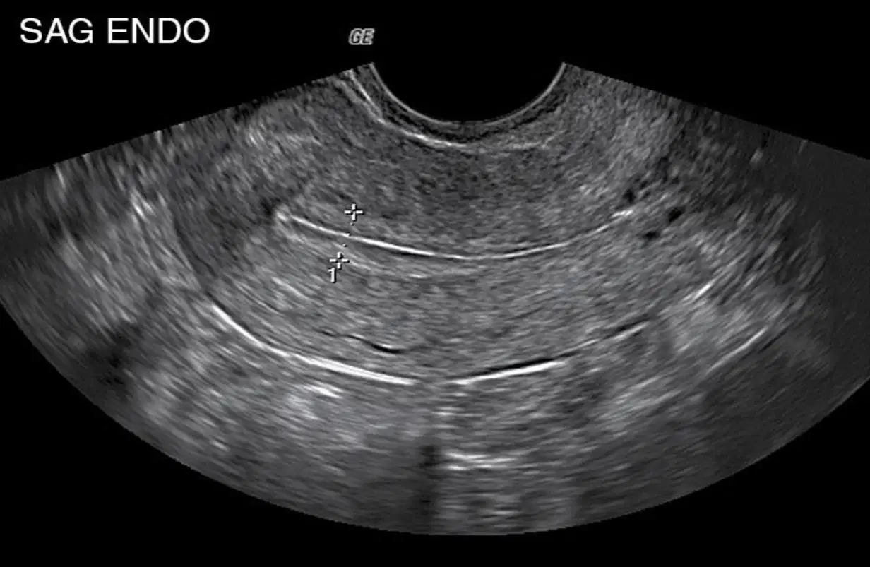
This image shows a transvaginal measurement of the (a)
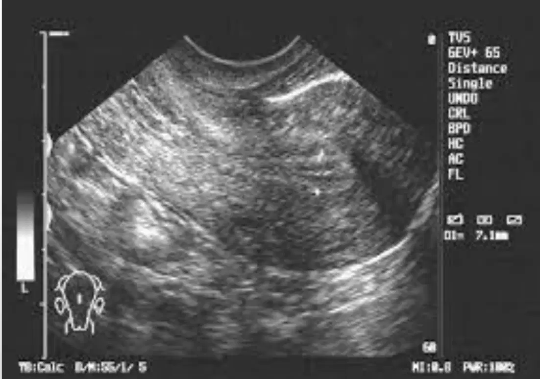
This image shows a transvaginal image of a (a) uterus
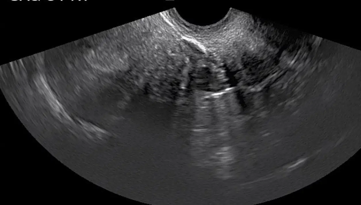
This image shows a transvaginal (a)

This image shows a transvaginal (a)

The probe is rotated 90 degrees to record the (a)
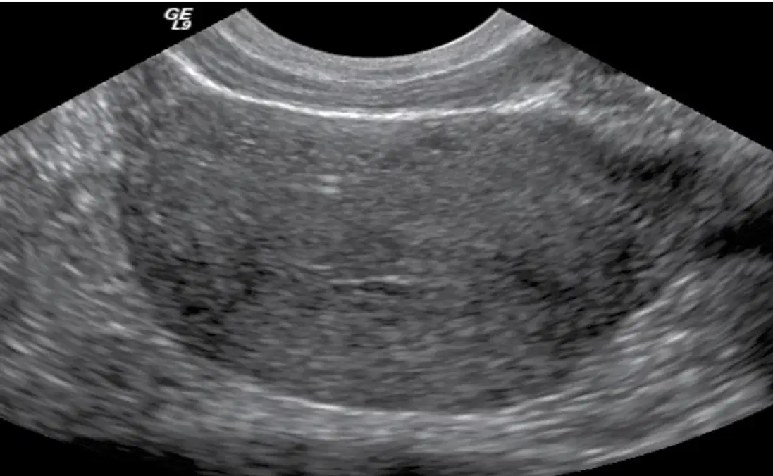
This image shows a transvaginal (a) view

This image is a transvaginal view of the (a) of the uterus, coronally
Rotate the probe 90 degrees; image the uterine fundus, body with the endometrial
stripe, and cervix.
(a)
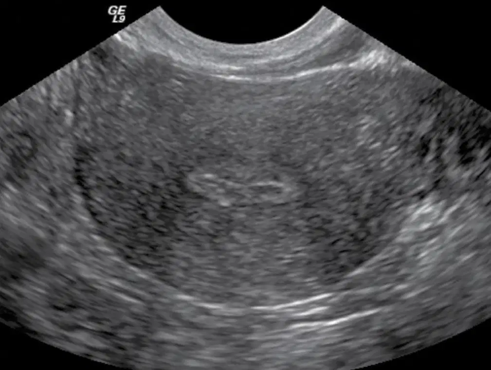
This shows a transvaginal coronal (a)

The probe is angled caudally to image the ___ __
(a)
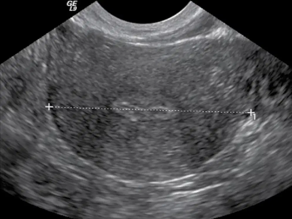
This shows a transvaginal (a) of the uterus MID measured

This image shows a transvaginal (coronal) (a)
The probe is angled slightly more caudal to image the (a)
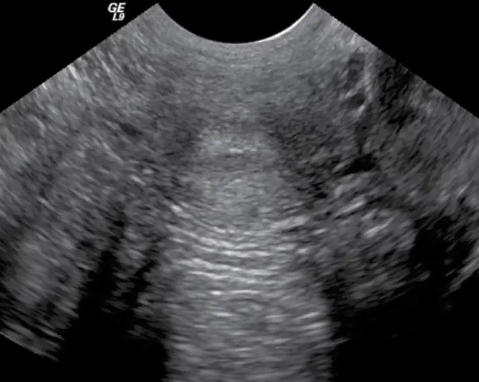
This image shows a (a)
The probe is withdrawn slightly to image the (a)

This image shows a measurement of a sag transvaginal (a)

This image shows a sagittal transvaginal view of the
(a)

this transvaginal image is of a (a)
After survey, standard views are obtained.
These include the following
(a)
Cervix, Endocervical canal
(a)
posterior cul-de-sac
(a)
Uterus (mid, right, left)
(a)
endometrium, right ovary and adnexa, left ovary and adnexa
(a)
Cervix and posterior cul-de-sac
(a)
LUS, uterine corpus and endometrium
(a)
uterine fundus, right ovary and adnexa, left ovary and adnexa
(a)
Which of the following is a coronal standard view only?
Cervix
Fundus
LUS
Uterus
1. Image from cervix to fundus, endometrial cavity
(a)
2. Measure long axis Ute length & A/P
(a)
3. Measure Endo thickness
(a)
4. Angle slowly to right of uterus
(a)
5. Angle slowly to left of uterus
(a)
6. Pull probe out slightly to image cervix
(a)
Rotate transducer 90 degrees; image uterine fundus, body with endometrial canal, LUS, and cervix
(a)
Look for free fluid surrounding uterine cavity
(a)
Follow fundus of uterus to area of cornu to image ovaries; internal iliac vessels serve as (a)
Color imaging may be used to separate vascular structures from ovary. Look for follicles surrounding periphery of ovary
(a)
Measure the long axis and A/P
(a)
Rotate transducer 90 degrees once sagittal plane of ovary
is obtained.
(a)
Measure width
(a)
Sonographically the sacrum may appear as a (a) with an overlying bowel shadow
(a) insert on pubic rami and are paired parasagittal straps in abdominal wall
Appear as hypoechoic structures with echogenic striations
(a)
separates sonographic appearance of rectus abdominis muscle from surrounding fat and bowel as a
bright linear echogenic reflector
(a)

This shows the
(a)
In lesser or true pelvis, the urinary bladder, reproductive organs, levator ani, obturator internus muscles are
identified
(a)
Sonographically, sections of obturator internus muscle are seen at __ __ corners of bladder at level of vagina and cervix
(a)
Muscle is hypoechoic, ovoid, surrounded by obturator fascia, and serves as tendinous attachment for (a) i muscle

The arrows point to
(a)
best visualized sonographically in transverse plane with caudal angulation at most inferior aspect of bladder
(a)
Hypoechoic, hammock-shaped area that is medial, caudal, posterior to obturator internus
(a)
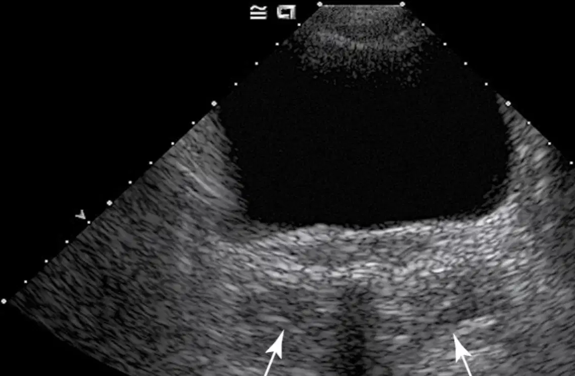
The arrow is pointing to the (a)
muscles are located deep, cranially, and posteriorly
(a)
Not routinely visualized
(a)
Can be seen in greater pelvis
(a)
is a combination of the iliacus muscle and the psoas major
(a)

the arrow is pointing to (a)
S/D ratio (or A/B ratio)
(a)
(RI) (A – B/A)
(a)
(PI) (A – B/mean)
(a)
A = Peak Systolic velocity
B = End Diastolic velocity
In (a) vascularity equations
Uterine wall is made up of (a) layers
serosa of uterus not visualized sonographically
(a)
of uterus is the myometrium
(a)
This layer should have homogeneous echotexture with smooth-walled borders
(a)
Inner layer of the uterus is the (a)
The (a) is thin, compact, and relatively hypo vascular
The endometrium is Hypoechoic and surrounds relatively echogenic endometrial stripe, creating (a) l halo

