
 Worksheets
Ch. 17 The Heart
Total questions: 77
Worksheet time: 39mins
Which of the following is NOT a component of the cardiovascular system?
Heart
Blood vessels
Lungs
Blood
What is the main function of the heart in the cardiovascular system?
To digest food
To pump blood
To filter waste
To produce hormones
Explain how the cardiovascular system supports the body's cells.
By transporting oxygen and nutrients to cells
By producing energy in the cells
By breaking down food in the stomach
By sending nerve signals to muscles
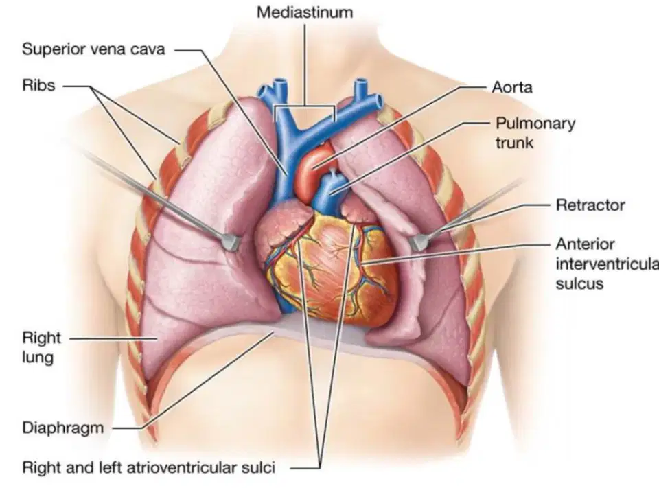
What is the approximate weight of the human heart?
250-350 grams
500-700 grams
100-150 grams
800-1000 grams
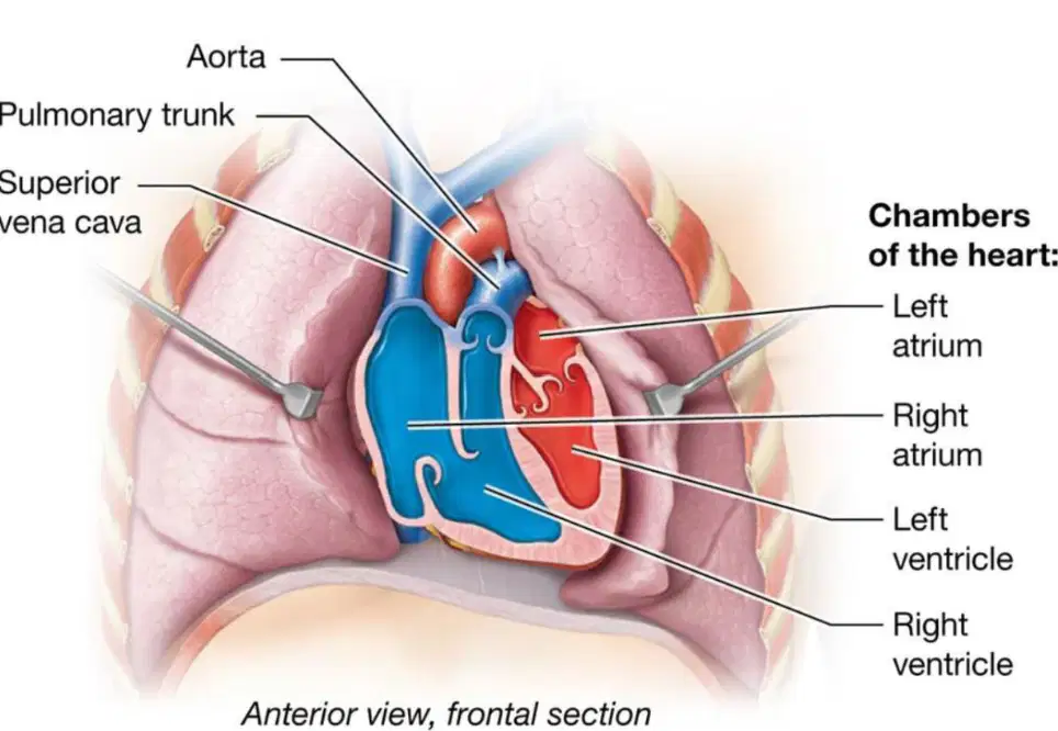
Which chambers of the heart are located superiorly?
Right and left atria
Right and left ventricles
Only the left ventricle
Only the right ventricle
What is the function of the atrioventricular sulcus in the heart?
It separates the right and left ventricles
It is a boundary between the atria and ventricles
It pumps blood to the lungs
It connects the atria to the aorta
Where is the interventricular sulcus located?
Between the atria and ventricles
Between the right and left atria
Between the right and left ventricles
Between the ventricles and the aorta

Which side of the heart is responsible for pumping blood into vessels leading to the lungs?
Right side
Left side
Both sides equally
Neither side
What is the main function of the left side of the heart?
To pump blood to the lungs
To receive deoxygenated blood from the body
To receive oxygenated blood from pulmonary veins and pump it to the rest of the body
To filter waste from the blood
The right side of the heart is also known as which of the following?
Systemic pump
Pulmonary pump
Coronary pump
Aortic pump
Explain why the left side of the heart is called the systemic pump and describe its role in circulation.
It pumps blood to the lungs for oxygenation.
It receives oxygenated blood from the lungs and pumps it to the rest of the body, creating the systemic circuit.
It filters blood before sending it to the kidneys.
It only circulates blood within the heart chambers.
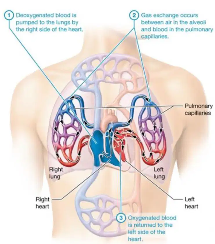
Which blood vessels deliver oxygen-poor and carbon dioxide-rich blood to the lungs in the pulmonary circuit?
Pulmonary arteries
Pulmonary veins
Coronary arteries
Aorta
Where does gas exchange occur in the lungs?
Between alveoli and pulmonary capillaries
Between bronchi and arteries
Between trachea and veins
Between diaphragm and heart
Explain the process of gas exchange in the pulmonary circuit and why it is important for the body.
Gas exchange allows oxygen to enter the blood and carbon dioxide to be removed, which is essential for cellular respiration and maintaining blood pH.
Gas exchange only removes waste from the digestive system.
Gas exchange increases the temperature of the blood.
Gas exchange is responsible for producing hormones in the lungs.
What happens to carbon dioxide in the pulmonary capillaries?
It diffuses from air in the alveoli into the blood.
It diffuses from blood in the pulmonary capillaries to air in the alveoli to be expired.
It is converted into oxygen in the alveoli.
It is absorbed by the heart.
Which side of the heart receives oxygen-rich blood from the pulmonary circuit?
Right side
Both sides
Left side
Neither side
Explain why the veins of the pulmonary circuit are unique compared to veins in the systemic circuit.
They carry deoxygenated blood to the heart.
They deliver oxygen-rich blood to the left side of the heart.
They transport nutrients instead of gases.
They only function during exercise.
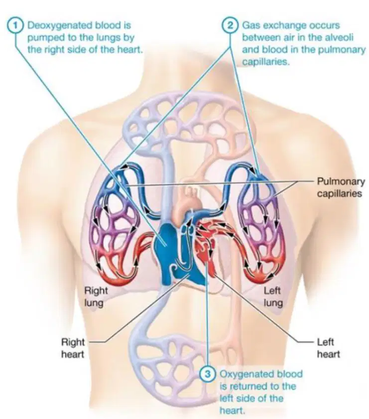
What is the main function of the pulmonary circuit?
To deliver oxygen to tissues and remove carbon dioxide
To digest food
To produce hormones
To filter waste from the kidneys
Explain how the pulmonary circuit supports cellular respiration in body tissues.
By delivering oxygen for energy production and removing carbon dioxide waste
By producing glucose for cells
By filtering toxins from the blood
By storing excess nutrients in the liver
Which circuit is described as a low-pressure circuit because it only pumps blood to the lungs?
Pulmonary circuit
Systemic circuit
Coronary circuit
Lymphatic circuit
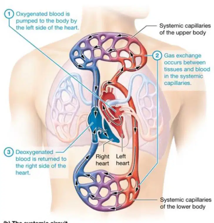
Why is the systemic circuit considered a high-pressure circuit?
Because it only pumps blood to the lungs
Because it pumps blood to the rest of the entire body
Because it pumps blood to the heart only
Because it pumps blood to the brain only
Which of the following is a primary function of the heart in maintaining homeostasis?
Regulating blood pressure
Producing digestive enzymes
Absorbing nutrients
Storing oxygen
What are the major factors that influence blood pressure and blood flow to organs?
The rate and force of the heart's contractions
The amount of oxygen in the blood
The size of the lungs
The number of red blood cells
Which hormone is produced by the atria of the heart and acts as an endocrine organ?
Atrial natriuretic peptide (ANP)
Insulin
Adrenaline
Thyroxine
How does atrial natriuretic peptide (ANP) lower blood pressure?
By decreasing sodium ion retention in the kidneys
By increasing heart rate
By constricting blood vessels
By increasing water absorption in the intestines
What is the main function of the fibrous pericardium?
Produces serous fluids
Anchors the heart to the diaphragm and great vessels
Pumps blood
Contracts to move blood
Which layer of the heart is responsible for producing serous fluids?
Fibrous pericardium
Serous pericardium
Myocardium
Endocardium
Why is low distensibility important for the heart?
It allows the heart to stretch and fill with more blood
It prevents overstretching and overfilling of the chambers with blood
It increases the heart's pumping efficiency
It helps the heart contract more forcefully

What is the pericardium?
The outer layer of the lungs
The membrane surrounding the heart
The muscle of the diaphragm
The blood vessels of the heart
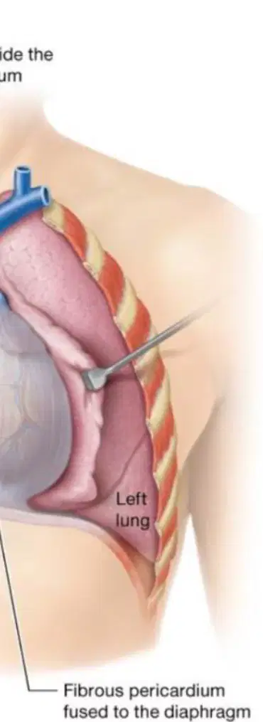
According to the diagram, where is the fibrous pericardium attached?
To the right lung
To the left lung
To the diaphragm
To the rib cage
Which layer of the pericardium is fused to the inner surface of the fibrous pericardium and encases the heart-like sac?
Parietal pericardium
Visceral pericardium
Myocardium
Endocardium
What is another name for the visceral pericardium, and what is its significance?
Epicardium; it is the most superficial layer of the heart wall
Myocardium; it is the thickest layer of the heart wall
Endocardium; it lines the heart chambers
Pericardial sac; it surrounds the lungs
What is the primary function of pericardial fluid?
Provides lubrication and decreases friction as the heart moves
Supplies oxygen to the heart muscle
Transports nutrients to the heart wall
Initiates electrical impulses in the heart
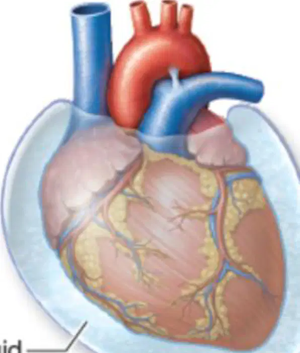
How does the structure of the pericardium contribute to the function of the heart?
The pericardium encases the heart, and its fluid reduces friction, allowing smooth heart movement during contraction and relaxation.
The pericardium produces hormones that regulate heart rate.
The pericardium absorbs nutrients for the heart muscle.
The pericardium generates electrical impulses for heartbeats.
Which layer of the heart is the second and thickest?
Endocardium
Epicardium
Myocardium
Pericardium
What are the two main components of the myocardium?
Endothelial cells and smooth muscle
Cardiac muscle cells (myocytes) and fibrous skeleton
Pericardial fluid and connective tissue
Nerve fibers and adipose tissue
What is the primary function of the fibrous skeleton in the heart?
To store energy for the heart
To provide a surface for blood flow
To give cardiac muscle cells something to pull on when contracting
To produce hormones
Which of the following is NOT a function of the fibrous skeleton of the heart?
Provides structural support
Acts as an insulator for the heart's electrical activity
Produces red blood cells
Gives cardiac muscle cells something to pull on when contracting
Which layer lines the lumen of the heart and is the third and deepest layer of the heart wall?
Myocardium
Epicardium
Endocardium
Pericardium
What type of tissue primarily composes the endocardium?
Stratified squamous epithelium
Simple squamous epithelium
Cuboidal epithelium
Transitional epithelium
Explain why the composition of the endocardium is important for its function in the heart.
It allows the heart to contract.
It provides a smooth lining that reduces friction and is continuous with blood vessels, aiding efficient blood flow.
It stores energy for the heart.
It produces hormones for the heart.
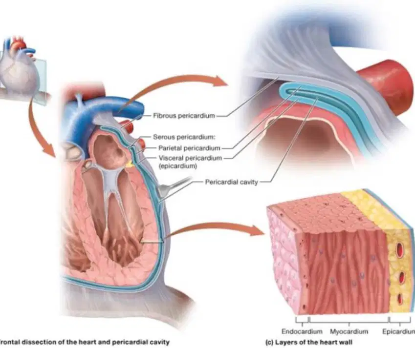
Which of the following is NOT a layer of the heart wall?
Epicardium
Myocardium
Endocardium
Peritoneum
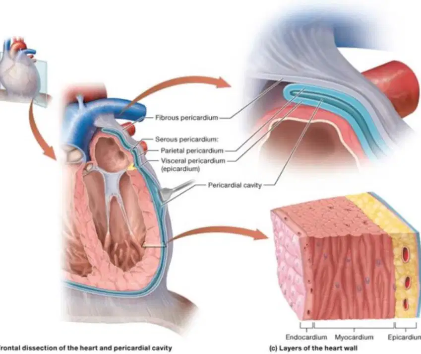
Arrange the following layers of the heart wall from outermost to innermost.
Endocardium, Myocardium, Epicardium
Epicardium, Myocardium, Endocardium
Myocardium, Endocardium, Epicardium
Epicardium, Endocardium, Myocardium
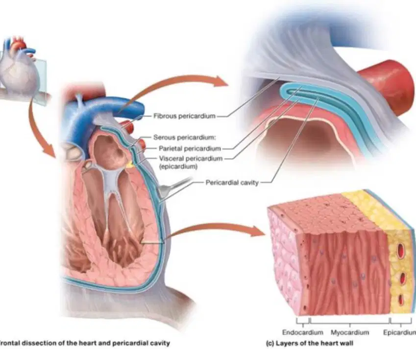
Which layer of the heart wall is primarily responsible for the contractile function of the heart?
Epicardium
Myocardium
Endocardium
Pericardium
Which chamber of the heart receives blood from veins and pumps it into the ventricles through valves?
Atria
Ventricles
Arteries
Capillaries
What is the primary function of the flaps in the heart valves?
To open and allow blood to flow backwards
To close when ventricles contract, preventing backflow of blood
To pump blood into the veins
To increase the speed of blood flow
When the ventricles contract, what happens next in the circulatory process?
Blood is ejected into the arteries
Blood is absorbed by the lungs
Blood flows back into the atria
Blood is filtered by the kidneys
Explain how the structure of heart valves contributes to the efficiency of blood flow through the heart.
The valves have flaps that close when ventricles contract, preventing blood from moving backwards and ensuring one-way flow.
The valves open to allow blood to flow in both directions.
The valves slow down the blood flow to allow for oxygen exchange.
The valves store blood for later use.
Which chambers of the heart have thicker walls and act as stronger pumps?
Ventricles
Right atrium
Left atrium
Auricles
How do the right and left atria differ from each other?
The right atrium is larger with a thinner wall, while the left atrium is smaller with a thicker wall.
The right atrium is smaller with a thicker wall, while the left atrium is larger with a thinner wall.
Both atria are the same size and thickness.
The left atrium is larger with a thinner wall, while the right atrium is smaller with a thicker wall.
What is the function of the auricles in the atria?
They expand to give the atria more space to hold blood.
They pump blood out of the heart.
They separate the ventricles.
They regulate heart rhythm.
Why are the ventricles considered stronger pumps compared to the atria?
Because they have much thicker walls.
Because they are smaller in size.
Because they contain auricles.
Because they are symmetrical.
Which ventricle of the heart has thinner walls and faces little resistance against which to pump?
Left ventricle
Right ventricle
Both ventricles have equal wall thickness
Neither ventricle
What is the primary reason for the size differences between the right and left ventricles of the heart?
Differences in oxygen content
Differences in blood volume
Pressure differences in the pulmonary and systemic circuits
Differences in heart rate
The left ventricle has walls that are approximately how many times thicker than the right ventricle?
2 times
3 times
4 times
5 times
Explain why the left ventricle has more muscle mass compared to the right ventricle.
It pumps blood to the lungs, which requires more force.
It pumps against greater resistance in the systemic circuit, requiring more muscle mass.
It receives more blood than the right ventricle.
It beats faster than the right ventricle.
Which structure in the heart is described as finger-like projections of muscle?
Trabeculae carneae
Papillary muscle
Chordae tendonae
Interventricular septum
What is the function of the interventricular septum in the heart?
It pumps blood into the atria
It separates the right and left ventricles and helps pump blood into the pulmonary trunk and aorta
It connects the atria to the ventricles
It produces red blood cells
Which of the following best describes chordae tendonae?
Ridged surfaces in the atria
Finger-like projections of muscle
Tendon-like cords in the ventricles
Muscular wall separating the atria
Explain how the interventricular septum contributes to the function of the heart during contraction.
It prevents blood from entering the atria
It contracts with the ventricular muscle to help pump blood into the pulmonary trunk and aorta
It produces hormones for heart function
It stores oxygen for the heart muscle
What are chordae tendineae?
Fibrous, tendon-like structures that attach to the anterior end of each cusp of the heart valves
Muscular tissues that form the walls of the ventricles
Blood vessels that supply the heart muscle
Nerves that control the heartbeat
To which structures are the chordae tendineae attached?
Papillary muscles
Aorta
Pulmonary veins
Sinoatrial node
What is the function of the tension created by the chordae tendineae?
Keeps the valve closed
Opens the valve wider
Increases blood pressure
Stimulates heart contractions
Explain how the chordae tendineae and papillary muscles work together to prevent backflow of blood in the heart.
Papillary muscles contract before the ventricles eject blood, creating tension on the chordae tendineae, which keeps the valve closed and prevents backflow.
Papillary muscles relax, allowing the chordae tendineae to open the valve and let blood flow backward.
Chordae tendineae contract to push blood through the valve, while papillary muscles keep the valve open.
Papillary muscles and chordae tendineae both relax, causing the valve to close automatically.

What is the function of the tricuspid valve in the heart?
Prevents backflow of blood from the right ventricle to the right atrium
Prevents backflow of blood from the left ventricle to the left atrium
Prevents blood from entering the pulmonary trunk
Prevents blood from entering the aorta

Which structure separates the left and right ventricles of the heart?
Interventricular septum
Chordae tendineae
Papillary muscle
Fossa ovalis

If the mitral (bicuspid) valve fails to function properly, which of the following is most likely to occur?
Blood may flow backward from the left ventricle to the left atrium
Blood may flow backward from the right ventricle to the right atrium
Blood may flow backward from the left atrium to the pulmonary veins
Blood may flow backward from the right atrium to the superior vena cava
Which of the following best describes the function of the great vessels of the heart?
They bring blood to and away from the heart.
They only bring oxygenated blood to the heart.
They only remove waste from the heart.
They produce red blood cells.
What is the primary function of the major systemic veins?
To drain the majority of the systemic circuit.
To supply oxygenated blood to the lungs.
To pump blood into the arteries.
To filter blood in the kidneys.
Which vessel drains deoxygenated blood from veins inferior to the diaphragm?
Inferior vena cava
Superior vena cava
Pulmonary vein
Aorta
The superior vena cava is responsible for draining deoxygenated blood from which area?
Veins superior to the diaphragm
Veins inferior to the diaphragm
The left ventricle
The pulmonary arteries
A patient has a blockage in the inferior vena cava. Based on your understanding of the great vessels, which part of the body would most likely be affected in terms of blood drainage?
The lower body, below the diaphragm
The upper body, above the diaphragm
The lungs
The brain
Which vessel receives deoxygenated blood from the right ventricle and sends it to the pulmonary arteries?
Pulmonary trunk
Aorta
Superior vena cava
Coronary artery
What is the primary function of the pulmonary trunk in the circulatory system?
To deliver oxygenated blood to the body
To receive deoxygenated blood from the right ventricle and send it to the lungs
To pump blood into the left atrium
To supply blood to the heart muscle
What happens to the blood after it reaches the lungs via the pulmonary arteries?
It becomes deoxygenated
It undergoes gas exchange and becomes oxygenated
It is filtered by the kidneys
It is stored in the liver
Which vessel returns oxygenated blood to the left atrium of the heart?
Pulmonary veins
Superior vena cava
Inferior vena cava
Pulmonary artery
After passing through the aortic valves, into which structure does the blood flow next?
Ascending aorta
Pulmonary artery
Right atrium
Superior vena cava

