
 Worksheets
Chapter 10: Bony Thorax-Sternum and Ribs Test Bank
Total questions: 67
Worksheet time: 1hrs 7mins
The suprasternal, manubrial, or jugular notch all correspond to the level of:
T2-3
T1
T4-5
C7
The xiphoid process corresponds to the vertebral level of:
T7.
T9-10.
T4-5.
L1-2.
What is the primary term for the superior margin of the sternum?
Sternal notch
Manubrial notch
Suprasternal notch
Jugular notch
At approximately what age does the xiphoid process become totally ossified?
12 years old
21 years old
40 years old
The xiphoid process never becomes ossified.
Which of the following structures connects the anterior aspect of the ribs to the sternum?
Costocartilage
Sternal tendons
Costovertebral joints
The tubercle portion of a typical rib connects the anterior end of the rib to the sternum.
True
False
The only bony connection between the shoulder girdle and the bony thorax is the acromioclavicular joint.
True
False
The sternal angle is a palpable landmark at the level of:
T4-5.
T2-3.
T7.
T9-10.
Which pair of ribs attaches to the sternum at the level of the sternal angle?
First
Second
Third
Fourth and fifth
In the erect adult bony thorax, the posterior portion of a typical rib is ______ higher or more superior to the anterior portion
1 to 2 inches (2.5 to 5 cm)
3 to 5 inches (7.5 to 13 cm)
6 to 8 inches (15 to 20 cm)
10 to 12 inches (25 to 30 cm)
Which of the following ribs is considered to be a false rib?
Seventh
First
Ninth
None of the above
Which of the following statements is true about floating ribs?
They do not possess a head.
They do not possess a costovertebral joint.
They do not possess costocartilage.
They are ribs 10 through 12.
The widest aspect of the thorax generally occurs at the level of:
the eleventh and twelfth ribs.
T7.
the sternoclavicular joints.
the eighth or ninth ribs.
What is the joint classification and type of movement for the sternoclavicular joints?
Cartilaginous with diarthrodial (ginglymus) movement
Synovial with diarthrodial (gliding) movement
Synovial with amphiarthrodial, limited movement
Cartilaginous with synarthrodial or no movement
What is the joint classification and type of movement for the costotransverse joint?
Cartilaginous with diarthrodial (ginglymus) movement
Synovial with diarthrodial (plane) movement
Synovial with amphiarthrodial, limited movement
Cartilaginous with synarthrodial or no movement
The part labeled i in this figure is called:
Stigma
Anther
Filament
Ovary

Refer to the diagram of the rib cage. What is the name of the part labeled iii?
Facet for the first rib
Body
Sternoclavicular joint
Sternal angle

Refer to the diagram of the rib cage. What is the name of the part labeled ii?
Xiphoid process
Body
Sternoclavicular joint
Sternal angle
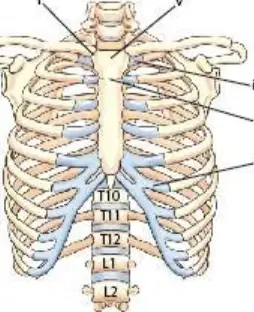
The structure labeled iii is costocartilage:
of the 10th rib.
portion of the first false rib.
of the last true rib.
of the sixth rib.

What is the name of the part labeled iv?
Facet for the sternum attachment
Head of the sternum
Facet for the second rib attachment
Costocartilage for the first rib attachment
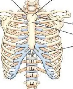
What is the name of the structure labeled v?
Head
Body
Manubrium
Xiphoid process
Which aspect of the rib articulates with the thoracic vertebral body?
Neck
Tubercles
Head
Facets
Which ribs are considered to be true ribs?
First and second ribs
First through seventh ribs
First through ninth ribs
11th and 12th ribs
The degree of rotation for the right anterior oblique (RAO) projection of the sternum is dependent on the size of the thoracic cavity.
True
False
The left anterior oblique (LAO) position of the sternum provides the best frontal image of the sternum with a minimal amount of distortion.
True
False
Which of the following techniques is most effective in preventing lung markings from obscuring the sternum on an oblique projection?
Use a high kV.
Oblique as much as needed to not superimpose the sternum over the hilum region.
Decrease the source image receptor distance (SID) to magnify the sternum.
Use an orthostatic (breathing) technique.
Why is the RAO sternum preferred to the LAO position?
The RAO produces less magnification of the sternum.
The RAO projects the sternum over the shadow of the heart.
The RAO reduces dose to the thyroid gland.
The RAO projects the sternum away from the hilum and heart.
Which of the following statements is true about radiography of ribs located above the diaphragm?
Suspend breathing upon inspiration.
Perform the study with the patient recumbent.
Use an analog kV range of 85 to 95.
Always include an anteroposterior (AP) projection as part of the routine.
Which of the following positions will best demonstrate the axillary portion of the left ribs?
AP
Posteroanterior (PA)
Which of the following positions will best demonstrate the axillary portion of the right ribs?
AP
PA
LAO
RAO
A right or left marker may be taped over the area of interest to indicate the location of the trauma to the ribs.
True
False
A recommended practice is to decrease the SID to less than 40 inches (102 cm) for the oblique sternum projection to increase the magnification and resultant unsharpness of overlying ribs.
True
False
Which of the following conditions may occur with trauma to the ribs?
Airway obstruction of the trachea
Pneumonia
Hemothorax
Pulmonary embolus
What is the recommended degree of obliquity for an RAO projection of the sternum for an asthenic type of patient?
20°
15°
30°
10°
Which position can replace the RAO of the sternum if the patient cannot lie prone?
LAO
Left lateral decubitus
LPO
RPO
What is the recommended SID for the lateral sternum position?
40 inches (102 cm)
44 inches (113 cm)
46 inches (117 cm)
60 to 72 inches (152 to 183 cm)
Where is the CR centered for a PA projection of the sternoclavicular joints?
At the level of the vertebra prominens (T1)
At the level of the sternal angle (T4-5)
Three inches (7 cm) distal to vertebra prominens (T2-3)
At the level of the thyroid cartilage (T9)
How much rotation and which oblique position are required to best demonstrate the left sternoclavicular joints?
10° to 15° LAO
35° to 45° LAO
10° to 15° RAO
5° to 10° RAO
Which two projections must be taken for an injury to the right anterior upper ribs?
PA and LAO
PA and RAO
AP and RAO
AP and LPO
Which two projections must be taken for an injury to the left posterior lower ribs?
AP and LPO
AP and RAO
PA and LPO
PA and RAO
Which analog kV range is recommended for an AP study of the ribs found below the diaphragm?
85 to 90 kV
60 to 65 kV
65 to 75 kV
70 to 80 kV
Which disease or condition may be associated with postoperative complications of open heart surgery?
Spondylitis
Osteoblastic metastases
Osteomyelitis
Flail chest
Fracture of adjacent ribs in two or more places with associated pulmonary injury is known as a(n)___ rib fracture.
compound
flail chest
acute
compression
The radiographic appearance of the erosion of bony rib margins is a possible indication of:
osteomyelitis.
osteoblastic metastases.
spondylolysis.
osteolytic metastases.
Which of the following conditions, if severe, requires a decrease adjustment of manual exposure factors?
Osteoblastic metastases
Osteomyelitis
Flail chest
None of the above
Both nuclear medicine and MRI (magnetic resonance imaging) studies can be performed to evaluate metastatic rib lesions before conventional rib radiographic examination.
True
False
A congenital defect characterized by anterior protrusion of the lower sternum and xiphoid process is termed:
pectus excavatum.
flail chest.
pectus carinatum.
sternal protrusion.
Multiple myeloma is seen often in the flat bones of the bony thorax.
True
False
Which condition of the sternum is often termed “funnel chest?”
Pectus excavatum
Flail chest
Pectus eruptus
Pectus deforminens
Both bony and soft tissue anatomy may be evaluated by CT for pathology involving the sternum or the sternoclavicular joints.
True
False
The condition, flail chest, is most commonly caused by:
pneumothorax.
emphysema.
blunt trauma.
congenital heart defect.
A radiograph of an RAO sternum reveals that it is partially superimposed over the spine. What must be done to eliminate this problem during the repeat exposure?
Perform an LPO projection instead of an RAO.
Angle CR 5° to 10° laterally to the sternum.
Increase rotation of the body.
Increase kV.
A radiograph of a lateral projection of the sternum reveals that the patient’s ribs are superimposed over the sternum. What needs to be done to correct this problem during the repeat exposure?
Increase the SID.
Angle the CR 5° anterior.
Ensure that the patient is not rotated.
Increase the kV.
A radiograph of an RAO projection of the sternum demonstrates excessive lung markings obscuring the sternum. A 1-second exposure time and an orthostatic (breathing) technique were used. Which of the following will produce a more diagnostic image of the sternum?
Ensure that the patient is not breathing during the exposure.
Increase the exposure time; decrease the mA.
Decrease the kV; increase the mA or time.
Initiate exposure on deeper inspiration.
A PA radiograph of the sternoclavicular (SC) joints demonstrates unequal distance from the SC joints to the midline of the spine. The left SC joint is farther from the sternum than the right. What specific positioning error is present on this radiograph?
Slight right rotation (right side toward the image receptor)
Slight left rotation (left side toward the image receptor)
Tilt of the upper thorax
Excessive angulation of the CR
A young female patient from the emergency department (ED) is brought to radiology for rib examination. She is able to sit up or stand for the procedure. She indicates that the region of pain is to the right anterior-to-mid axillary region. Which rib projections should be performed to minimize the effective dose to this patient?
PA and RPO
AP and RPO
PA and RAO
PA and LAO
A patient enters the ED with an injury to the left anterior lower ribs. Which of the following projections should be taken to demonstrate the involved area?
AP and LAO
PA and RAO
AP and LPO
PA and LAO
An ambulatory patient enters the ED with a possible injury to the right upper posterior ribs. Which of the following routines should be taken to demonstrate the involved area?
Erect PA and LPO
Erect AP and RPO
Recumbent AP and RPO
Erect PA and LAO
58. A patient enters the ED with blunt trauma to the sternum. The patient is in great pain and cannot lie prone on the table or stand erect. Which of the following routines would be best for the sternum examination in this situation?
Perform the examination with the patient in the lateral decubitus position
Perform the examination with the patient standing erect
Perform the examination with the patient lying prone
Perform the examination with the patient in the Trendelenburg position
A patient enters the ED with trauma to the bony thorax. The initial radiographs reveal that there are fractured ribs and a possible pneumothorax of the left thorax. The physician orders a chest study to confirm the pneumothorax; however, the patient cannot stand. Which of the following positions would best demonstrate the pneumothorax?
Left lateral decubitus
Right lateral decubitus
Ventral decubitus
Dorsal decubitus
Initial PA projections of the SC joints indicate a possible defect involving the left SC joint. The vertebral column is preventing a clear view of it. Which of the following projections will demonstrate the right SC joint without superimposition over the spine?
Horizontal beam lateral
LAO
RAO
Erect lateral projection
The use of 125 kV is recommended for AP and PA projections of the ribs to reduce skin dose.
True
False
What is the minimum number of ribs that must be demonstrated for a unilateral rib study above the diaphragm?
Ribs 1 through 6
Ribs 1 through 8
Ribs 1 through 10
All ribs must be demonstrated.
A lateral projection of the sternum requires that respiration be suspended on expiration.
True
False
A radiograph of an RAO projection of the ribs demonstrates the left axillary ribs are foreshortened, whereas the right side is elongated. Which of the following is the most likely reason for this radiographic outcome?
The patient requires more rotation to the right.
An LAO was performed rather than the RAO position.
The technologist should have performed a PA projection to demonstrate the left axillary ribs, not an RAO.
CR angulation was incorrect.
A patient with metastatic disease in the ribs comes to radiology following a nuclear medicine scan. The radiologist orders a right, upper posterior rib study performed. Which of the following positioning factors should be followed for this specific study?
Perform positions erect if the patient’s condition permits.
Exposure on full expiration.
Include the RPO position as part of the positioning routine.
Both A and C are correct.
Which of the following positioning considerations does NOT apply for a study of the lower ribs?
Perform positions recumbent.
Use a digital kV range between 65 and 70 kV.
Exposure on full expiration.
Both A and B are incorrect.

