
NEW
Font size
 Worksheets
Bone Physiology
Total questions: 74
Worksheet time: 37mins
Fill in the blank: One of the functions of bones is _________
Support
Digestion
Photosynthesis
Evaporation
Fill in the blank: Bones help in _________
Movement
Digestion
Photosynthesis
Evaporation
Fill in the blank: Bones provide _________ to vital organs.
Protection
Energy
Digestion
Movement
Fill in the blank: Bones are important for _________ storage.
Mineral
Water
Fatty acid
Protein
Fill in the blank: Bones are involved in blood-cell formation and _________ storage.
Energy
Protein
Oxygen
Calcium
According to the classification of bones, which of the following is NOT a type of bone?
Long bones
Short bones
Flat bones
Round bones
Fill in the blank: There are 4 types of bones: Long bones, Short bones, Flat bones, and _________
Irregular bones
Round bones
Circular bones
Triangular bones
Which of the following is a type of bone according to the classification of bones?
Irregular bones
Circular bones
Triangular bones
Oval bones
How many types of bones are there according to the classification of bones?
4
2
5
3
Which of the following best describes long bones?
They are wider than they are long
They are considerably longer than they are wide
They are flat and thin
They are irregularly shaped
Long bones consist of a shaft and two distinct ________ ends.
expanded
narrow
flat
curved
Most bones of the limbs are what type of bones?
Short bones
Flat bones
Long bones
Irregular bones
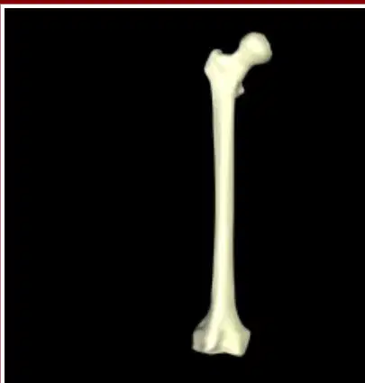
Look at the image of the bone below. What type of bone is shown?
Short bone
Flat bone
Long bone
Irregular bone
Which of the following best describes short bones?
A) Long and thin
B) Flat and broad
C) Roughly cube shaped
D) Irregularly shaped
Where do short bones primarily occur?
Skull and spine
Wrist and ankle
Ribs and sternum
Arm and leg
What is a sesamoid bone?
A) A type of long bone
B) A special type of short bone that forms within a tendon
C) A bone found only in the skull
D) A bone that forms in the rib cage
Name two locations in the body where short bones are found.
Wrist and ankle
Skull and ribcage
Femur and humerus
Pelvis and clavicle
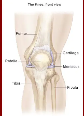
Identify the bone labeled 'Patella' in the knee diagram.
Patella
Femur
Tibia
Fibula
What are the characteristics of flat bones?
A) Long and cylindrical
B) Thin, flattened and usually somewhat curved
C) Short and round
D) Irregularly shaped
Which of the following is an example of a flat bone?
Femur
Ribs
Vertebrae
Carpals
Fill in the blank: Flat bones include the _______, sternum, scapula, and most bones of the skull.
ribs
femur
vertebrae
phalanges

The image shows a part of the human skeleton. Which type of bones are highlighted in the image?
Long bones
Short bones
Flat bones
Irregular bones
What are irregular bones?
Bones with a regular shape
Bones that have various shapes that do not fit into the previous categories
Bones that are only found in the arms
Bones that are only found in the legs
Which of the following is an example of an irregular bone? Choose the correct answer.
Femur
Vertebrae
Tibia
Radius
Name two examples of irregular bones mentioned in the passage.
Vertebrae, Hip bones (also acceptable: sphenoid and ethmoid bones of the skull)
Femur, Tibia
Scapula, Clavicle
Radius, Ulna

In the diagram, what is labeled as the canal through which the spinal cord passes?
Spinous Process
Vertebral Canal
Hip Bone
Ethmoid Bone
Bone is made up of: ________ tissue (Fill in the blank with one of the components listed in the image)
Bone tissue
Muscle tissue
Nervous tissue
Epithelial tissue
Bone is made up of: ________ connective tissue (Fill in the blank with one of the components listed in the image)
Fibrous connective tissue
Adipose connective tissue
Cartilaginous connective tissue
Areolar connective tissue
Bone is made up of: ________ (Fill in the blank with one of the components listed in the image)
Cartilage
Muscle
Fat
Blood
Bone is made up of: ________ tissue (Fill in the blank with one of the components listed in the image)
Vascular tissue
Muscular tissue
Nervous tissue
Epidermal tissue
Bone is made up of: ________ tissue (Fill in the blank with one of the components listed in the image)
Lymphatic tissue
Muscular tissue
Epithelial tissue
Nervous tissue
Bone is made up of: ________ tissue (Fill in the blank with one of the components listed in the image)
Adipose tissue
Muscle tissue
Nervous tissue
Epithelial tissue
Bone is made up of: ________ tissue (Fill in the blank with one of the components listed in the image)
Nervous tissue
Muscle tissue
Epithelial tissue
Connective tissue
What is another name for Compact Bone?
Cortical Bone
Spongy Bone
Trabecular Bone
Cancellous Bone
Which of the following is a characteristic of Compact Bone?
Loose deposits of minerals
Dense deposits of minerals
No mineral deposits
Arranged randomly
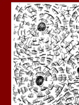
What is the central structure around which the compact bone is arranged in concentric circles?
Haversian canal
Osteocyte
Periosteum
Trabeculae
Fill in the blank: ________ are long cylindrical structures that contain blood vessels and nerve fibers that supply the bone cells with nutrients.
Osteon or Haversian Canals
Canaliculi
Lacunae
Trabeculae
Fill in the blank: Each tube concentric matrix that surrounds each Haversian Canal is called a ________.
Lamella
Osteon
Canaliculi
Trabeculae
Fill in the blank: ________ lie perpendicular to the Haversian Canals and connect blood and nerve supply of the periosteum to that of the Haversian Canals and the marrow cavity.
Perforating canals or Volkman's canals
Canaliculi
Lacunae
Lamellae
Fill in the blank: ______ are mature bone cells that are spider-shaped. (Anatomy of Compact Bone)
Osteocytes
Osteoblasts
Chondrocytes
Fibroblasts
Fill in the blank: ______ are small cavities in the solid matrix where the osteocytes reside. (Anatomy of Compact Bone)
Lacunae
Canaliculi
Lamellae
Osteons
Fill in the blank: ______ are thin tubes that run through the matrix, connecting the neighboring lacunae to one another and to the nearest capillaries. (Anatomy of Compact Bone)
Canaliculi
Osteons
Lamellae
Trabeculae

Identify the part labeled 'Concentric lamellae' in the diagram of the compact bone.
Concentric lamellae are the circular layers of bone matrix found in compact bone.
Haversian canal is the central channel containing blood vessels and nerves.
Lacunae are small spaces housing osteocytes.
Canaliculi are tiny channels connecting lacunae.

What is the function of the 'Periosteum' in the compact bone?
The periosteum is a membrane that covers the outer surface of all bones, providing nourishment and serving as an attachment for tendons and muscles.
It is the central cavity of bone shafts where red bone marrow is stored.
It is the hard, dense layer that gives bones their strength.
It is the spongy tissue found at the ends of long bones.

Which part of the compact bone contains blood vessels?
Periosteal blood vessels are found in the periosteum of the compact bone.
Osteocytes are found in the lacunae of the compact bone.
Collagen fibers are found in the matrix of the compact bone.
Canaliculi are found between the lacunae of the compact bone.

The spongy bone is found inside the compact bone and is less dense than the compact bone.
True
False

Fill in the blank: The ________ is the cavity within the bone that contains bone marrow.
Medullary (marrow) cavity
Periosteum
Epiphyseal plate
Articular cartilage

Which layer of the periosteum is closer to the bone?
Inner layer
Outer layer
Middle layer
Fibrous layer
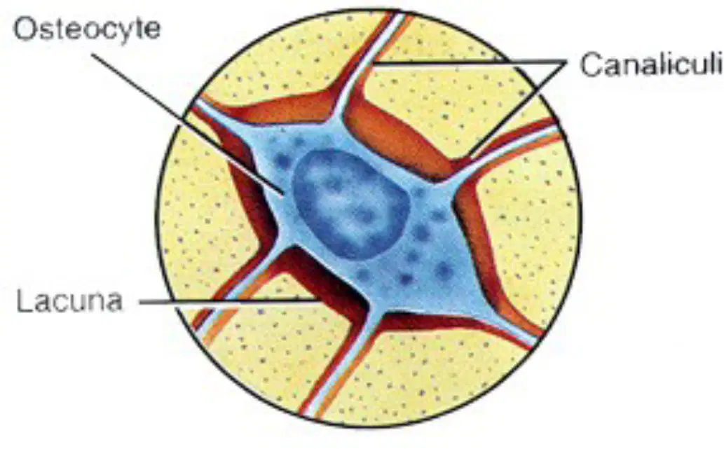
In the diagram of the compact bone, what is the name of the cell located in the center?
Osteocyte
Chondrocyte
Erythrocyte
Fibroblast

In the diagram of the compact bone, what are the small channels radiating from the center called?
Canaliculi
Osteons
Lacunae
Lamellae
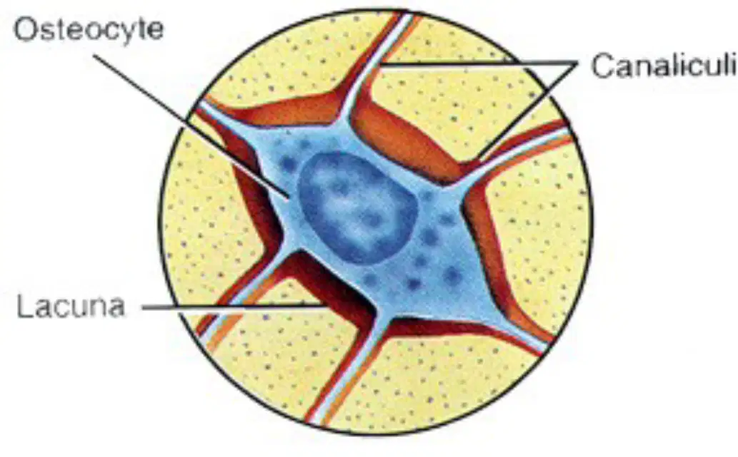
In the diagram of the compact bone, what is the small space surrounding the osteocyte called?
Lacuna
Canaliculus
Haversian canal
Lamella
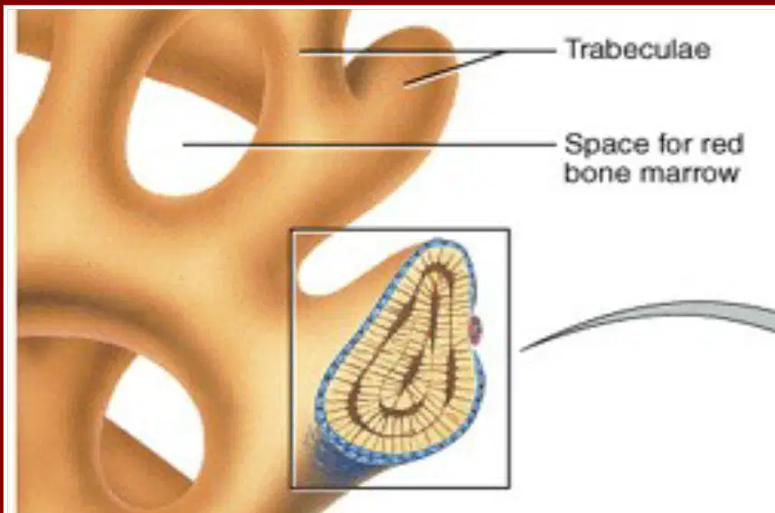
What is another name for spongy bone?
Compact bone
Trabecular bone
Cartilage
Marrow bone
Fill in the blank: The small needle-like or flat pieces in spongy bone are called ________.
trabeculae
osteons
lamellae
canaliculi
Fill in the blank: The spaces in spongy bone are filled with ________.
red bone marrow
yellow bone marrow
cartilage
synovial fluid
Which type of bone is less complex: spongy bone or compact bone?
Spongy bone
Compact bone
Both are equally complex
Neither
Each trabecula in spongy bone contains several layers of ______ and osteocytes.
lamellae
canaliculi
periosteum
marrow
Each trabecula in spongy bone has its own osteons and blood vessels.
True
False
How does spongy bone receive nutrients?
From osteons within the trabecula
From capillaries in the endosteum surrounding the trabecula
From the periosteum
From cartilage

What is the outer dense layer of the bone called?
compact bone
spongy bone
marrow
periosteum
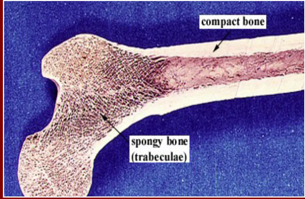
What is the inner porous region of the bone called?
spongy bone (trabeculae)
compact bone
periosteum
bone marrow
Fill in the blank: Bone consists of cells separated by and ________ matrix.
extracellular
intracellular
vascular
muscular
Which of the following is NOT a component of bone matrix?
Mineral crystals
Collagen fibers
Ground substances
Blood cells
Tissue fluid
Bone matrix contains mineral crystals, collagen fibers, ground substances, and a small amount of tissue fluid.
True
False
What is the function of Osteoblasts?
Osteoblasts are bone-forming cells responsible for synthesizing and mineralizing bone during both initial bone formation and later bone remodeling.
Osteoblasts are cells that break down bone tissue during bone resorption.
Osteoblasts are responsible for transmitting nerve impulses in bone tissue.
Osteoblasts are involved in the production of red blood cells within the bone marrow.
What is the function of Osteocytes?
Osteocytes are mature bone cells that maintain the bone matrix and communicate with other bone cells to regulate bone remodeling.
Osteocytes are responsible for producing red blood cells in the bone marrow.
Osteocytes break down bone tissue during bone resorption.
Osteocytes form the initial cartilage template during bone development.
What is the function of Osteoclasts?
Osteoclasts are bone-resorbing cells responsible for breaking down bone tissue during bone remodeling and healing.
Osteoclasts are responsible for producing red blood cells in the bone marrow.
Osteoclasts synthesize collagen fibers for bone strength.
Osteoclasts store calcium and phosphate in the bone matrix.
In the diagram, what is the role of Mesenchyme?
Mesenchyme is a type of connective tissue from which bone and other connective tissues develop.
Mesenchyme is a type of muscle tissue responsible for contraction.
Mesenchyme is a type of epithelial tissue that lines organs.
Mesenchyme is a type of nervous tissue involved in signal transmission.
What is the function of Canaliculi?
Canaliculi are small channels that connect osteocytes and allow for the exchange of nutrients and waste.
Canaliculi are responsible for producing red blood cells in bone marrow.
Canaliculi store calcium and phosphate ions in the bone matrix.
Canaliculi are large cavities that house bone marrow cells.
What is the main function of osteoblasts?
Break down bone tissue
Synthesize and secrete collagen fibers and other organic components of bone matrix
Store calcium
Transport oxygen
Fill in the blank: Osteoblasts __________ and secrete collagen fibers and other organic components of bone matrix.
synthesize
absorb
destroy
inhibit

Which cell type is responsible for synthesizing and secreting collagen fibers and other organic components of bone matrix?
A) Osteoclasts
B) Osteoblasts
What are osteocytes? (Fill in the blank) Osteocytes are _________.
Mature bone cells
Immature bone cells
Cartilage cells
Blood cells
What is the main function of osteocytes? Osteocytes are responsible for _________.
maintaining bone tissue
producing red blood cells
initiating muscle contraction
transmitting nerve impulses
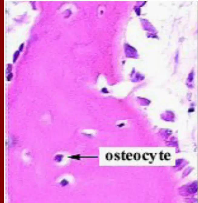
Look at the diagram. What type of cell is labeled in the image?
Osteoblast
Osteoclast
Osteocyte
Chondrocyte

