
 Worksheets
L03 Circulatory System Quiz 1
Total questions: 100
Worksheet time: 50mins
Which type of blood vessels carry blood away from the heart?
Veins
Capillaries
Arteries and Arterioles
Venules
What is the primary function of capillaries in the circulatory system?
Carry blood away from the heart
Return blood to the heart
Facilitate exchange of oxygen, nutrients, carbon dioxide, and wastes
Provide structural support to blood vessels
Which layer of the blood vessel wall is composed of smooth muscle and elastic tissue?
Tunica adventitia
Tunica media
Tunica intima
Endothelium
What is the function of the tunica adventitia in blood vessels?
Provides a smooth surface for blood flow
Facilitates exchange of gases and nutrients
Offers support and protection with its fibrous collagen layer
Contracts to regulate blood flow
Which layer of the blood vessel wall is made up of a single-cell endothelial lining?
Tunica adventitia
Tunica media
Tunica intima
Basement membrane
What is the primary reason arterial walls are thicker than venous walls?
To allow for greater elasticity
To withstand higher pressure
To store more blood
To reduce blood flow
What characteristic of arterial bleeding makes it hard to stop?
It is slow and steady
It is pulsatile and matches the heartbeat
It clots quickly
It has low pressure
What is the diameter range of the largest arteries?
0.5–1 cm
1–2.5 cm
2.5–5 cm
5–10 cm
What type of tissue is abundant in the tunica media of the largest arteries?
Smooth muscle
Elastic tissue
Connective tissue
Collagen fibers
Why are arteries near the heart called elastic arteries?
They have a high amount of smooth muscle
They stretch and recoil to push blood forward
They are resistant to pressure changes
They regulate systemic blood pressure
What is the primary composition of the tunica media in arterioles?
Elastic tissue
Smooth muscle
Collagen fibers
Endothelial cells
What happens to the composition of arteries as they branch into smaller vessels?
Elastic tissue increases, smooth muscle decreases
Elastic tissue decreases, smooth muscle increases
Both elastic tissue and smooth muscle increase
Both elastic tissue and smooth muscle decrease
What is the role of arterioles in regulating blood flow and pressure?
They store blood for later use
They contract and relax smooth muscle to regulate diameter
They act as elastic reservoirs
They prevent blood from flowing backward
Why are arterioles referred to as resistance vessels?
They have thick walls to resist pressure
They primarily determine systemic blood pressure
They store blood during diastole
They are located near the heart
What are anastomoses in the circulatory system?
Direct links between veins supplying different areas
Direct links between arteries supplying the same area
Indirect links between arteries and veins
Indirect links between arteries supplying different areas
What is the primary function of anastomoses in the circulatory system?
To increase blood pressure
To provide collateral circulation as a backup route if blockage occurs
To decrease oxygen levels in the blood
To prevent the formation of blood clots
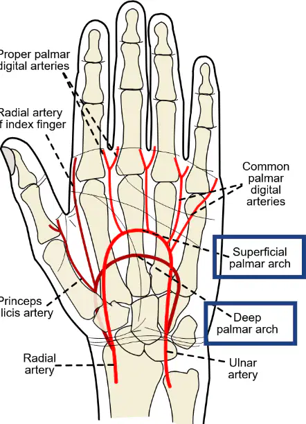
Which arteries are involved in the formation of the superficial and deep palmar arches in the hand?
Radial artery and ulnar artery
Brachial artery and femoral artery
Carotid artery and subclavian artery
Axillary artery and popliteal artery
What is the sole source of blood supply to a tissue called?
End-arteries
Collateral arteries
Capillaries
Venules
What happens if an end-artery is blocked?
Tissue death (infarction) due to no alternative supply
Blood reroutes through collateral arteries
Increased blood flow to the tissue
Formation of new arteries immediately
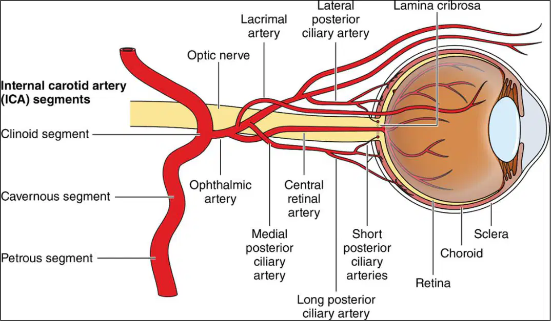
Which artery is labeled as the central artery of the retina in the diagram?
Central retinal artery
Lacrimal artery
Ophthalmic artery
Short posterior ciliary artery
What is the primary function of capillaries in the circulatory system?
Transport blood cells and plasma proteins directly to tissues
Exchange nutrients, oxygen, and waste between blood and tissue fluid
Store oxygen for later use by tissues
Prevent the passage of water and small molecules into tissues
Which structural feature of capillaries allows them to be permeable to water and small molecules?
Thick muscular walls
Single endothelial layer and thin basement membrane
Presence of valves
Large diameter
What is the approximate diameter of capillaries, which requires red blood cells to bend to pass through?
10–12 µm
3–4 µm
7–8 µm
1–2 µm
Which body areas lack capillaries, making them exceptions to their presence near all body cells?
Brain and spinal cord
Skin surface and cornea
Heart and lungs
Liver and kidneys
What is the special characteristic of capillaries in the blood-brain barrier?
They are highly permeable to all molecules
They are less permeable to protect neural tissue
They have a larger diameter to allow faster blood flow
They are absent in the brain and spinal cord
Where are sinusoids primarily found in the human body?
Heart and lungs
Liver and bone marrow
Kidneys and pancreas
Brain and spinal cord
What is a key structural characteristic of sinusoids compared to normal capillaries?
Thicker walls
Wider and leakier with incomplete walls
Narrower and less permeable
Completely sealed walls
How does the direct contact of blood with cells in sinusoids affect exchange processes?
Slows down exchange
Prevents exchange
Leads to faster exchange
Causes irregular exchange
What is the approximate diameter of liver sinusoids?
100 µm
170 µm
250 µm
300 µm
What is the sequence of structures in the low-pressure return system of veins?
Arteries → arterioles → capillaries
Capillaries → venules → veins
Veins → venules → capillaries
Arterioles → veins → capillaries
How do the walls of veins compare to those of arteries?
Veins have thicker walls than arteries.
Veins have thinner walls than arteries but the same three layers.
Veins have the same wall thickness as arteries but fewer layers.
Veins have no walls, unlike arteries.
What is the primary function of valves in veins?
To increase blood pressure.
To prevent backflow of blood.
To reduce the flow of blood to the heart.
To increase the oxygen content of blood.
Where are valves most commonly found in the venous system?
In the veins of the thorax and abdomen.
In the veins of the head and neck.
In the veins of the lower limbs.
In the arteries of the lower limbs.
Why are veins referred to as capacitance vessels?
They have thick walls that resist expansion.
They hold approximately two-thirds of the blood volume and are compliant.
They have valves that prevent backflow.
They maintain high blood pressure during blood loss.
What happens to venous pressure during bleeding, and why is hemostasis easier in veins than in arteries?
Venous pressure increases, and walls collapse, making hemostasis harder.
Venous pressure decreases, and walls collapse, making hemostasis easier.
Venous pressure remains constant, and walls remain rigid.
Venous pressure decreases, and walls expand, making hemostasis harder.
What mechanism helps veins return blood to the heart against gravity?
The contraction of arterial walls.
The skeletal muscle pump combined with valves.
The high pressure within veins.
The absence of valves in the venous system.
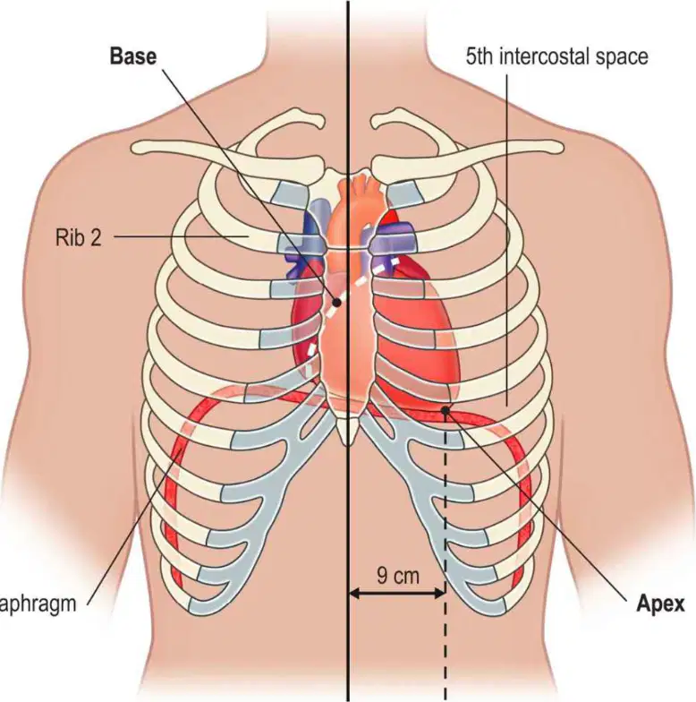
Where is the heart located within the thoracic cavity?
In the abdominal cavity
Within the mediastinum (between lungs)
In the cranial cavity
Behind the diaphragm
What direction does the heart slant within the thoracic cavity?
Slightly right
Slightly left
Straight down
Straight up
At what level is the apex of the heart located?
2nd intercostal space
5th intercostal space
7th intercostal space
10th intercostal space
How far is the apex of the heart located from the midline?
~5 cm
~7 cm
~9 cm
~12 cm
What is the shape of the base (superior surface) of the heart?
Rounded and pointed
Broad and flattened
Narrow and elongated
Circular and elevated
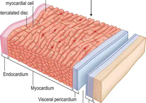
What are the three layers that compose the heart wall?
Pericardium, Myocardium, Endocardium
Epidermis, Dermis, Hypodermis
Endothelium, Mesothelium, Epithelium
Peritoneum, Pleura, Pericardium
Which layer of the heart wall is the thickest and primarily composed of cardiac muscle tissue?
Pericardium
Myocardium
Endocardium
Epicardium
What is the outermost layer of the heart wall called?
Endocardium
Myocardium
Pericardium
Epicardium
Which layer of the heart wall lines the inner chambers of the heart and is in direct contact with blood?
Pericardium
Myocardium
Endocardium
Epicardium
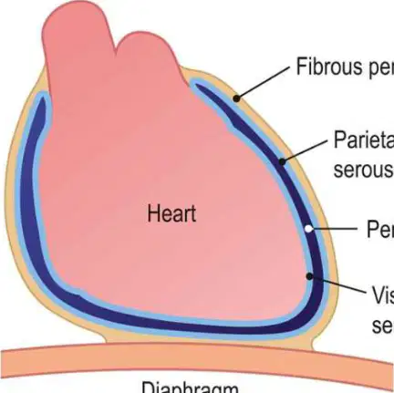
What is the outermost covering of the heart called?
Myocardium
Endocardium
Pericardium
Epicardium
Which type of tissue is the fibrous pericardium made of?
Elastic tissue
Dense fibrous, inelastic tissue
Loose connective tissue
Adipose tissue
What is the function of the fibrous pericardium?
Pumps blood through the heart
Protects the heart and prevents over-distension
Produces hormones for heart function
Stores oxygen for the heart
Which layer of the serous pericardium is firmly attached to the myocardium?
Parietal layer
Visceral layer
Fibrous pericardium
Pericardial cavity
What is the approximate amount of fluid in the pericardial cavity?
10 mL
20 mL
50 mL
100 mL
What is the purpose of the fluid in the pericardial cavity?
To store nutrients for the heart
To provide lubrication and friction-free beating
To transport oxygen to the heart
To regulate blood pressure
What type of muscle is the myocardium?
Skeletal muscle
Smooth muscle
Cardiac muscle
Voluntary muscle
Which of the following best describes the fibers of the myocardium?
Non-striated, involuntary, multinucleated
Striated, voluntary, multinucleated
Striated, involuntary, single nucleus
Non-striated, voluntary, single nucleus
What structure allows myocardial cells to join end-to-end and behave like a single sheet?
Sarcomeres
Intercalated discs
Myofibrils
Axons
What is the primary function of intercalated discs in cardiac muscle tissue?
To store calcium ions
To allow impulses to spread cell-to-cell for coordinated contraction
To generate ATP for muscle contraction
To anchor the muscle to bones
Why is the myocardium mitochondria-rich?
To store oxygen for muscle contraction
To provide energy for continuous and efficient contraction
To regulate calcium levels in the muscle
To produce proteins for muscle repair
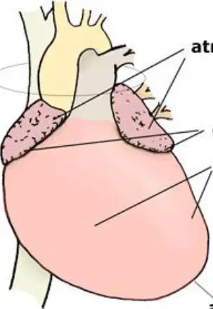
What part of the myocardium is the thickest?
Base
Atria
Apex
Ventricles
What is the function of the specialized conducting fiber network in the myocardium?
To pump blood out of the heart
To carry the heart's electrical signals
To produce atrial natriuretic peptide (ANP)
To regulate blood pressure
What do atrial muscle cells secrete?
Adrenaline
Atrial natriuretic peptide (ANP)
Insulin
Dopamine

What is the primary function of the very smooth surface of the endocardium?
To increase blood pressure
To minimize friction as blood flows over it
To absorb nutrients from the blood
To regulate heart rate

What type of cells make up the endocardium?
Cuboidal epithelial cells
Flattened epithelial cells
Columnar epithelial cells
Stratified epithelial cells

The endocardium is continuous with which structure throughout the circulatory system?
Vessel endothelium
Myocardium
Pericardium
Pulmonary artery

Where is the endocardium located in the heart?
Surrounding the heart muscle
Lining all heart chambers and valves
Covering the outer surface of the heart
Between the atria and ventricles
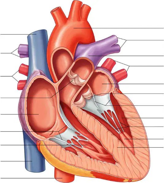
What structure separates the right and left sides of the heart?
Atria
Ventricles
Septum
AV valve
What is the function of the AV valve in the heart?
To separate the right and left sides of the heart
To connect each atrium to its corresponding ventricle
To pump blood out of the heart
To supply oxygen to the myocardium
Which layer of the heart covers the myocardium and forms the septum?
Endocardium
Epicardium
Pericardium
Myocardium itself
What is the role of the myocardium in the heart's septum?
It pumps blood to the lungs.
It separates the right and left sides of the heart.
It connects the atria to the ventricles.
It regulates the heart's electrical signals.
Which atrioventricular (AV) valve has three cusps?
Left mitral valve
Right tricuspid valve
Aortic valve
Pulmonary valve
What is the primary function of the chordae tendineae and papillary muscles during systole?
To open the semilunar valves
To anchor the cusps and prevent prolapse into the atria
To close the atrioventricular valves
To increase blood flow to the ventricles
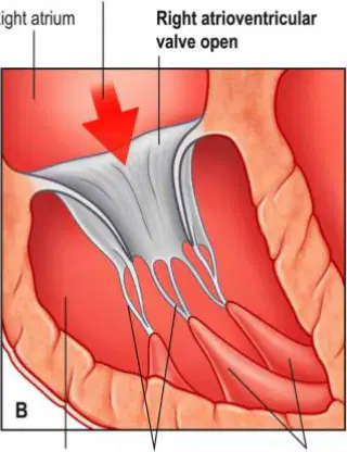
When do the atrioventricular (AV) valves open?
When ventricular pressure is greater than atrial pressure
When atrial pressure is greater than ventricular pressure
During ventricular systole
During atrial diastole
What is the primary purpose of an echocardiogram (ultrasound)?
To measure blood pressure in the arteries
To provide a real-time view of the heart's chambers and valves, and evaluate pumping, valve function, and blood flow
To detect the electrical activity of the heart
To measure oxygen levels in the blood
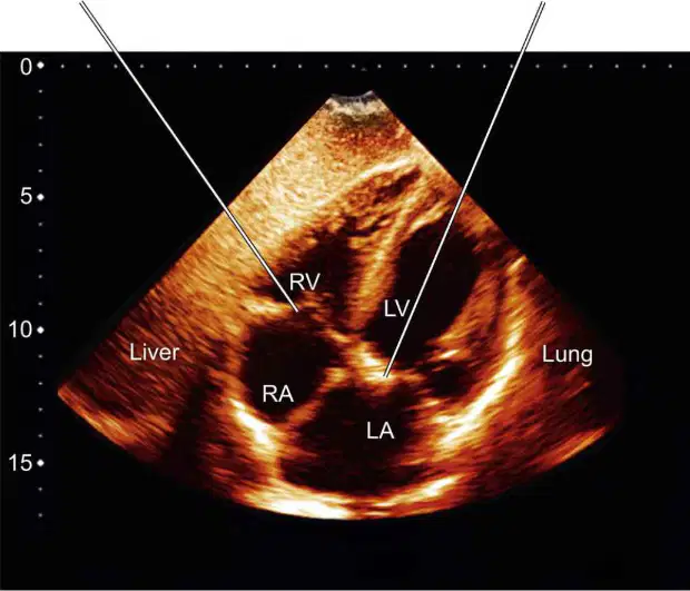
Which heart valve is labeled as the "Right atrioventricular (tricuspid) valve" in an echocardiogram?
Mitral valve
Pulmonary valve
Tricuspid valve
Aortic valve
What does the echocardiogram image help diagnose?
Lung infections
Heart pumping efficiency, valve function, and blood flow issues
Liver diseases
Bone fractures
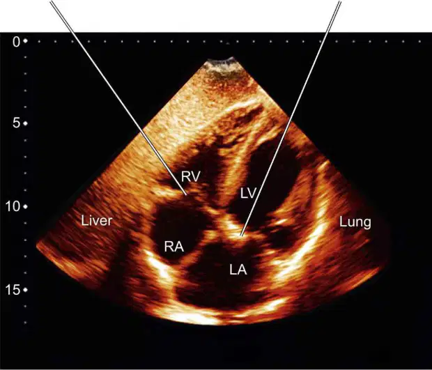
Which chamber of the heart is labeled as "RV" in the echocardiogram image?
Right atrium
Right ventricle
Left atrium
Left ventricle
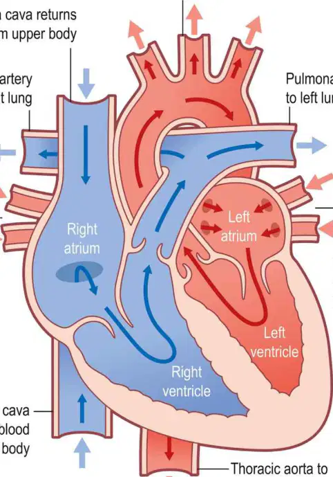
What is the correct sequence of blood flow through the right side of the heart?
SVC & IVC → Right atrium → Tricuspid valve → Right ventricle → Pulmonary valve → Pulmonary arteries → Lungs
Pulmonary veins → Left atrium → Mitral valve → Left ventricle → Aortic valve → Aorta → Systemic circulation
Right atrium → Pulmonary arteries → Pulmonary valve → Right ventricle → SVC & IVC → Lungs
Aorta → Systemic circulation → Pulmonary veins → Left atrium → Mitral valve → Left ventricle
Which valve is responsible for preventing backflow of blood between the right atrium and right ventricle?
Tricuspid valve
Pulmonary valve
Mitral valve
Aortic valve
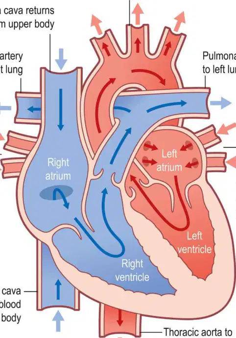
What is the primary function of the lungs in the blood flow process?
Gas exchange: CO₂ out, O₂ in
Pumping blood to the systemic circulation
Filtering blood from the left ventricle
Transporting oxygenated blood to the right atrium
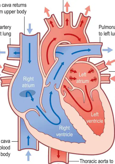
What is the correct sequence of blood flow through the left side of the heart?
Pulmonary veins → Left atrium → Mitral valve → Left ventricle → Aortic valve → Aorta → Systemic circulation
SVC & IVC → Right atrium → Tricuspid valve → Right ventricle → Pulmonary valve → Pulmonary arteries → Lungs
Left atrium → Pulmonary arteries → Mitral valve → Left ventricle → Pulmonary veins → Systemic circulation
Aorta → Systemic circulation → Pulmonary veins → Left atrium → Mitral valve → Left ventricle
Which valve is responsible for preventing backflow of blood between the left atrium and left ventricle?
Mitral valve
Tricuspid valve
Pulmonary valve
Aortic valve
What is the primary function of the pulmonary and aortic valves?
To pump blood into the ventricles
To prevent backflow of blood
To oxygenate the blood
To regulate heart rate
How many cusps do the semilunar valves have?
2
3
4
5
What is the contraction pattern of the heart?
Ventricles contract first, then atria contract together
Atria contract together, then ventricles contract together
Atria and ventricles contract simultaneously
Only the ventricles contract
Why are the walls of the atria thinner compared to the ventricles?
Atria have less workload and are aided by gravity
Atria pump blood at a higher pressure
Atria require more muscle for contraction
Atria handle systemic circulation
Which ventricle has a thicker wall, and why?
Right ventricle, because it handles systemic circulation
Left ventricle, because it handles systemic circulation
Right ventricle, because it handles pulmonary circulation
Left ventricle, because it handles pulmonary circulation
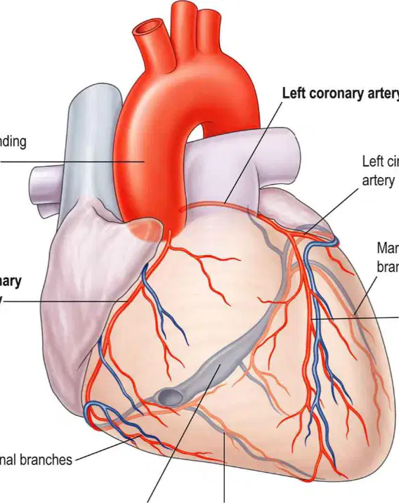
Which arteries branch from the aorta just beyond the aortic valve to supply blood to the heart?
Pulmonary arteries
Coronary arteries
Carotid arteries
Subclavian arteries
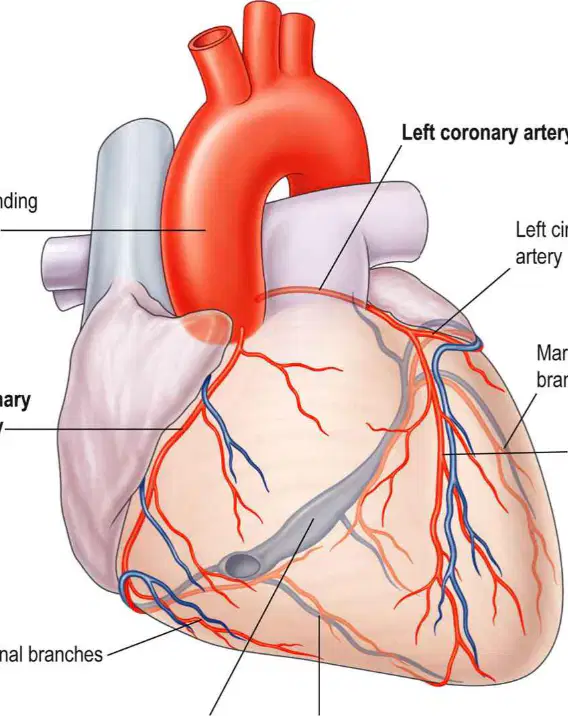
What does the left anterior descending (LAD) artery primarily supply?
Right ventricle and atrium
Left ventricle and atrium
SA and AV nodes
Pulmonary arteries
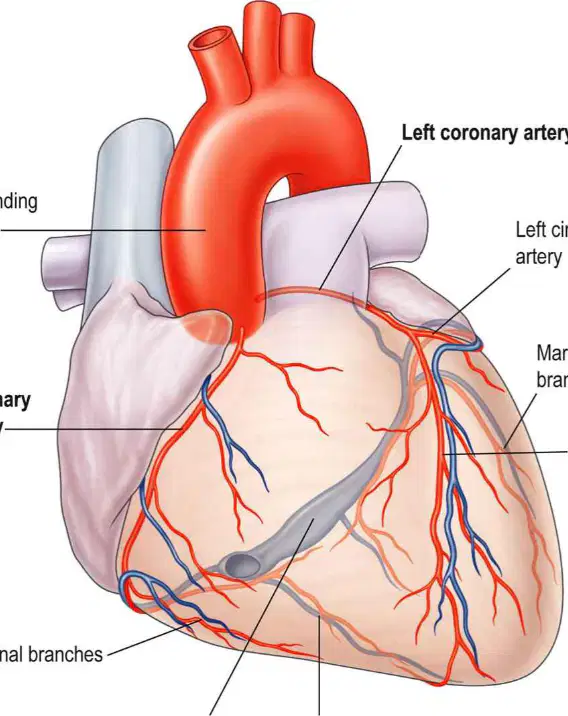
Which part of the heart is primarily supplied by the right coronary artery?
Left ventricle and atrium
Right ventricle, right atrium, SA and AV nodes
Pulmonary arteries
Aortic valve
What percentage of cardiac output is received by coronary circulation?
10%
5%
15%
0.5%
Which part of the heart receives the majority of the blood supply due to its highest workload?
Right atrium
Left ventricle
Right ventricle
SA node
What does "autorhythmicity" of the heart mean?
The heart relies on nerves and hormones to generate impulses.
The heart generates its own impulses independent of nerves and hormones.
The heart requires external stimulation to contract.
The heart's impulses are controlled solely by the brain.
Which type of fibers increase the heart rate under autonomic control?
Parasympathetic fibers
Sympathetic fibers
Neuromuscular fibers
Hormonal fibers
What is the effect of parasympathetic fibers on the heart rate?
Increase heart rate
Decrease heart rate
No effect on heart rate
Stop the heart rate completely
Which hormones influence the heart rate?
Insulin and glucagon
Adrenaline (epinephrine) and thyroxine
Dopamine and serotonin
Cortisol and melatonin
What is the role of the conduction system in the heart?
To pump blood directly into the arteries
To initiate and conduct impulses for coordinated contraction
To regulate blood pressure in the body
To control the oxygen levels in the blood
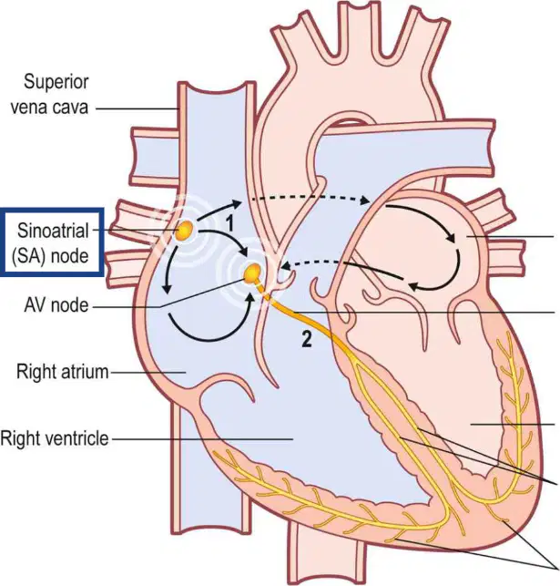
Where is the sinoatrial (SA) node located in the heart?
Wall of the left atrium, near the opening of the inferior vena cava.
Wall of the right atrium, near the opening of the superior vena cava.
Wall of the left ventricle, near the apex of the heart.
Wall of the right ventricle, near the tricuspid valve.
What is the primary function of the sinoatrial (SA) node?
To pump blood into the ventricles.
To generate regular impulses due to electrical instability.
To act as a barrier between the atria and ventricles.
To regulate blood pressure in the arteries.
At what rate does the sinoatrial (SA) node depolarize to initiate each heartbeat?
30–50 times per minute.
60–90 times per minute.
100–120 times per minute.
10–20 times per minute.
Why is the sinoatrial (SA) node referred to as the pacemaker of the heart?
It is the only structure that contracts the heart muscles.
It is the fastest discharging site, setting the heart rate.
It prevents the backflow of blood into the atria.
It regulates the blood flow through the coronary arteries.
What is the sequence of the cardiac cycle initiated by the sinoatrial (SA) node?
Contraction → Depolarisation → Repolarisation → Rapid repeat.
Depolarisation → Contraction → Repolarisation → Rapid repeat.
Repolarisation → Contraction → Depolarisation → Rapid repeat.
Contraction → Repolarisation → Depolarisation → Rapid repeat.
How long does it take for the impulse from the sinoatrial (SA) node to cross the atria and trigger atrial contraction?
~0.5 seconds.
~0.1 seconds.
~1 second.
~2 seconds.

