
 Worksheets
Physio Practice 3
Total questions: 93
Worksheet time: 2hrs 20mins
Which event directly exposes the binding sites on actin filaments during muscle contraction?
ATP binds to the myosin head
Calcium binds to troponin
Myosin detaches from actin
Tropomyosin binds to myosin
What causes the myosin head to detach from the actin filament?
Release of calcium from the sarcoplasmic reticulum
Hydrolysis of ATP
Binding of a new ATP molecule to myosin
Movement of tropomyosin back into place
Which of the following describe roles of ATP in muscle contraction?
(Select all that apply)
binds to myosin to release it from actin after a powerstroke
ATP hydrolysis "cocks" myosin head
directly opens calcium channels in SR
powers calcium pumps that return Ca2+ to SR for relaxation
binds to actin to cause powerstroke
Which statements about troponin and tropomyosin are true?
(Select all that apply)
Troponin is responsible for hydrolyzing ATP during contraction
Tropomyosin blocks actin’s active sites in resting muscle
Tropomyosin directly binds to calcium
Troponin physically moves tropomyosin when bound to calcium
neuromuscular junction (NMJ) is best described as:
site where motor neurons release calcium to trigger contraction
synapse where motor neuron communicates with muscle fiber to start contraction
gap between muscle fibers where sodium enters cell
location where AChE breaks down neurotransmitters
protein chain that holds thick filaments in place
M line
myofibril
transverse (T) tubules
sarcoplasmic reticulum (SR)
Z disc
contractile organelle of muscle fibers
M line
myofibril
transverse (T) tubules
sarcoplasmic reticulum (SR)
Z disc
extension of sarcolemma that brings action potential into cell
M line
myofibril
transverse (T) tubules
sarcoplasmic reticulum (SR)
Z disc
organelle that stores, releases, and takes up calcium ions
myofibril
transverse (T) tubules
sarcoplasmic reticulum (SR)
Z disc
sarcomere
protein structure that binds sarcomeres to each other
myofibril
transverse (T) tubules
sarcoplasmic reticulum (SR)
Z disc
sarcomere
myofibril functional unit
myofibril
transverse (T) tubules
sarcoplasmic reticulum (SR)
Z disc
sarcomere
Which statements accurately describe the role of calsequestrin in muscle cells?
(SELECT TWO)
binds and “hides” Ca²⁺ within the SR to store for contraction
hydrolyzes ATP to provide energy for the myosin power stroke
blocks actin binding sites at rest to prevent contraction
is required so that Ca²⁺ can be transported from the sarcoplasm back into SR
a single motor neuron and all the muscle fibers it innervates
sarcomere
motor unit
neuromuscular junction (NMJ)
myofibril
Muscles that require precise, fine movements, such as the muscles controlling the fingers for writing, typically have motor units with:
large number of muscle fibers per motor neuron
small number of muscle fibers per motor neuron
equal numbers of muscle fibers in all motor units
no relationship between fiber number and precision
Muscles that produce strong, powerful movements, such as the quadriceps during a jump, typically have motor units with:
large number of muscle fibers per motor neuron
small number of muscle fibers per motor neuron
equal numbers of muscle fibers in all motor units
no relationship between fiber number and precision
process by which the weakest motor units are used first, followed by progressively stronger motor units, to increase muscle force during a contraction
recruitment
adaptation
unfused tetanus
fused tetanus
serial contraction
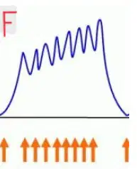
sustained muscle contraction in which the muscle fibers partially relax between stimuli, producing a wavering contraction
recruitment
adaptation
unfused tetanus
fused tetanus
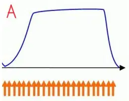
sustained contraction without any relaxation between stimuli, producing a smooth, continuous contraction
recruitment
adaptation
unfused tetanus
fused tetanus
Muscle tone keeps skeletal muscles firm.
How do muscles maintain muscle tone?
small groups of motor units are alternatively active and inactive
all motor units contract simultaneously at a low level
muscles remain fully relaxed until voluntary contraction occurs
calcium is continuously released from the sarcoplasmic reticulum without regulation

muscle contraction in which the muscle changes length while tension remains constant, producing movement
isotonic contraction
isometric contraction
muscle contraction in which the muscle develops tension but does not change length, producing no movement
isotonic contraction
isometric contraction
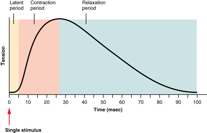
delay between stimulus and muscle contraction as AP moves over sarcolemma, down T-tubules, and Ca2+ is released
Latent Period
Contraction Period
Relaxation Period
Refractory Period
Ca2+binds to troponin exposing myosin binding sites on actin
myosin heads interact with actin and pull towards M-line generating tension and the muscle shortens
Latent Period
Contraction Period
Relaxation Period
Refractory Period
Ca2+ pumped back into SR, myosin binding sites covered by tropomyosin, myosin heads detach from actin
Latent Period
Contraction Period
Relaxation Period
Refractory Period
short time following a muscle fiber’s action potential during which it cannot respond to another stimulus
Latent Period
Contraction Period
Relaxation Period
Refractory Period
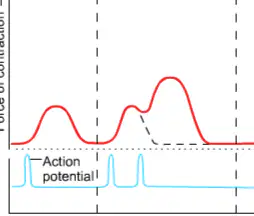
second stimulus arrives after the Refractory period, but before the muscle has completely relaxed from the first
Results in second contraction stronger than first contraction
wave summation
tetanus
recruitment
twitch contraction
process that enables ATP production during low oxygen by converting pyruvate to lactate in muscles; the lactate is sent to the liver, turned into glucose, and returned to the muscles for energy
krebs cycle
lactate (cori) cycle
oxidative phosphorylation
electron transport chain
which two processes describe how lactate is recycled after being produced in muscle cells during anaerobic metabolism?
(select TWO)
transported to liver to go through gluconeogenesis for future ATP production
excreted by the kidneys to maintain acid–base balance
converted back into glucose within muscle cells if oxygen is restored quickly
converted into glycogen within the mitochondria for long-term storage
high-energy molecule in muscle cells that quickly donates a phosphate group to ADP, regenerating ATP during the first few seconds of contraction
glycogen
creatine phosphate
myoglobin
hemoglobin
ATP synthase
oxygen-binding protein in muscle cells that stores and releases oxygen to support aerobic ATP production during contraction
glycogen
creatine phosphate
myoglobin
hemoglobin
ATP synthase
Which events occur during rigor mortis?
(select all that apply)
SR becomes leaky to Ca2+, allowing sustained cross-bridge formation, results in fused tetanus
new ATP molecules are continually produced to maintain muscle tone
ATP reserves are quickly depleted, preventing detachment of myosin from actin
actin and myosin remain linked until muscle proteins begin to decompose
nerve impulses continue stimulating the muscle for several hours after death
pacemaker of the heart, located in the right atrium, spontaneously generates action potentials to set rate and rhythm of cardiac contractions; initiates impulses that spread through the atria causing atrial contraction
Sinoatrial (SA) Node
Atrioventricular (AV) Node
Atrioventricular (AV) Bundle
Right and Left bundle branches
Purkinje Fibers
located between the atria and ventricles; receives impulses from the SA node and provides a critical delay to ensure the atria fully contract and fill the ventricles before ventricular contraction begins.
Is backup pacemaker if SA node fails
Sinoatrial (SA) Node
Atrioventricular (AV) Node
Atrioventricular (AV) Bundle
Right and Left bundle branches
Purkinje Fibers
electrical connection between the atria and ventricles; its smaller-diameter fibers and fewer gap junctions slow conduction so atrial contraction completes before ventricular contraction; transmits impulses from the AV node down the interventricular septum
Sinoatrial (SA) Node
Atrioventricular (AV) Node
Atrioventricular (AV) Bundle
Right and Left bundle branches
Purkinje Fibers
fibers that branch from the AV bundle and carry electrical impulses along the interventricular septum toward the apex of the heart, ensuring coordinated spread of depolarization to both ventricles
Sinoatrial (SA) Node
Atrioventricular (AV) Node
Atrioventricular (AV) Bundle
Right and Left bundle branches
Purkinje Fibers
large-diameter fibers that rapidly conduct impulses throughout the ventricular myocardium from apex to base, producing a coordinated and powerful contraction of the ventricles
Sinoatrial (SA) Node
Atrioventricular (AV) Node
Atrioventricular (AV) Bundle
Right and Left bundle branches
Purkinje Fibers
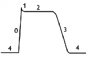
Phases of Cardiac Muscle AP:
Resting MP (-90mV)
equal, steady movement of K+ and Na+ across membrane, some K+ outflow
Phase 4
Phase 0
Phase 1
Phase 2
Phase 3
Phases of Cardiac Muscle AP:
Rapid depolarization, rapid Na+ outflow
Phase 4
Phase 0
Phase 1
Phase 2
Phase 3
Phases of Cardiac Muscle AP:
Slight dip in AP, fast Na+ gates close, K+ open causing dip
some K+ outflow
Phase 4
Phase 0
Phase 1
Phase 2
Phase 3
Phases of Cardiac Muscle AP:
Plateau. Balance between slow voltage-gate Ca2+ channels (inward) and some voltgage-gate K+ channels (outward).
Increased Ca2+ in sarcoplasm triggers contraction, long depolarization
Phase 4
Phase 0
Phase 1
Phase 2
Phase 3
Phases of Cardiac Muscle AP:
Repolarization. Ca2+ channels close. More K+ channels open
large K+ outflow
Phase 4
Phase 0
Phase 1
Phase 2
Phase 3
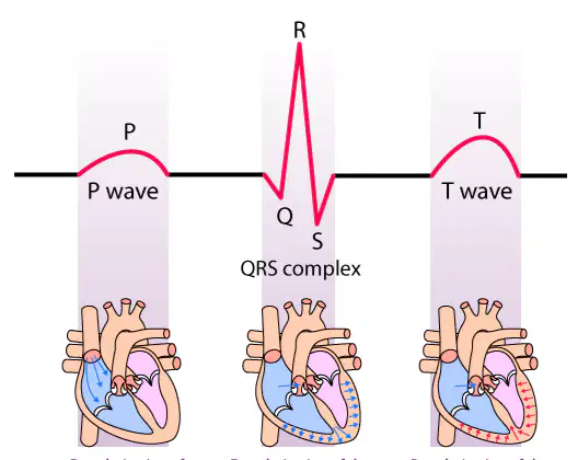
What is represented by P Wave?
depolarization of both Atria
depolarization of both ventricles (repolarization of Atria hidden here)
repolarization of both ventricles
What is represented by QRS complex?
depolarization of both Atria
depolarization of both ventricles (repolarization of Atria hidden here)
repolarization of both ventricles
What is represented by T wave?
depolarization of both Atria
depolarization of both ventricles (repolarization of Atria hidden here)
repolarization of both ventricles
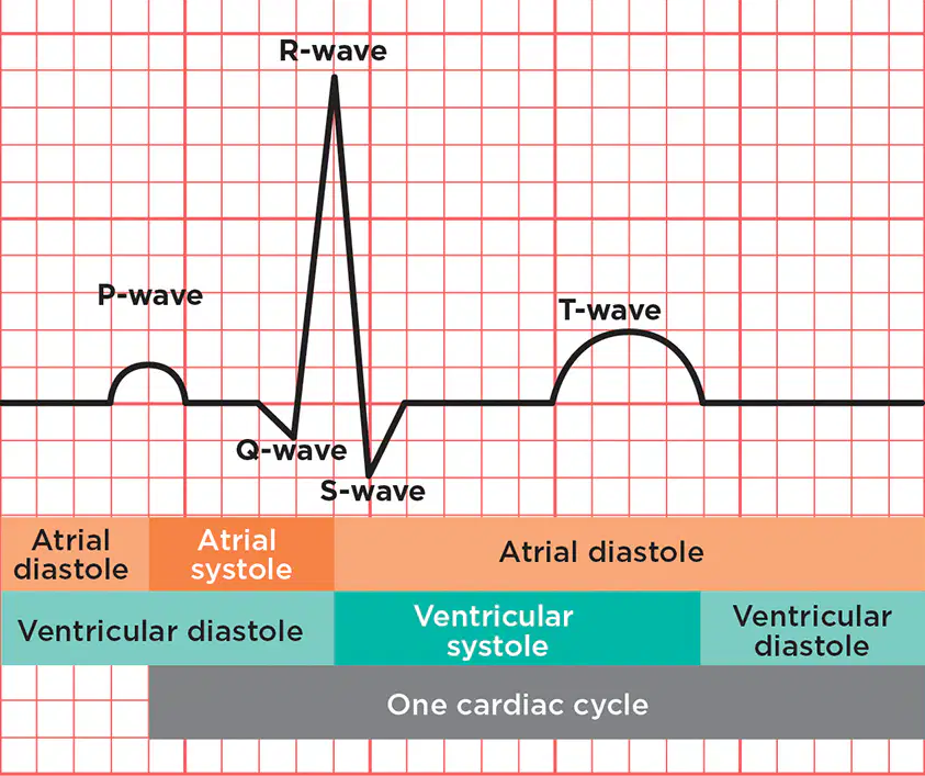
Ventricular Filling
period between the T wave of one heartbead and the P wave of the next
AV valves open, SL valves closed; Atria/Ventricles are in diastole
AV valves open, SL valves closed; Atria in systole, Ventricles in diastole
AV valves closed, SL valves closed; Atria in diastole, Ventricles in systole, ventricular pressure↑, volume no change
AV valves closed, SL valves closed; Atria/Ventricles in diastole; ventricles relax but volume no change
AV valves closed, SL valves open; Atria in diastole, Ventricles in systole; blood is ejected into the arteries
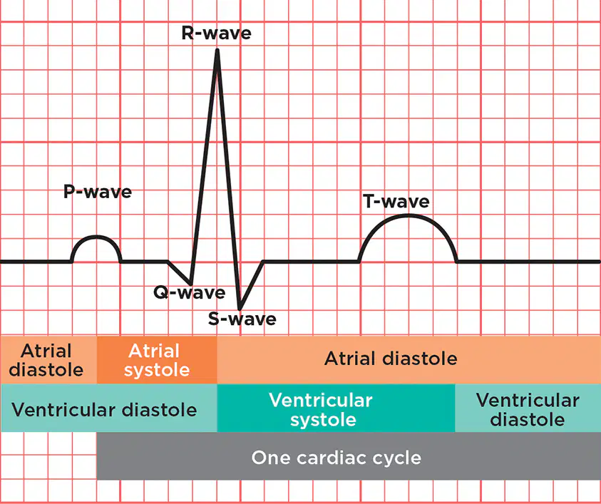
Atrial Contraction
P Wave
AV valves open, SL valves closed; Atria/Ventricles are in diastole
AV valves open, SL valves closed; Atria in systole, Ventricles in diastole
AV valves closed, SL valves closed; Atria in diastole, Ventricles in systole, ventricular pressure↑, volume no change
AV valves closed, SL valves closed; Atria/Ventricles in diastole; ventricles relax but volume no change
AV valves closed, SL valves open; Atria in diastole, Ventricles in systole; blood is ejected into the arteries
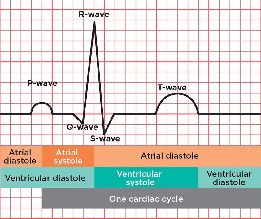
Isovolumetric Contraction
start of the QRS complex
AV valves open, SL valves closed; Atria/Ventricles are in diastole
AV valves open, SL valves closed; Atria in systole, Ventricles in diastole
AV valves closed, SL valves closed; Atria in diastole, Ventricles in systole, ventricular pressure↑, volume no change
AV valves closed, SL valves closed; Atria/Ventricles in diastole; ventricles relax but volume no change
AV valves closed, SL valves open; Atria in diastole, Ventricles in systole; blood is ejected into the arteries
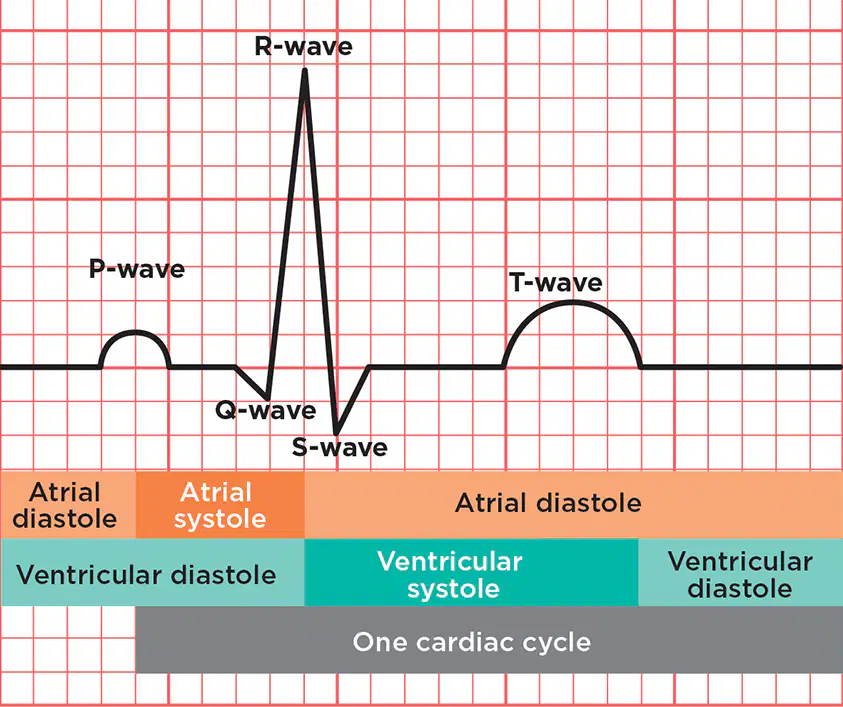
Isovolumetric Relaxation
T wave
AV valves open, SL valves closed; Atria/Ventricles are in diastole
AV valves open, SL valves closed; Atria in systole, Ventricles in diastole
AV valves closed, SL valves closed; Atria in diastole, Ventricles in systole, ventricular pressure↑, volume no change
AV valves closed, SL valves closed; Atria/Ventricles in diastole; ventricles relax but volume no change
AV valves closed, SL valves open; Atria in diastole, Ventricles in systole; blood is ejected into the arteries
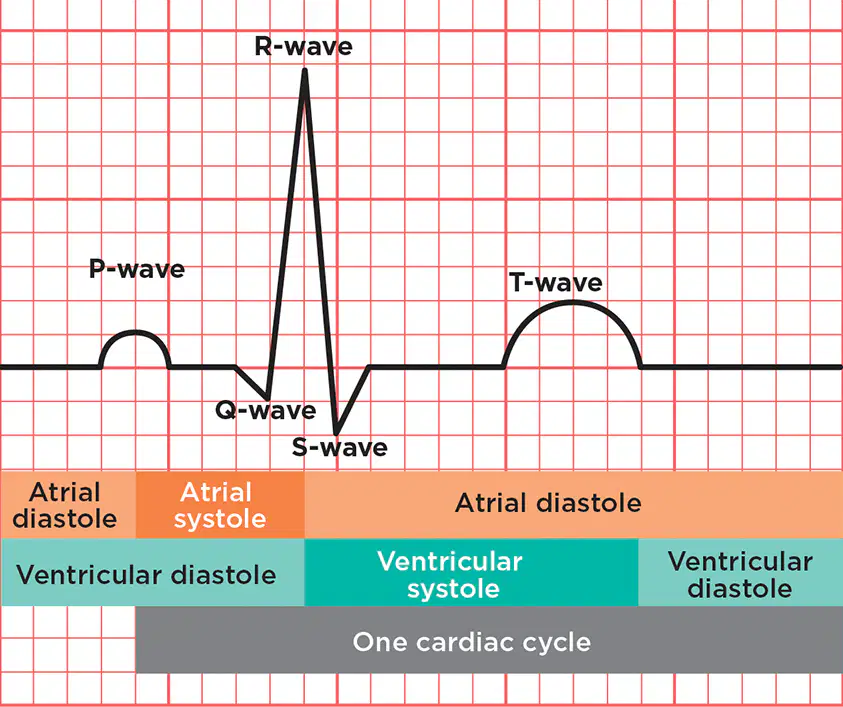
Ventricular Ejection
end of the QRS complex
AV valves open, SL valves closed; Atria/Ventricles are in diastole
AV valves open, SL valves closed; Atria in systole, Ventricles in diastole
AV valves closed, SL valves closed; Atria in diastole, Ventricles in systole, ventricular pressure↑, volume no change
AV valves closed, SL valves closed; Atria/Ventricles in diastole; ventricles relax but volume no change
AV valves closed, SL valves open; Atria in diastole, Ventricles in systole; blood is ejected into the arteries
During a cardiac cycle, a patient’s end-diastolic volume (EDV) is measured at 135 mL, and the end-systolic volume (ESV) is 65 mL.
What is the stroke volume (SV) of the patient’s heart?
60mL
70mL
75mL
-70mL
A person has a stroke volume (SV) of 70 mL and a heart rate (HR) of 75 beats per minute.
What is this person’s cardiac output (CO)?
4.25 L/min
5.25 L/min
5.75 L/min
6.25 L/min
A person has an end-diastolic volume (EDV) of 140 mL and an end-systolic volume (ESV) of 70 mL.
Their cardiac output (CO) is measured at 4.9 L/min.
What is this person’s heart rate (HR)?
60 bpm
70 bpm
80 bpm
90 bpm
usually harmless; felt as heart 'skipping a beat'
premature atrial contractions
atrial fibrillation
bradycardia
atrial tachycardia
ventricle tachycardia
problem with autorhythmic fibers overwhelming SA node
premature atrial contractions
atrial fibrillation
bradycardia
atrial tachycardia
ventricle tachycardia
heartbeat under 60 bpm; caused by SA node misfiring
premature atrial contractions
atrial fibrillation
bradycardia
atrial tachycardia
ventricle tachycardia
heartbeat above 100 bpm; felt as palpitations
atrial tachycardia
ventricle tachycardia
ventricular fibrillation
premature ventricular contractions
premature atrial contractions
life-threatening, rapid heartbeat arising in inferior chambers
atrial tachycardia
ventricle tachycardia
ventricular fibrillation
premature ventricular contractions
premature atrial contractions
life-threatening, ventricles stop pumping blood chambers quiver and beat ineffectively
requires the heart to be reset by an electric shock.
atrial tachycardia
ventricle tachycardia
ventricular fibrillation
premature ventricular contractions
premature atrial contractions
often called heart palpitations
caused by high BP, low blood oxygen, or heart attack
atrial tachycardia
ventricle tachycardia
ventricular fibrillation
premature ventricular contractions
premature atrial contractions
What factors regulate stroke volume and help ensure that the left and right ventricles pump equal volumes of blood?
(select all that apply)
preload — degree of stretch of ventricular muscle fibers at end of diastole
afterload — pressure that must be overcome before semilunar valves open
contractility — forcefulness of ventricular contraction at a given preload
refractory period — time during which muscle cell cannot respond to another stimulus
increase strength of contraction, often by promoting Ca2+ inflow; increases stroke volume & cardiac output.
examples include norepinephrine, epinephrine, digitalis
positive inotropic agent
negative inotropic agent
decrease strength of contraction by lowering intracellular Ca2+ levels or blocking β-adrenergic receptors;
reduces stroke volume & cardiac output.
Examples include: anaesthetics, beta blockers
positive inotropic agent
negative inotropic agent
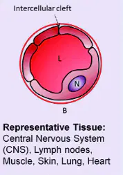
Least permeable, exchange of gases, small solutes,
only gaps are intercellular clefts
cell membrane of endothelial cells forms a tube
Continuous Capillaries
Fenestrated Capillaries
Sinusoid Capillaries
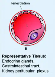
Moderately permeable, allows passage of larger molecules,
rapid exchange of fluids/larger solutes
has fenestrations covered by thin diaphragm
Continuous Capillaries
Fenestrated Capillaries
Sinusoid Capillaries
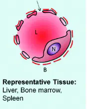
Highly permeable, allows passage of large proteins/cells,
free exchange between blood and tissue
large fenestrations w/out diaphragm
Gaps between endothelial cells, discontinuous lining
Continuous Capillaries
Fenestrated Capillaries
Sinusoid Capillaries
most important method of capillary exchange; substances move down concentration gradient (o₂ and nutrients from blood to tissues, co₂ and wastes from tissues to blood);
occurs through intercellular clefts, fenestrations, or directly through endothelial cells
Diffusion
Transcytosis
Bulk Flow
involves substances enclosed in vesicles that enter endothelial cells; important mainly for large, lipid-insoluble molecules such as albumin and insulin that cannot cross capillary walls by other means
Diffusion
Transcytosis
Bulk Flow
passive process where large numbers of ions, molecules, or particles move together in the same direction due to pressure gradients; main mechanism regulating blood and interstitial fluid volumes, including filtration (out of capillaries) and reabsorption (into capillaries)
Diffusion
Transcytosis
Bulk Flow
pressure inside the capillaries and pushes fluid out of the capillary into the interstitial fluid; it is generated by the pumping action of the heart and promotes filtration.
Blood Hydrostatic Pressure
(BHP)
Interstitial Fluid Osmotic Pressure
(IFOP)
Blood Colloid Osmotic Pressure
(BCOP)
Interstitial Fluid Hydrostatic Pressure
(IFHP)
pressure inside the capillaries and pulls fluid into the capillary from the interstitial space;is caused by the presence of plasma proteins that are too large to leave the capillary, and promotes reabsorption
Blood Hydrostatic Pressure
(BHP)
Interstitial Fluid Osmotic Pressure
(IFOP)
Blood Colloid Osmotic Pressure
(BCOP)
Interstitial Fluid Hydrostatic Pressure
(IFHP)
pressure is in the interstitial fluid and pulls fluid out of the capillary toward solutes in the interstitial space; it is created by proteins and other solutes in the interstitial fluid and promotes filtration
Blood Hydrostatic Pressure
(BHP)
Interstitial Fluid Osmotic Pressure
(IFOP)
Blood Colloid Osmotic Pressure
(BCOP)
Interstitial Fluid Hydrostatic Pressure
(IFHP)
pressure of the interstitial fluid that pushes fluid away; located outside capillaries but is ZERO and tends to promote reabsorption
Blood Hydrostatic Pressure
(BHP)
Interstitial Fluid Osmotic Pressure
(IFOP)
Blood Colloid Osmotic Pressure
(BCOP)
Interstitial Fluid Hydrostatic Pressure
(IFHP)
Net filtration pressure (NFP) represents the balance between pressures that promote filtration and those that promote reabsorption across capillary walls. Which of the following correctly expresses this relationship?
NFP = (BHP + BCOP) – (IFOP + IFHP)
NFP = (BHP + IFOP) – (BCOP + IFHP)
NFP = (BCOP + IFHP) – (BHP + IFOP)
NFP = (IFHP + IFOP) – (BHP + BCOP)
As blood moves from the arterial end to the venous end of a capillary, net filtration pressure (NFP) changes. Which of the following statements correctly describes this change?
arterial end, NFP is negative and fluid moves into the capillary; venous end, NFP is positive and fluid leaves the capillary.
arterial end, NFP is positive and fluid leaves the capillary; venous end, NFP is negative and fluid moves into the capillary.
NFP is constant along the length of capillary, always promoting filtration
At both ends, NFP is negative, and all fluid is reabsorbed into the capillary
During Bulk Flow not all fluid filtered out of capillaries is reabsorbed. Which statement correctly describes what happens to the excess fluid?
About 15% of filtered fluid enters lymphatic capillaries (~3 L/day) and is eventually returned to the blood
All filtered fluid is immediately reabsorbed at the venous end of capillaries
Excess filtered fluid is excreted directly into urine by the kidneys.
None of the filtered fluid returns to the circulation
At the arterial end of a capillary:
BHP = 35 mmHg
IFOP = 2 mmHg
BCOP = 25 mmHg
IFHP = 0 mmHg
What is the net filtration pressure (NFP)?
9 mmHg
10 mmHg
11 mmHg
12 mmHg
At the venous end of a capillary:
BHP = 23 mmHg
IFOP = -2 mmHg
BCOP = 39 mmHg
IFHP = 0 mmHg
What is the net filtration pressure (NFP)?
-17 mmHg
-18 mmHg
-19 mmHg
-20 mmHg
The value that reflects how well blood flows through the body and major organs is:
cardiac output
(CO)
stroke volume
(SV)
mean arterial pressure
(MAP)
pulse pressure
The value calculated as the difference between systolic and diastolic pressure, representing the force the heart generates each time it contracts, is:
cardiac output
(CO)
stroke volume
(SV)
mean arterial pressure
(MAP)
pulse pressure
A patient’s blood pressure is measured at 120/90 mmHg. What is their mean arterial pressure (MAP)?
(Hint: MAP = diastolic pressure + ⅓(systolic − diastolic))
95 mmHg
100 mmHg
105 mmHg
110 mmHg
A patient’s blood pressure is measured at 120/90 mmHg. What is their pulse pressure?
210 mmHg
30 mmHg
100 mmHg
60 mmHg
The pressure in the arteries when the ventricles are relaxed and filling with blood is called:
systolic pressure
diastolic pressure
The pressure in the arteries during ventricular contraction, representing the peak force of blood against the artery walls, is called:
systolic pressure
diastolic pressure
What factors affect the distribution of Cardiac Output.
(how the total amount of blood pumped by the heart (CO) is divided among different organs and tissues)
(select all that apply)
Blood Pressure
Vascular Resistance
Venous Return
Velocity of Blood Flow
What does vascular resistance refer to, and what factors determine it?
ease of blood flow through vessels; depends on HR and blood volume
opposition to blood flow caused by friction between blood and vessel walls; depends on lumen size, blood viscosity, and vessel length
amount blood ejected per heartbeat; depends on SV and arterial pressure
pressure difference between arteries and veins; depends on vessel elasticity and venous return
What is venous return and what primarily drives it?
flow of blood from heart to the arteries; driven by left atrial contraction
volume of blood flowing back to heart through systemic veins; driven by pressure generated when left ventricle contracts
rate blood flow through pulmonary circuit; driven by right ventricular contraction
pressure difference between arteries and arterioles; driven by arterial recoil
Which condition would most likely reduce venous return?
decreased pressure in right atrium
dilation of systemic veins
increased pressure in the right atrium due to leaky tricuspid valve
increased contraction strength of left ventricle
What is velocity of blood flow, and what determines it?
rate which blood moves through a vessel; decreases as cross-sectional area of vessels increases
total volume of blood pumped per minute; determined by stroke volume and heart rate
resistance blood encounters in vessels; determined by viscosity and vessel length
pressure difference between arteries and veins; increases with vessel diameter
What does circulation time refer to?
time it takes for a red blood cell to move from artery to vein within a single tissue
time required for one heartbeat
time it takes for blood to move from the heart, through the body, and back to the heart
time between ventricular systole and diastole
How are velocity of blood flow and circulation time related to cardiac output?
higher cardiac output increases blood flow velocity and decreases circulation time
higher cardiac output decreases blood flow velocity and increases circulation time
cardiac output does not affect either blood flow velocity or circulation time
both increase as total vessel cross-sectional area increases
In response to sympathetic stimulation, these hormones are released by the adrenal medulla to raise CO by increasing rate and force of heart contractions
Epinephrine and Norepinephrine
Antidiuretic Hormone (ADH) or Vasopressin
Atrial Natriuretic
Peptide (ANP) and Brain-type Natriuretic
Peptide (BNP)
Produced by hypothalamus and released by posterior pituitary, these hormones increase BP by retaining water
Epinephrine and Norepinephrine
Antidiuretic Hormone (ADH) or Vasopressin
Atrial Natriuretic
Peptide (ANP) and Brain-type Natriuretic
Peptide (BNP)
These hormones lower blood pressure by causing vasodilation and promoting loss of salt and water in urine
Epinephrine and Norepinephrine
Antidiuretic Hormone (ADH) or Vasopressin
Atrial Natriuretic
Peptide (ANP) and Brain-type Natriuretic
Peptide (BNP)

