
Font size
 Worksheets
Histology Full Unit Review
Total questions: 86
Worksheet time: 57mins

Identify this type of ET.
stratified columnar ciliated
pseudostratified columnar ciliated
simple columnar ciliated
pseudostratified transitional ciliated

Identify these cells.
columnar cells
brush border
transitional cells
goblet cells
dome cells

Identify this type of ET.
stratified cuboidal
simple transitional
simple squamous
simple cuboidal

Identify the supportive structure at the yellow arrows.
(a)

The yellow arrows are pointing to the (a) surface of this sample.

Identify the space marked as X.
(a)

Identify this type of ET.
simple transitional
stratified transitional
simple squamous
simple cuboidal

Identify this type of ET.
stratified cuboidal
stratified squamous non keratinized
stratified squamous keratinized
stratified transitional

Identify the apical cells of this ET type.
(a)

Identify the ET shown at the center of the image.
stratified transitional
simple cubodial
stratified cuboidal
stratified squamous

Identify the type of ET shown here.
stratified transitional
stratified squamous
stratified squamous keratinized
simple squamous
stratified cuboidal

Identify the type of ET shown here.
stratified transitional
stratified squamous
stratified squamous keratinized
simple squamous
stratified cuboidal
CHOOSE ALL THAT APPLY:
Which types of ET may have cilia?
stratified columnar
simple columnar
stratified cuboidal
stratified squamous
pseudostratified columnar
Label the diagram provided. Zoom in for clarity.
connective tissue
apical surface
basal surface
epithelial tissue
basement MB
Which type of intercellular adhesion protein provides a strong, physical connection between neighboring cells to hold the tissue together against wear and tear?
cilia
desmosome
microvilli
tight junction
Which type of intercellular adhesion protein prevents any leakage between adjacent epithelial cells?
cilia
desmosome
microvilli
tight junction
Transitional cells....
shape change from cuboidal at the basal surface to squamous at the apical surface
change shape from cuboidal when relaxed to squamous when stretched
Epithelial tissue lacks blood vessels. The term for this is:
(a)
Epithelial cells that are thin and flat are called
(a)
Match the following vocabulary to the correct description.
the "filling" between the fibers and the cells
ground substance
all materials found outside of the cells
extracellular matrix
the strongest/largest/most abundant fiber
collagen fiber
this fiber is able to stretch and recoil
elastic fiber
this fiber forms a net-like scaffolding arrangement
reticular fiber
Match the following cells to their role in connective tissue.
fibroblasts
produce the extracellular matrix of CT
adipocytes
fat cells; energy storage
plasma cells
make protein antibodies
mast cells
release histamine for local inflammation
macrophages
engulf foreign substances/invaders
All types of mature connective tissue originate (arise) from embryonic connective tissue known as :
(a)

What type of tissue is this? .
bone
blood
Elastic Cartilage
Fibrocartilage

What type of tissue is this? Look carefully.
dene regular CT
elastic CT
Elastic Cartilage
Fibrocartilage

What type of tissue is this? Look carefully.
dene regular CT
elastic CT
Elastic Cartilage
Fibrocartilage

reticular CT
Hyaline Cartilage
Elastic Cartilage
Fibrocartilage

dense regular CT
elastic CT
Fibrocartilage
Elastic Cartilage

What type of tissue is this?
hyaline cartilage
reticular CT
elastic CT
elastic cartilage

Adipose Connective Tissue
Blood
Bone
Simple Cuboidal Epithelium
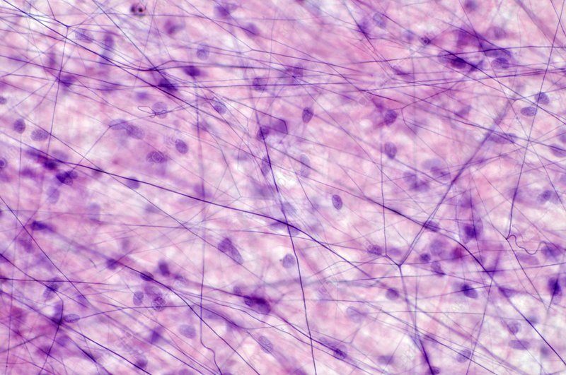
What type of tissue is this?
areolar CT
dense irregular CT
Elastic CT
hyaline cartilage
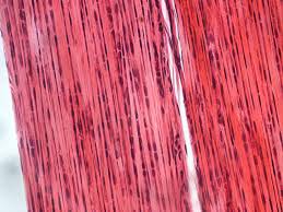
What type of tissue is this?
Dense regular CT
Elastic CT
Dense irregular CT
Hyaline cartilage

reticular CT
dene irregular CT
dense regular CT
Areolar CT

elastic cartilage
fibrocartilage
hyaline cartilage

Hyaline cartilage
reticular Ct

What type of tissue is this?
reticular CT

What type of tissue is this?
reticular CT
elastic cartilage
hyaline cartilage
fibrocartilage
What type of tissue is this?
reticular CT
elastic cartilage
hyaline cartilage
fibrocartilage

What type of tissue is this?
fibrocartilage
reticular CT
adipose CT
areolar CT

What type of tissue is this?
Hyaline cartilage
Elastic cartilage

What type of tissue is this?
Dense Irregular CT
Dense Regular CT
Elastic CT
Areolar CT

What tissue is this?
Hyaline cartilage
What type of tissue is this?
reticular CT
Hyaline cartilage
reticular Ct
elastic cartilage
fibrocartilage
hyaline cartilage
reticular CT
dene irregular CT
dense regular CT
Areolar CT
What type of tissue is this?
Dense regular CT
Elastic CT
Dense irregular CT
Hyaline cartilage
What type of tissue is this?
areolar CT
dense irregular CT
Elastic CT
hyaline cartilage
Adipose Connective Tissue
Blood
Bone
Simple Cuboidal Epithelium

What type of tissue is this?
Blood
dense regular CT
reticular CT
dense irregular CT
What type of tissue is this?
hyaline cartilage
reticular CT
elastic CT
elastic cartilage
dense regular CT
elastic CT
Fibrocartilage
Elastic Cartilage
reticular CT
Hyaline Cartilage
Elastic Cartilage
Fibrocartilage
What type of tissue is this? Look carefully.
dene regular CT
elastic CT
Elastic Cartilage
Fibrocartilage
What type of tissue is this? Look carefully.
dene regular CT
elastic CT
Elastic Cartilage
Fibrocartilage
What type of tissue is this? .
bone
blood
Elastic Cartilage
Fibrocartilage

What type of tissue is this? .
reticular CT
blood
Elastic Cartilage
Fibrocartilage

What type of tissue is this? .
dense regular CT
dense irregular CT
fibrocartilage
elastic CT

What type of tissue is this? .
dense regular CT
dense irregular CT
fibrocartilage
elastic CT

The lining of a joint that secretes a slippery fluid into the joint space.
synovial membrane
serous membrane
mucous membrane
cutaneous membrane
What is the function of the synovial membrane?
protects any passageway open to the surface from invasion
protects body from infection, chemicals and physical wear/tear
reduces friction around organs that move constantly (heart, lungs, GI tract)
produces fluid which lubricates the joint, absorbs shock and nourishes cartilage.
Which one of the following places in the body would you expect to find mucous membranes?
covering the heart
covering the external surface of the body
respiratory tract
around your knee bones

Which body membrane has 2 layers?
synovial
serous
cutaneous
mucous
Which body membrane will you find surrounding the entire body?
Serous
Mucous
Synovial
Cutaneous
In which type of membrane would a goblet cell be found?
serous membrane
synovial membrane
cutaneous membrane
mucous membrane
Muscle tissue that is multinucleate.
Smooth muscle tissue
Skeletal muscle tissue
Cardiac muscle tissue
Muscle tissue composed of branching cells with intercalated discs.
Smooth muscle tissue
Skeletal muscle tissue
Cardiac muscle tissue
Muscle tissue that moves food through the digestive system.
Smooth muscle tissue
Skeletal muscle tissue
Cardiac muscle tissue
Muscle tissue that is NOT striated.
Smooth muscle tissue
Skeletal muscle tissue
Cardiac muscle tissue
Muscle tissue cells that are tapered at both ends.
Smooth muscle tissue
Skeletal muscle tissue
Cardiac muscle tissue
Which type of muscle tissue is involuntary?
Smooth only
Cardiac only
Smooth and skeletal
Cardiac and smooth

Skeletal MT
cardiac MT
smooth MT

Name the tissue:
Skeletal muscle tissue
Cardiac muscle tissue
Smooth muscle tissue
Nervous tissue

Name the tissue:
Skeletal muscle tissue
Cardiac muscle tissue
Smooth muscle tissue
nervous tissue

Name the tissue:
Skeletal muscle tissue
Cardiac muscle tissue
Smooth muscle tissue
nervous tissue

Name the tissue:
Skeletal muscle tissue
Cardiac muscle tissue
Smooth muscle tissue
nervous tissue

What type of muscle tissue is this?
Cardiac
Smooth
Skeletal
bone tissue

Where would this type of muscle be found?
Heart
Internal Organs
Attached to the Skeleton

What type of tissue is this?
Muscle
Epithelial
Connective
Nervous

Name the type of tissue shown in this image.
Answer should be two words: _____ _____
(a)
Match the tissue to its function.
nervous tissue
sends electrical messages through body
connective tissue
supports and connects other tissues
epithelial tissue
forms coverings, linings and glands
muscle tissue
create movement through contraction
membrane
ET+CT forming flexible, protective cover
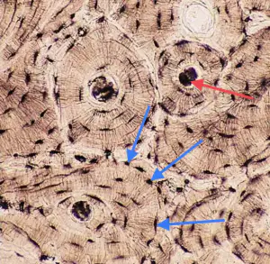
What cells are located at the tips of the blue arrows? (Use plural form)
(a)
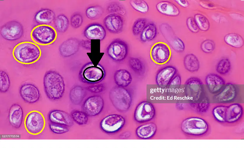
Identify the cell located at the tip of the black arrow (circled in black).
(use singular form)
(a)
Identify the SPACES circled in yellow.
(use plural form)
(a)
Label this image of CT.
ground substance
red blood cells
nuclei
collagen fiber
white blood cells
platelets
Label the image of CT.
ground substance
lacuna
osteocyte
collagen fibers
elastic fibers
reticular fibers
chondrocyte
Study the following images carefully and then match the images to their name.

bone CT

hyaline cartilage CT

elastic cartilage CT

fibrocartilage CT

blood CT

