
Font size
 Worksheets
The Meninges
Total questions: 132
Worksheet time: 1hrs 13mins
Which meningeal layer is described as thin, innermost, and bound tightly to the brain surface?
Dura mater
Arachnoid mater
Pia mater
Periosteum
Which statement best characterizes the arachnoid mater based on the description provided?
Outermost tough membrane in contact with cranial bones
Middle membrane that contains the subarachnoid space
Innermost membrane tightly adherent to brain gyri and sulci
Highly vascular membrane forming the venous sinuses
Identify the correct pairing of meningeal layer and position relative to the skull.
Dura mater – innermost next to brain
Arachnoid mater – outermost under the scalp
Pia mater – middle between dura and brain
Dura mater – outermost in contact with cranial bones
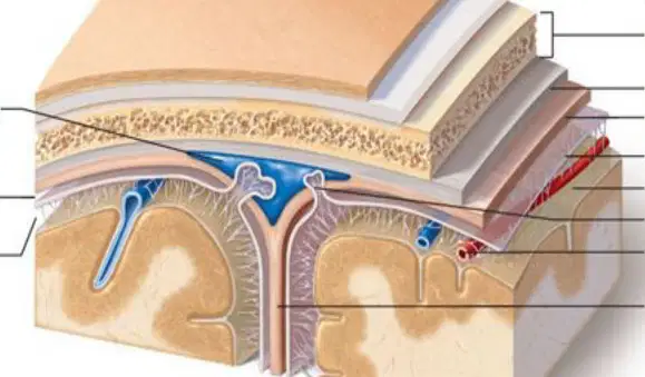
According to the labeled cross-sectional diagram of the cranial wall and meninges, which space lies between the arachnoid mater and pia mater?
Subdural space
Epidural space
Subarachnoid space
Superior sagittal sinus
Which structure is the site where cerebrospinal fluid (CSF) diffuses into venous blood?
Subarachnoid space
Arachnoid granulations
Superior sagittal sinus
Falx cerebri
Which statement about the subarachnoid space is accurate?
It contains cerebrospinal fluid (CSF)
It houses venous blood returning to the heart
It is a potential space between skull and dura mater
It is formed by the periosteal layer of the dura
The superior sagittal sinus primarily functions to:
Produce CSF from choroid plexuses
Receive venous blood from the brain
Provide arterial supply to meninges
Anchor the pia mater to the cortex
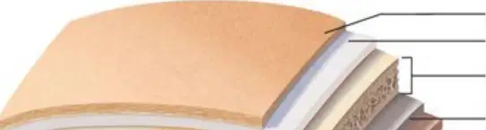
Which sequence correctly orders the layers encountered moving from the scalp toward the brain, as indicated in the diagrams?
Scalp → bone of skull → dura mater → arachnoid mater → pia mater → brain
Scalp → dura mater → bone of skull → arachnoid mater → pia mater → brain
Scalp → arachnoid mater → bone of skull → dura mater → pia mater → brain
Scalp → bone of skull → pia mater → arachnoid mater → dura mater → brain
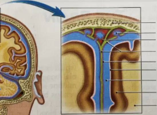
In the detailed view of Figure 15.1, which labeled structure corresponds to the meningeal layer directly contacting brain tissue?
Dura mater
Arachnoid mater
Pia mater
White matter
Which pairing correctly matches a component with its role within the meninges?
Arachnoid granulations – produce CSF
Superior sagittal sinus – receives venous blood
Subdural space – contains CSF
Pia mater – outermost protective layer
Which description best distinguishes dura mater from the other meningeal layers?
It is vascular and loosely attached to the brain
It is the tough outermost layer in contact with cranial bones
It forms the subarachnoid space
It is a delicate membrane tightly bound to brain tissue
A blockage of arachnoid granulations would most directly impair which process?
CSF production within ventricles
Venous blood flow from scalp veins
CSF diffusion into venous blood
Arterial perfusion of dura mater
If the subarachnoid space were reduced in volume, which immediate consequence would be most expected?
Decreased CSF surrounding the brain and spinal cord
Accumulation of arterial blood within the dura mater
Separation of the periosteal dura from the skull
Increased production of CSF by arachnoid granulations

Based on Figure 15.1, which labeled region contains CSF and lies deep to the arachnoid mater?
Subdural space
Subarachnoid space
Superior sagittal sinus
Gray matter
Which structure is correctly identified as part of the venous drainage pathway associated with the meninges?
Pia mater
Subarachnoid space
Superior sagittal sinus
Periosteum
Which structure produces cerebrospinal fluid (CSF) within the ventricles of the brain?
Arachnoid villi
Choroid plexus
Cerebral aqueduct
Dura mater
Identify the location of the third ventricle based on the instructional text: it lies primarily within which brain region?
Cerebellum
Diencephalon
Pons
Medulla oblongata

According to the lateral brain diagram, which channel connects the third ventricle to the fourth ventricle for CSF passage?
Interventricular foramen
Cerebral aqueduct
Median aperture
Lateral aperture
How many lateral ventricles are present in the brain, and where are they located?
One, in the midline
Two, one in each cerebral hemisphere
Three, surrounding the diencephalon
Four, distributed across brainstem regions
Which statement best describes the anatomical surroundings of the fourth ventricle?
Embedded within the cerebral hemispheres
Situated in the diencephalon
Surrounded by the pons, medulla, and cerebellum
Located within the central canal of the spinal cord

Place the initial steps of CSF circulation in order, starting from secretion. Which option lists the correct first three steps?
Secretion by arachnoid villi → flow through cerebral aqueduct → addition by choroid plexus in fourth ventricle
Secretion by choroid plexus in lateral ventricles → flow through interventricular foramina → third ventricle adds more CSF
Secretion by choroid plexus in third ventricle → flow through median aperture → entry to lateral ventricles
Secretion by dura mater → flow through lateral apertures → entry to subarachnoid space

Which openings allow CSF to exit the fourth ventricle into the subarachnoid space?
Interventricular foramina
Cerebral aqueduct only
Two lateral apertures and one median aperture
Sagittal sinus and central canal
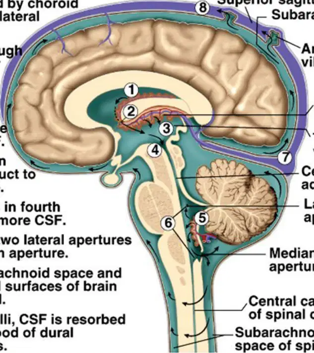
Where is CSF ultimately resorbed into the venous circulation according to the flow diagram?
Choroid plexus
Arachnoid villi into dural venous sinuses
Central canal of the spinal cord
Cerebral aqueduct

Which structure directly precedes the fourth ventricle in the CSF pathway?
Interventricular foramen
Median aperture
Cerebral aqueduct
Arachnoid granulations
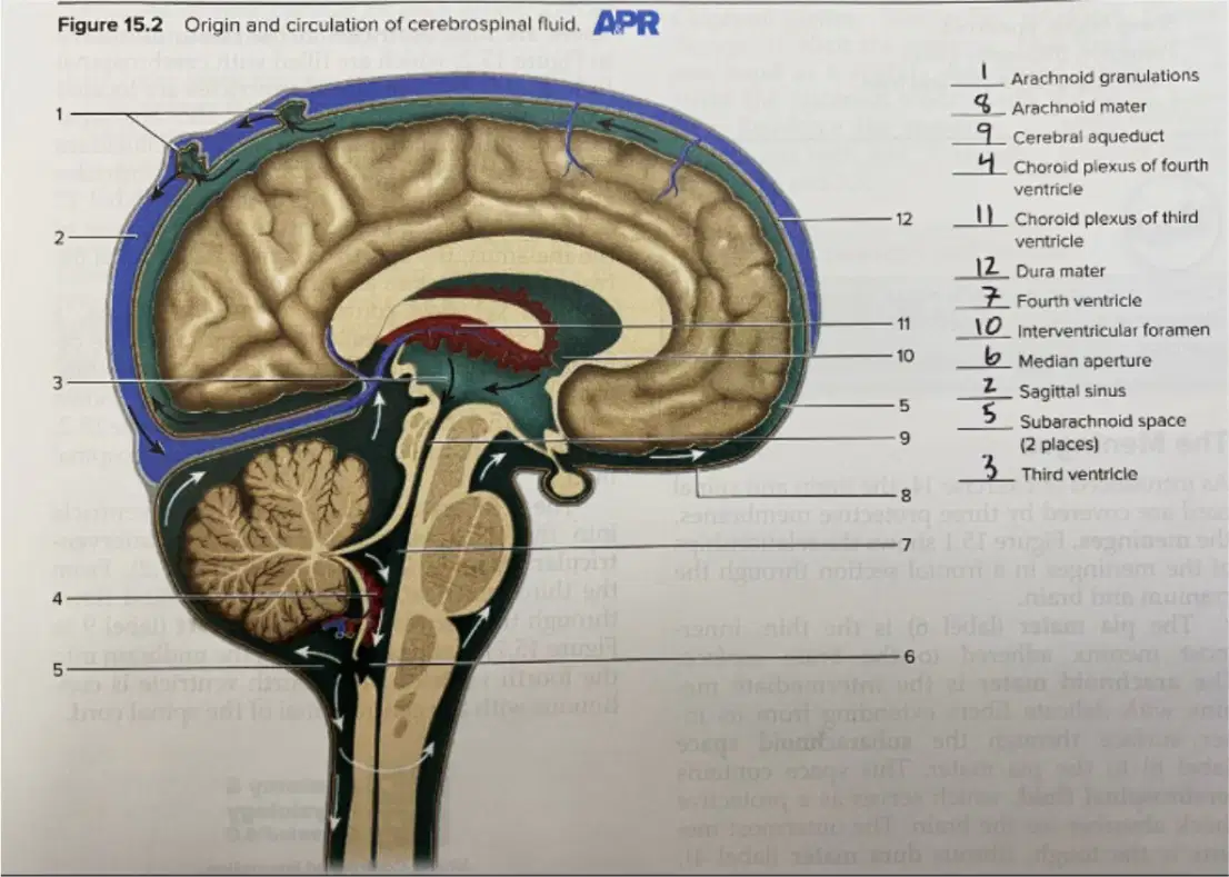
According to Figure 15.2, which labeled feature is the space into which CSF circulates around the brain and spinal cord?
Dura mater
Subarachnoid space
Sagittal sinus
Interventricular foramen

In Figure 15.2, which labeled structure is responsible for CSF production within the third ventricle?
Arachnoid granulations
Choroid plexus of third ventricle
Median aperture
Cerebral aqueduct
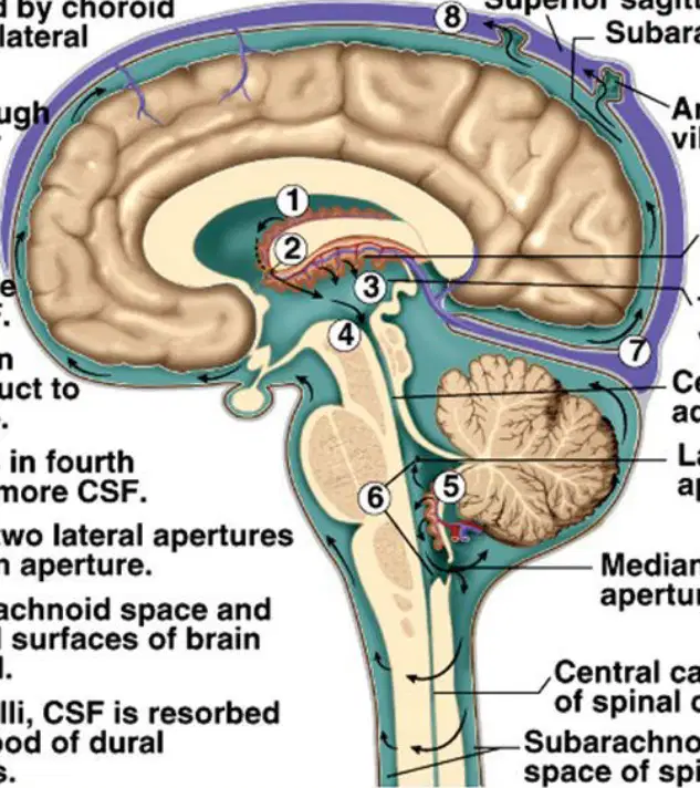
Which route correctly describes how CSF can reach the spinal cord according to the diagrams?
Lateral ventricles → sagittal sinus → central canal of spinal cord
Third ventricle → lateral apertures → cerebral aqueduct → central canal
Fourth ventricle → median aperture → subarachnoid space → central canal of spinal cord
Interventricular foramen → dura mater → arachnoid villi → spinal canal
Which major brain region primarily integrates and interprets incoming sensory impulses as sensations and initiates conscious motor responses?
Cerebellum
Brainstem
Cerebrum
Diencephalon
In the cerebrum, what structure separates the right and left cerebral hemispheres?
Central sulcus
Longitudinal fissure
Lateral fissure
Corpus callosum
Which pairing correctly matches a sulcus or fissure with the lobes it separates?
Central sulcus separates temporal from frontal lobes
Lateral fissure separates frontal from parietal lobes
Central sulcus separates frontal and parietal lobes
Longitudinal fissure separates frontal and temporal lobes
What is the function of the cerebellum as introduced in the section?
Primary relay for sensory information and autonomic control
Enables muscular coordination, balance, and posture
Relays information between the body and the cerebrum
Initiates conscious thought processes and memory
Which statement about gyri and sulci in the cerebrum is accurate?
Gyri are grooves between sulci; sulci are folds that increase surface area
Gyri are folds that increase surface area; sulci are grooves between gyri
Gyri and sulci both refer to lobes of the cerebral cortex
Sulci form the primary motor cortex; gyri form the sensory cortex
The central sulcus lies between which two cortical gyri?
Precentral gyrus and postcentral gyrus
Superior temporal gyrus and inferior frontal gyrus
Cingulate gyrus and parahippocampal gyrus
Angular gyrus and supramarginal gyrus
What is the relationship between cerebral hemispheres and control of body sides?
Each hemisphere controls the same side of the body
Right hemisphere controls the right side; left controls the left side
Right hemisphere controls the left side; left controls the right side
Control alternates depending on task complexity
Which brain region is described as having numerous functions and relaying information between the body and the cerebrum and cerebellum?
Diencephalon
Brainstem
Cerebrum
Cerebellum
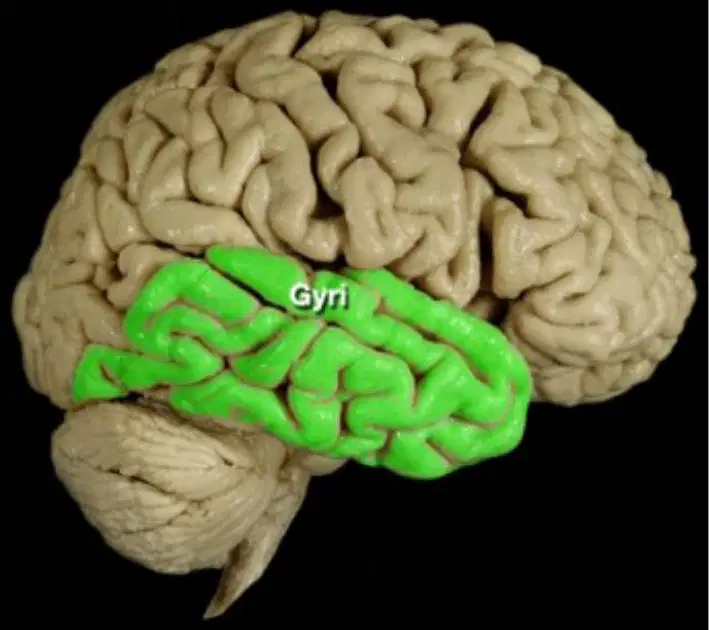
Refer to the left lateral brain diagram highlighting green regions labeled “Gyri.” Which statement best defines gyri as shown?
Raised folds of the cerebral cortex
Deep grooves separating lobes
Membranous coverings of the brain
Bundles of white matter tracts
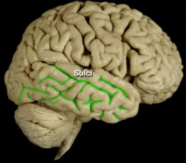
Using the right lateral brain diagram highlighting green lines labeled “Sulci,” what are sulci?
Raised ridges on the cortical surface
Shallow and deep grooves between gyri
Openings leading into ventricles
Thick bands of myelinated fibers
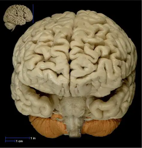
Which feature is indicated by the midline separation in the superior view image where the two cerebral hemispheres are divided?
Lateral fissure
Central sulcus
Longitudinal fissure
Transverse sinus
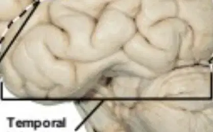
In the labeled cerebrum diagram, which lobe lies inferior to the lateral fissure and anterior to the occipital lobe?
Frontal lobe
Temporal lobe
Parietal lobe
Insular cortex
Which pairing correctly matches surface feature to its morphology based on the diagrams?
Gyrus — groove; Sulcus — fold
Gyrus — fold; Sulcus — groove
Gyrus — fissure; Sulcus — ridge
Gyrus — ventricle; Sulcus — gyrus
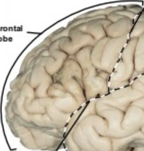
According to the lobe boundaries in the cerebrum diagram, which structure demarcates the frontal and parietal lobes on the lateral surface?
Central sulcus
Longitudinal fissure
Lateral fissure
Transverse fissure
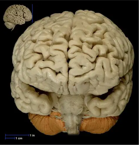
A lesion confined along a deep midline cleft visible in the superior view would most directly affect communication between hemispheres. Which cleft is involved?
Lateral fissure
Longitudinal fissure
Calcarine sulcus
Precentral sulcus
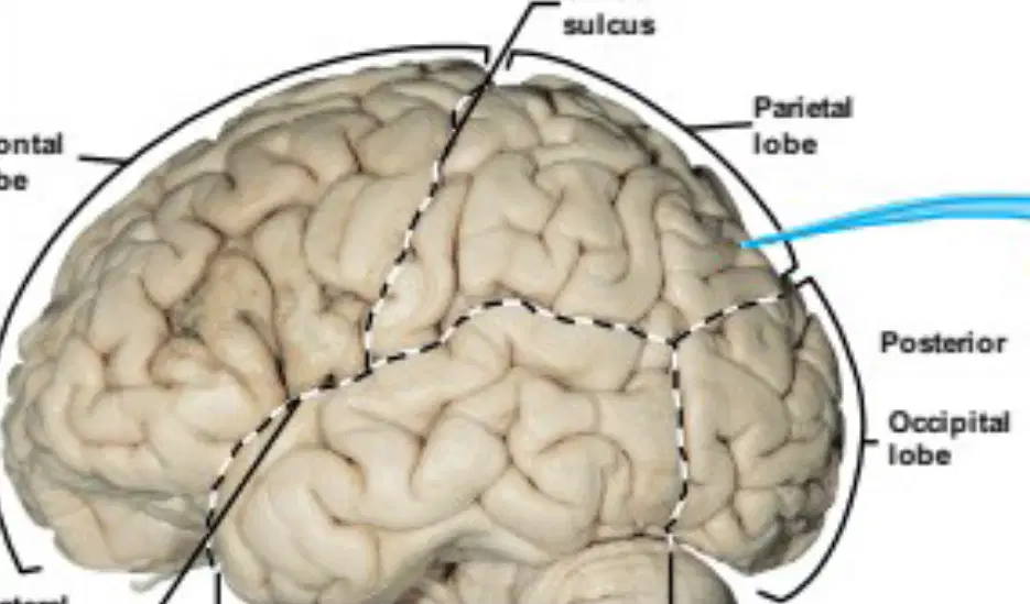
In the labeled cerebrum, which lobe is most posterior and inferior on the lateral surface?
Frontal lobe
Parietal lobe
Temporal lobe
Occipital lobe
Consider cortical folding shown in the gyri/sulci images. Which functional inference best explains the presence of many gyri and sulci on the cerebrum’s surface?
They reduce blood flow to minimize heat production
They increase surface area allowing more cortical neurons
They prevent hemispheric communication by forming barriers
They serve primarily to anchor meninges to skull bones

In the lateral view diagram of the human brain, which landmark separates the frontal lobe from the parietal lobe?
Lateral sulcus
Central sulcus
Longitudinal fissure
Parieto-occipital sulcus
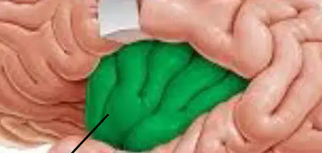
In the insula illustration where the lateral fissure is retracted, which buried cortical region becomes visible?
Cingulate gyrus
Insula
Hippocampus
Calcarine cortex
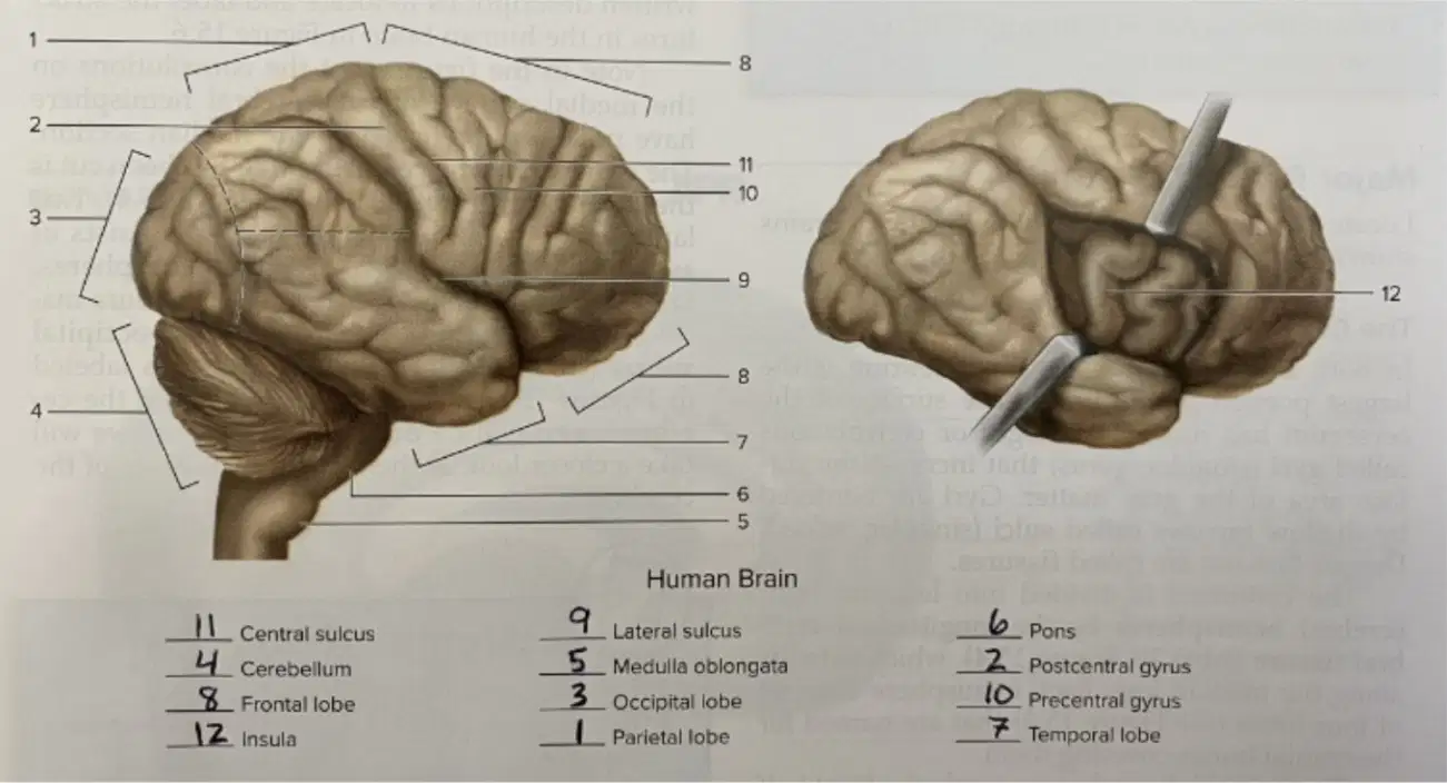
Which groove marks the boundary between the frontal and temporal lobes in the labeled brain diagram?
Central sulcus
Lateral sulcus
Parieto-occipital sulcus
Precentral sulcus

According to the labeled figure, which gyrus lies immediately anterior to the central sulcus?
Postcentral gyrus
Precentral gyrus
Superior temporal gyrus
Cingulate gyrus
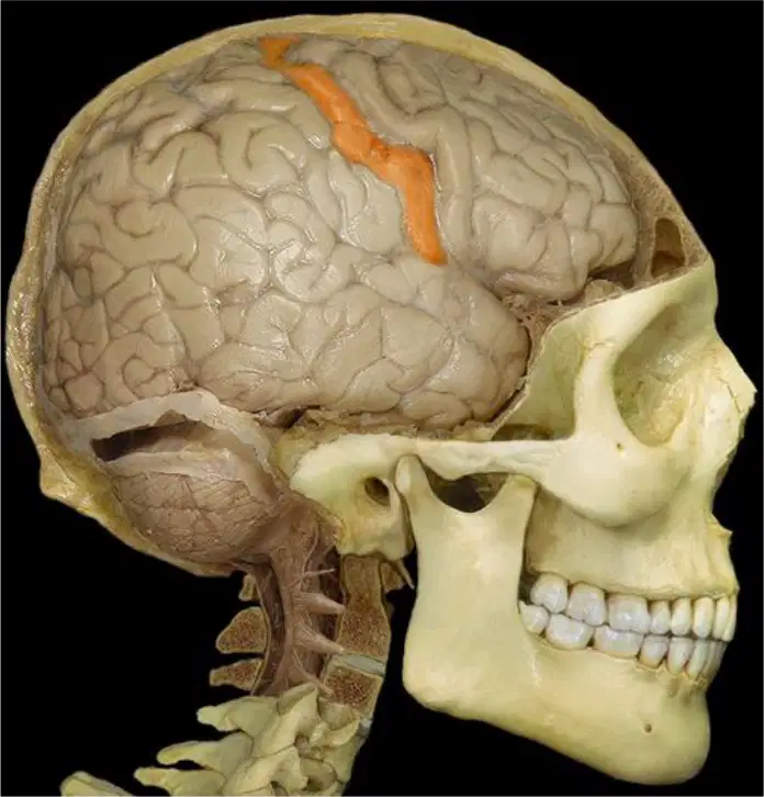
In the skull-brain overlay images highlighting orange cortical ridges, the ridge posterior to the central sulcus corresponds to which structure?
Precentral gyrus
Postcentral gyrus
Superior frontal gyrus
Angular gyrus
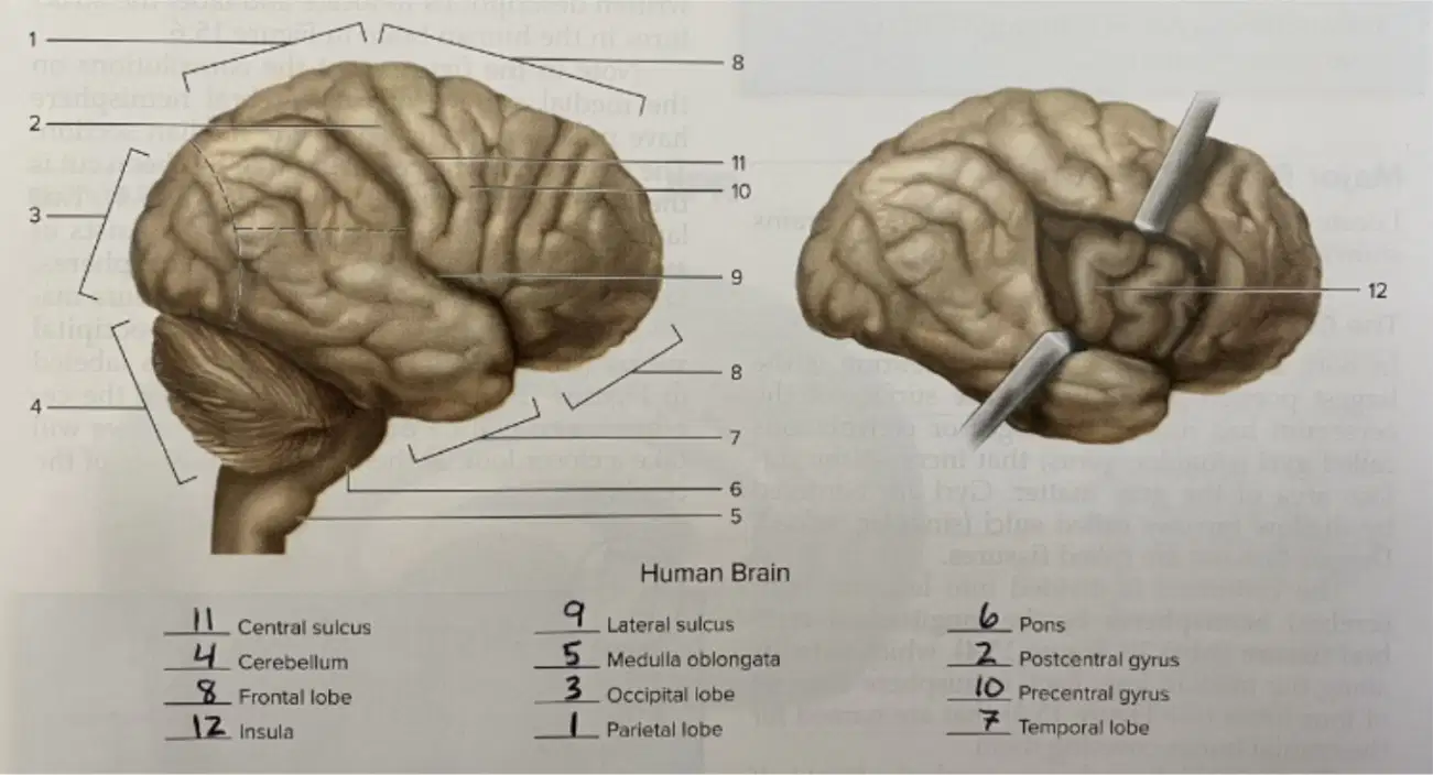
From the labeled lateral brain figure, which lobe occupies the most posterior portion of the cerebrum?
Frontal lobe
Temporal lobe
Parietal lobe
Occipital lobe
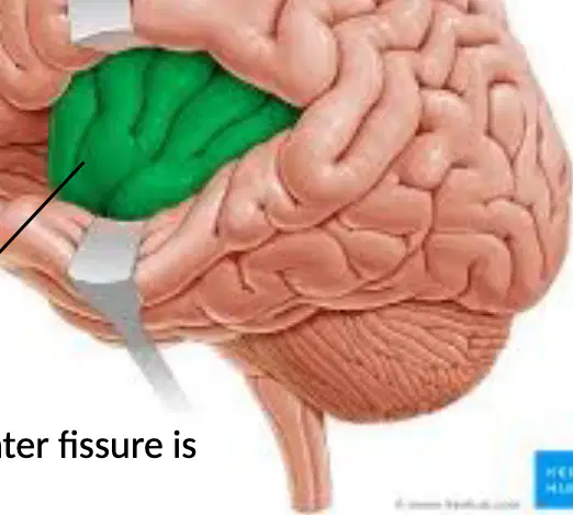
When identifying the insula, which surrounding sulcus must be opened or retracted to expose it in the illustration?
Central sulcus
Lateral sulcus
Calcarine sulcus
Parieto-occipital sulcus
Which statement best matches the spatial relationship shown between the precentral and postcentral gyri across the central sulcus?
They are continuous ridges within the frontal lobe.
They lie on opposite sides of the central sulcus, with the precentral anterior and postcentral posterior.
Both are posterior to the lateral sulcus within the temporal lobe.
They are hidden within the insula and only visible when the lateral sulcus is opened.

In the labeled cerebrum figure, which feature is indicated deep within the lateral sulcus rather than on the outer surface?
Cerebellum
Insula
Pons
Medulla oblongata
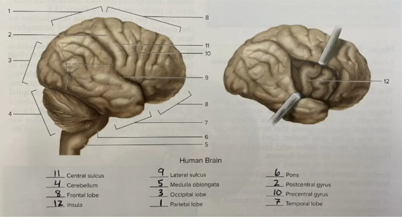
Using the labeled diagram, which groove runs roughly horizontally on the lateral surface and separates the frontal and parietal regions from the temporal lobe?
Central sulcus
Lateral sulcus
Interhemispheric fissure
Transverse fissure
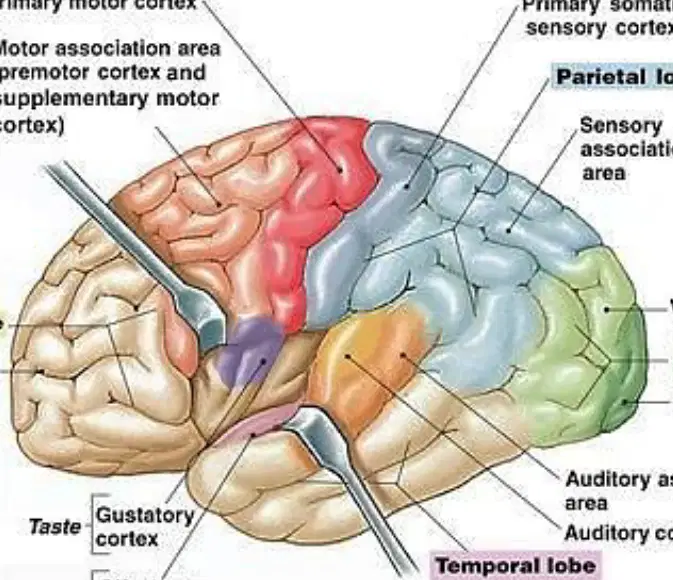
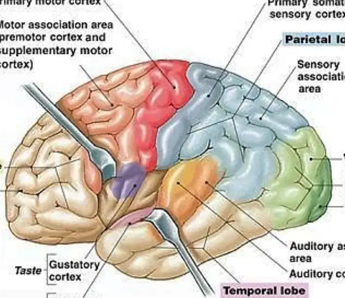
According to the labeled brain diagram, which lobe primarily contains the primary motor cortex responsible for initiating voluntary skeletal muscle movement?
Frontal lobe
Parietal lobe
Temporal lobe
Occipital lobe
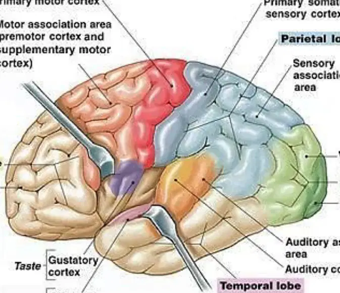
Which cortex receives sensory information from skin, musculoskeletal system, viscera, and taste buds and lies in the parietal lobe?
Primary somatic sensory cortex
Primary motor cortex
Visual cortex
Auditory cortex
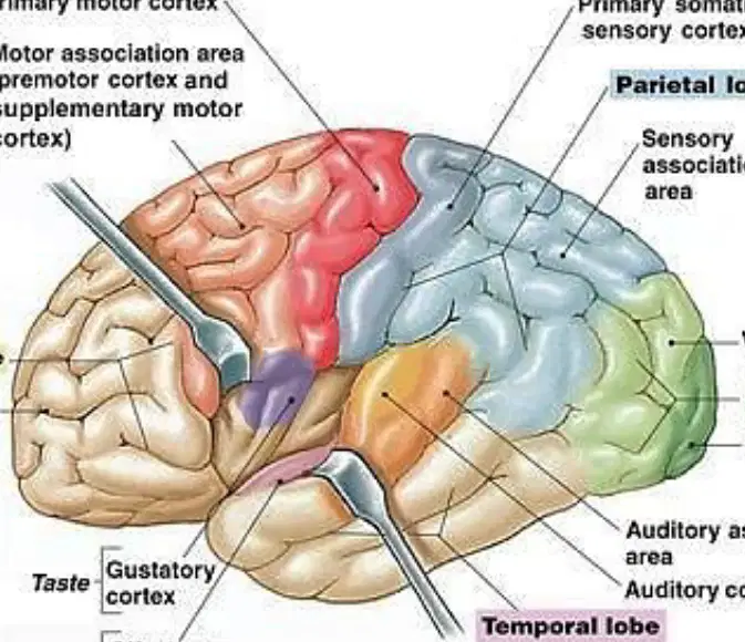
Which statement best characterizes the occipital lobe based on the figure?
It houses the visual cortex and visual association area for processing vision.
It coordinates reasoning skills and behavior through the prefrontal association area.
It integrates auditory information in the primary auditory cortex.
It controls gustation and smell via the gustatory and olfactory cortices.
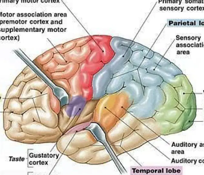
In the diagram, the prefrontal association area is noted to coordinate information from other association areas and control some behaviors. Which lobe is this area located in?
Frontal lobe
Parietal lobe
Temporal lobe
Occipital lobe
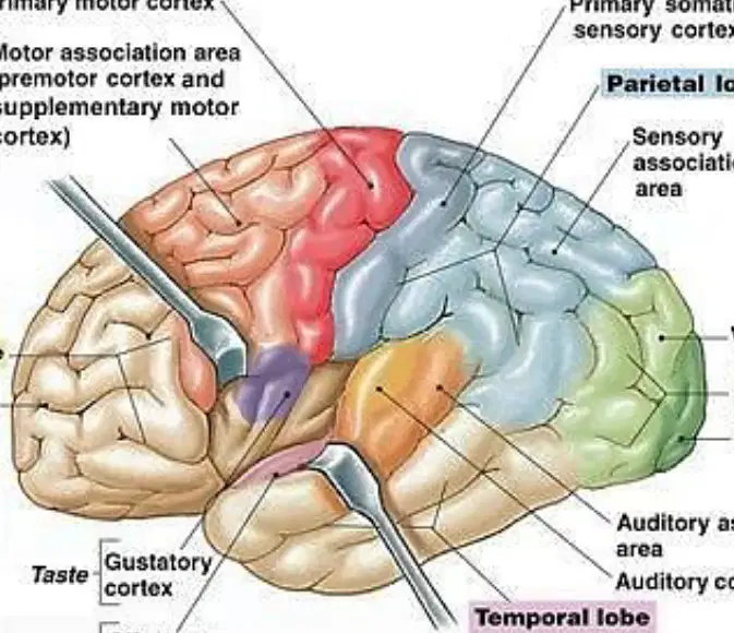
Which pairing correctly matches a lobe with a primary sensory cortex shown in the figure?
Temporal lobe — primary auditory cortex
Parietal lobe — primary visual cortex
Occipital lobe — primary gustatory cortex
Frontal lobe — primary somatic sensory cortex
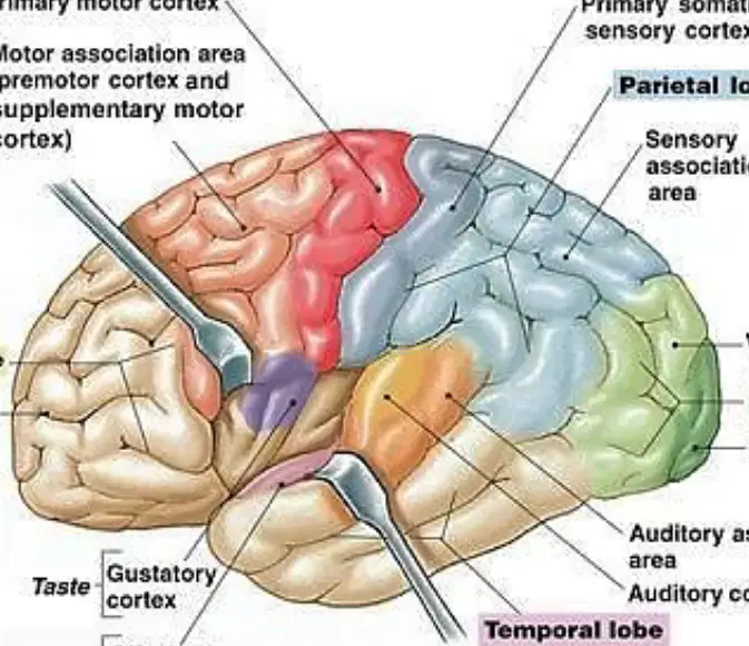
The motor association area (premotor and supplementary motor cortex) is shown adjacent to the primary motor cortex. What is its principal role?
Planning and coordinating complex voluntary movements
Detecting touch and proprioception from the body
Interpreting visual stimuli from the retina
Processing auditory signals from the cochlea
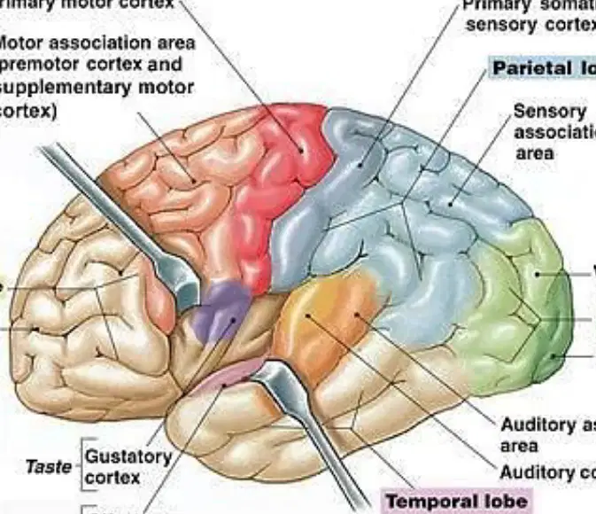
Which cortical area is linked with taste (gustation) in the provided illustration?
Primary gustatory cortex
Primary visual cortex
Primary motor cortex
Primary somatic sensory cortex
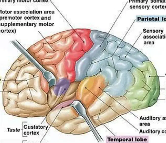
Identify the area that handles smell (olfaction) as indicated in the figure.
Olfactory cortex
Auditory association area
Somatosensory association area
Visual association area
Which region directly processes auditory input and is located in the temporal lobe?
Primary auditory cortex
Motor speech area
Somatosensory association area
Common integrative area
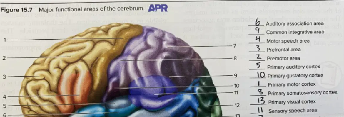
Which numbered label in Figure 15.7 corresponds to the primary visual cortex?
Label 12
Label 10
Label 8
Label 5
In Figure 15.7, which area is identified as the somatosensory association area involved in interpreting touch and proprioceptive information?
Label 13
Label 11
Label 7
Label 3
Based on the functional map, which sequence best traces the flow for visual information within the occipital lobe?
Primary visual cortex to visual association area
Visual association area to primary motor cortex
Primary somatic sensory cortex to visual association area
Auditory cortex to primary visual cortex
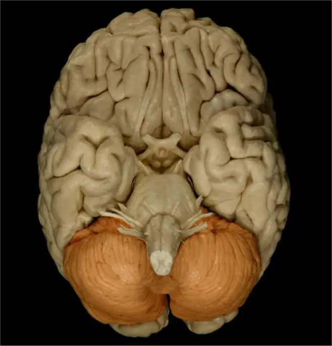
Refer to the brain images showing the cerebellum shaded in orange. Which statement best identifies the cerebellum’s anatomical position in relation to the cerebrum in these views?
Anterior and superior to the cerebrum
Posterior and inferior to the cerebrum
Deep within the lateral ventricles of the cerebrum
Directly continuous with the corpus callosum
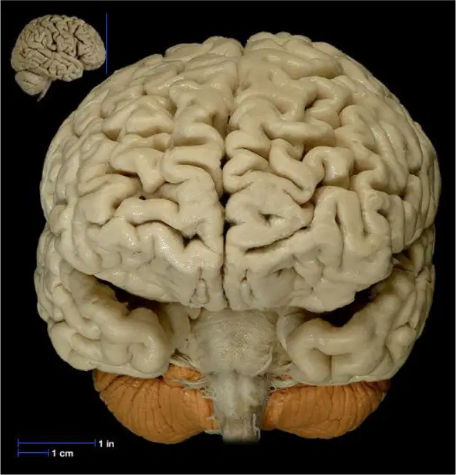
In the diagrams where the cerebellum is highlighted in orange, which brain region is shown superior to the cerebellum and comprises the wrinkled outer tissue?
Cerebrum
Medulla oblongata
Spinal cord
Olfactory bulbs
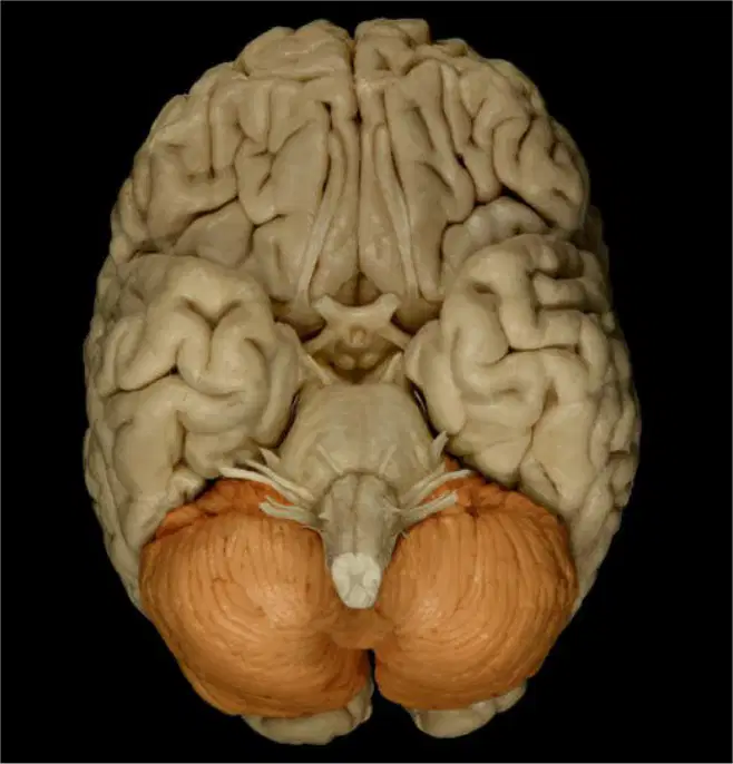
A lesion is depicted that affects the orange-highlighted structure in the images. Based on its location, which functional deficit is most consistent with damage to this region?
Loss of conscious sensation across the body
Impaired coordination and balance during movement
Complete loss of language comprehension
Failure of autonomic breathing control
According to the labeled medial view diagram of internal brain anatomy, which structure is explicitly identified as part of the diencephalon?
Pons
Thalamus
Cerebellum
Medulla oblongata
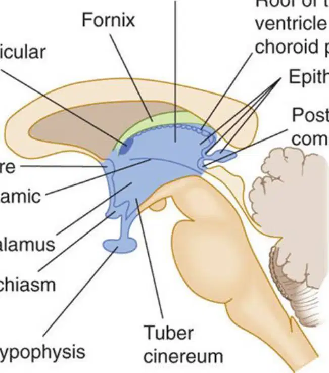
Which statement best defines the diencephalon as presented: it is a region of the brain that derives from the prosencephalon and is located near which area?
The lateral surface of the brain
The midline of the brain
The posterior-most spinal cord
The ventral horn of the cerebrum
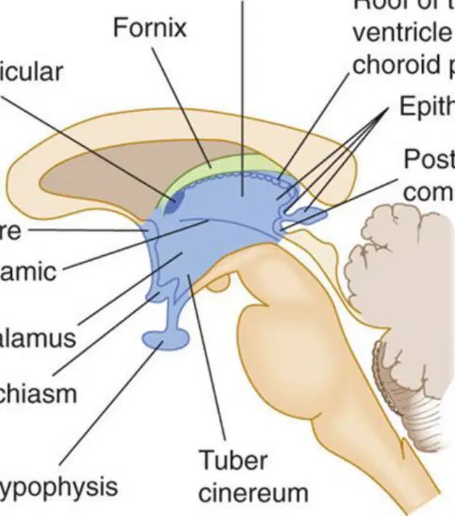
Which list correctly identifies components contained within the diencephalon as enumerated in the material?
Fornix, Thalamus, Hypothalamus, Third ventricle
Corpus callosum, Midbrain, Pons, Optic nerve
Cerebellar cortex, Medulla, Basal nuclei, Pituitary stalk
Frontal lobe, Parietal lobe, Occipital lobe, Temporal lobe
In the internal brain anatomy diagram, which label marks the large commissural fiber bundle superior to the diencephalon?
Optic chiasm
Corpus callosum
Fornix
Posterior commissure

Which structure shown adjacent to the hypothalamus in the schematic is involved in crossing of visual pathways?
Anterior commissure
Optic chiasm
Interventricular foramen
Tuber cinereum
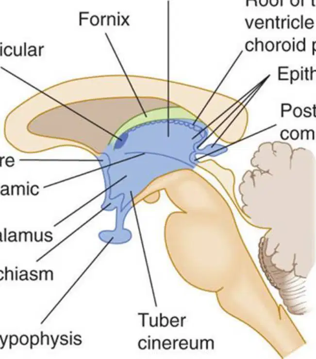
Which functional role is most accurately associated with the thalamus as a component of the diencephalon?
Primary center for motor reflex arcs of the spinal cord
Relay and integration hub for sensory information en route to the cerebrum
Main regulator of cerebrospinal fluid formation in the fourth ventricle
Principal site of voluntary motor neuron cell bodies

Which description aligns with the hypothalamus within the diencephalon based on its anatomical context and typical functions?
Cortical region responsible for high-level language processing
Autonomic and endocrine regulation center including homeostatic control
Segment of the cerebellar peduncles coordinating fine motor output
Primary tract carrying olfactory signals to the limbic system
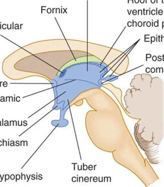
Identify the ventricular space specifically listed as part of the diencephalon.
Lateral ventricle
Fourth ventricle
Third ventricle
Cerebral aqueduct
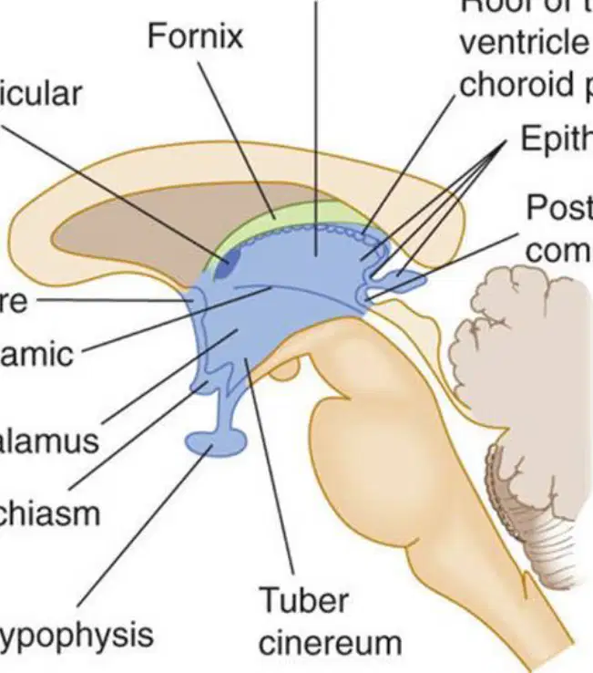
Which structure in the schematic arches near the thalamus and is part of limbic circuitry, and is enumerated among diencephalic contents?
Fornix
Pons
Medulla oblongata
Cerebral aqueduct
Considering spatial orientation in the medial view diagram, which pairing correctly matches region and orientation?
Cerebrum — anterior only
Cerebellum — anterior to the cerebrum
Brainstem — inferior to the cerebrum
Corpus callosum — inferior to the cerebellum

Which cerebral lobe is this?
frontal
parietal
occipital
temporal

Which lobe of the brain is responsible for processing visual information from the eyes and is also responsible for processing auditory information?
Occipital Lobe
Frontal Lobe
Parietal Lobe
Temporal Lobe

What information does the temporal lobe process?
motor
touch
visual
auditory

What part of the brain is shown in Purple?
Frontal Lobe
Temporal Lobe
Parietal Lobe
Occipital Lobe
What information does the occipital lobe process?
motor
touch
visual
auditory

What is the green part of the brain called
Frontal
Temporal
Occipital
Pariental
What is the pink part of the brain called
Frontal
Temporal
Occipital
Pariental
What is the yellow part of the brain called
Temporal
Frontal
Occipital
Parietal
What is the blue part of the brain called
Parietal
Occipital
Frontal
Temporal
Facial
Trigeminal
Glossopharngeal
Hypoglossal
Bell's Palsy causing weakness or paralysis of facial muscles would be associated with which cranial nerve?

Which of the following cranial nerve controls the movement of the eyeball, the opening of the eyelid, pupillary dilation, and lens accommodation?
Optic
Oculomotor
Vagus
Trigeminal
Which of the following does not effect the eyes
Optic
Trochlear
Vagus
Abducens

optic nerve
oculomotor nerve
trochlear nerve

accessory nerve
vestibulocochlear nerve
vagus nerve

Oculomotor
Optic
Olfactory
Trochlear

Abducens
Accessory
Facial
Trochlear

Vestibulocochlear
Vagus
Abducens
Glossopharyngeal

Hypoglossal
Abducens
Trochlear
Trigeminal

Abducens
Vagus
Accessory
Facial
Oculomotor
Optic
Olfactory
Trochlear

Olfactory
Oculomotor
Trigeminal
Optic

Oculomotor
Optic
Trochlear
Trigeminal

Trochlear
Abducens
Trigeminal
Facial
Hypoglossal
Abducens
Trochlear
Trigeminal
Abducens
Accessory
Facial
Trochlear
Abducens
Vagus
Accessory
Facial
Vestibulocochlear
Vagus
Abducens
Glossopharyngeal

Hypoglossal
Glossopharyngeal
Vagus
Facial

Accessory
Abducens
Vestibulocochlear
Vagus

Hypoglossal
Accessory
Abducens
Vagus

Accessory
Hypoglossal
Vagus
Facial
Hearing and balance
Oculomotor
Accessory
Vestibuolocochlear
Hypoglossal
Smell
Optic
Glossopharyngeal
Hypoglossal
Olfactory
Vision
Oculomotor
Optic
Trochlear
Trigeminal
Eye movement
Eyelid opening
Pupillary Constriction
Visual focus
Optic
Trochlear
Oculomotor
Abducens
Eye movement
Facial
Trochlear
Accessory
Optic
Facial sensation
Tongue sensation
Open/close jaw
Trochlear
Facial
Vagus
Trigeminal
Eye movement
Abducens
Trigeminal
Hypoglossal
Vestibulocochlear
Facial Expression
Crying
Taste
Salivation
Facial
Trigeminal
Abducens
Accessory
Tongue sensation
Taste
Swallowing
Trigeminal
Glossopharyngeal
Vagus
Hypoglossal
Taste
Swallowing
Talking
Coughing
Glossopharyngeal
Facial
Vagus
Trigeminal
Shoulder shrugging
Head turning
Accessory
Hypoglossal
Vagus
Abducens
Tongue movement
Facial
Vagus
Glossopharyngeal
Hypoglossal

