
 Worksheets
Functions of the Respiratory System
Total questions: 164
Worksheet time: 1hrs 27mins
Which process of the respiratory system is responsible for breathing?
Pulmonary ventilation
External respiration with additional oxygen transport
Internal respiration with carbon dioxide removal
Alveolar gas exchange
What is the term for the process where gas diffusion occurs across alveoli, allowing oxygen to move from alveolar air to blood and carbon dioxide to move from blood to alveolar air?
External respiration
Internal respiration
Pulmonary ventilation
Cellular respiration
Which process describes the movement of gases in the blood?
Transport of respiratory gases
Pulmonary ventilation
External respiration
Internal respiration
During which process does gas exchange occur at the tissues, with oxygen moving from blood to cells and carbon dioxide moving from cells to blood?
Internal respiration
External respiration
Pulmonary ventilation
Alveolar gas exchange
Which function of the respiratory system is altered by changing blood carbon dioxide levels?
Regulation of blood pH
Production of sound and speech
Olfaction
Protection against microorganisms
What is the function of the respiratory system that involves movement of air past vocal folds to produce sound and speech?
Production of sound and speech
Regulation of blood pH
Olfaction
Protection against microorganisms
Which function of the respiratory system allows smell to occur when airborne molecules are drawn into the nasal cavity?
Olfaction
Regulation of blood pH
Production of sound and speech
Protection against microorganisms
What is the function of the respiratory system that acts against microorganisms by preventing their entry and removing them from respiratory surfaces?
Protection against microorganisms
Olfaction
Regulation of blood pH
Production of sound and speech
Which of the following is NOT one of the seven principal organs of the respiratory system?
Nose
Pharynx
Heart
Diaphragm
The pharynx is divided into three regions. Which region is located closest to the nasal cavity?
Nasopharynx
Oropharynx
Laryngopharynx
Bronchi
Which organ serves as the main passageway for air to enter the lungs after leaving the larynx?
Trachea
Bronchi
Diaphragm
Nose
Select the correct sequence of airflow through the respiratory system starting from the nose.
Nose → Pharynx → Larynx → Trachea → Bronchi → Lungs
Nose → Larynx → Pharynx → Trachea → Bronchi → Lungs
Nose → Trachea → Pharynx → Larynx → Bronchi → Lungs
Nose → Bronchi → Trachea → Larynx → Pharynx → Lungs
Which organ is primarily responsible for the exchange of gases between air and blood?
Lungs
Diaphragm
Trachea
Nose
Which part of the respiratory system contracts and relaxes to help move air in and out of the lungs?
Diaphragm
Larynx
Bronchi
Pharynx
Which type of alveolar cell covers 90-95% of the surface area in the lungs?
Type I alveolar cell
Type II alveolar cell with additional surfactant secretion
Dust cell (macrophage)
Type III alveolar cell
What is the primary function of dust cells (macrophages) in the lungs?
Phagocytize dust particles
Secrete pulmonary surfactant and repair epithelium
Cover 90-95% of the alveolar surface
Transport oxygen across the alveolar membrane
Which cell type is responsible for secreting pulmonary surfactant in the lungs?
Type II alveolar cell
Type I alveolar cell with extended surface coverage
Dust cell (macrophage)
Type III alveolar cell
Refer to the diagram showing the alveolar sac and associated blood vessels. Which vessel carries oxygen-poor blood from the heart to the lungs?
Pulmonary artery
Pulmonary vein with oxygen-rich blood
Capillary with mixed blood
Bronchiole
Which statement best describes the function of pleural fluid in the lungs?
Provides lubrication between pleural layers
Transports oxygen to the alveoli
Repairs the epithelium of the lung surface
Phagocytizes dust particles in the pleural cavity
Which pleural layer adheres to the mediastinum, inner surface of the rib cage, and superior surface of the diaphragm?
Parietal pleura
Visceral pleura covering lung fissures
Pleural fluid layer
Alveolar pleura
Which pleural layer covers the surface of the lung and extends to fissures between lobes?
Visceral pleura
Parietal pleura with rib cage adherence
Pleural fluid layer
Alveolar pleura
Which law explains why air moves in and out of the lungs by stating that gas pressure is inversely proportional to volume at a constant temperature?
Boyle's Law
Charles's Law with temperature held constant
Dalton's Law with added temperature effects
Henry's Law with variable solubility
According to Boyle's Law, what happens to the pressure of a gas if the volume of the container increases while temperature remains constant?
The pressure decreases
The pressure increases due to more space for molecules
The pressure remains unchanged
The pressure fluctuates unpredictably
If the volume of the lungs decreases, what will happen to the pressure inside the lungs according to Boyle's Law?
The pressure will increase
The pressure will decrease and air will rush in
The pressure will remain the same
The pressure will become zero
Which formed element makes up 95% of the volume of formed elements and is specialized for oxygen transport?
Red blood cells
White blood cells with multi-lobed nuclei
Platelets from megakaryocytes
Lymphocytes and monocytes
Which type of cell is responsible for defense in the blood?
White blood cells
Red blood cells
Platelets
Megakaryocytes
Which type of white blood cell has cytoplasm containing large granules and multi-lobed nuclei, and includes neutrophils, eosinophils, and basophils?
Granulocytes
Lymphocytes
Monocytes
Platelets
Which type of white blood cell has cytoplasm containing small granules and nuclei that are not lobed, and includes lymphocytes and monocytes?
Agranulocytes
Granulocytes
Red blood cells
Platelets
What is the function of platelets in the blood?
Form platelet plugs and release chemicals necessary for blood clotting
Transport oxygen throughout the body
Defend against pathogens with multi-lobed nuclei
Carry nutrients to tissues
Which of the following is NOT a type of granulocyte?
Monocyte
Neutrophil
Eosinophil
Basophil
Which cell fragment is derived from megakaryocytes and is involved in blood clotting?
Platelet
Red blood cell
Lymphocyte
Neutrophil
Which of the following statements best describes agranulocytes?
They have cytoplasm with small granules and nuclei that are not lobed, and include lymphocytes and monocytes.
They have multi-lobed nuclei and large granules, including neutrophils, eosinophils, and basophils.
They are specialized for oxygen transport and make up 95% of formed elements.
They are cell fragments that form platelet plugs and release chemicals for blood clotting.
Which blood cell type is responsible for transporting oxygen and carbon dioxide in the blood?
Red blood cells
Neutrophils with additional immune functions
Platelets that only help in clotting
Basophils that release histamine
Which blood cell type phagocytizes microorganisms and other substances?
Neutrophils
Eosinophils that attack parasites
Lymphocytes that produce antibodies
Platelets that form clots
Which blood cell type attacks certain worm parasites and releases chemicals that modulate inflammation, negatively impacting airways during asthma attacks?
Eosinophils
Basophils that release histamine
Monocytes that become macrophages
Neutrophils that phagocytize bacteria
Which blood cell type releases histamine, promoting inflammation, and heparin, which prevents clot formation?
Basophils
Eosinophils that attack parasites
Lymphocytes that regulate immunity
Platelets that help clotting
Which blood cell type produces antibodies and other chemicals responsible for destroying microorganisms, contributes to allergic reactions, graft rejection, tumor control, and regulation of the immune system?
Lymphocytes
Neutrophils that phagocytize bacteria
Monocytes that become macrophages
Basophils that release histamine
Which blood cell type is a phagocytic cell in the blood that leaves the blood and becomes a macrophage, which phagocytizes bacteria, dead cells, cell fragments, and other debris within tissues?
Monocytes
Neutrophils that remain in the blood
Lymphocytes that produce antibodies
Platelets that form clots
Which formed element of the blood is responsible for forming platelet plugs and releasing chemicals necessary for blood clotting?
Platelets
Red blood cells that transport gases
Basophils that release histamine
Monocytes that become macrophages
Which blood cell type contributes to allergic reactions and tumor control?
Lymphocytes
Eosinophils that attack parasites
Neutrophils that phagocytize bacteria
Platelets that form clots
Which cell type is illustrated as a round cell with a biconcave disc shape and is responsible for gas transport? Refer to the diagram showing a red, disc-shaped cell.
Red blood cell
Neutrophil with segmented nucleus
Eosinophil with granules
Basophil with purple granules
Which cell type is illustrated as a cell with a multi-lobed nucleus and small granules, and is responsible for phagocytizing microorganisms? Refer to the diagram showing a cell with a segmented nucleus.
Neutrophil
Eosinophil with large granules
Lymphocyte with round nucleus
Monocyte with kidney-shaped nucleus
Which cell type is illustrated as a cell with large red granules and a bilobed nucleus, and is responsible for attacking worm parasites and modulating inflammation? Refer to the diagram showing a cell with red granules.
Eosinophil
Basophil with purple granules
Neutrophil with segmented nucleus
Lymphocyte with round nucleus
Which cell type is illustrated as a cell with large purple granules and is responsible for releasing histamine and heparin? Refer to the diagram showing a cell with purple granules.
Basophil
Eosinophil with red granules
Neutrophil with segmented nucleus
Monocyte with kidney-shaped nucleus
Which cell type is illustrated as a small cell fragment and is responsible for forming platelet plugs and releasing chemicals for clotting? Refer to the diagram showing small, irregularly shaped fragments.
Platelet
Red blood cell with biconcave shape
Lymphocyte with round nucleus
Monocyte with kidney-shaped nucleus
A patient is experiencing an allergic reaction and increased inflammation. Which blood cell type is most likely responsible for releasing histamine and promoting these symptoms?
Basophils
Eosinophils that attack parasites
Neutrophils that phagocytize bacteria
Platelets that form clots
A person with a parasitic worm infection is likely to have increased numbers of which blood cell type?
Eosinophils
Basophils that release histamine
Lymphocytes that produce antibodies
Monocytes that become macrophages
What is the process called in which all formed elements of blood are produced?
Hematopoiesis
Osteogenesis, which also produces bone cells
Hemolysis, which breaks down blood cells
Leukopoiesis, which only produces white blood cells
In infants, which of the following sites is NOT involved in blood cell production?
Heart
Yolk sac of embryo
Liver
Spleen
After birth, where does blood cell production primarily occur in adults?
Red bone marrow in the axial skeleton
Yellow bone marrow in the appendicular skeleton
Liver and spleen
Lymph nodes only
All formed elements in blood are derived from a single population of which type of stem cell?
Hemocytoblasts
Erythroblasts, which only produce red blood cells
Megakaryoblasts, which only produce platelets
Lymphoblasts, which only produce lymphocytes
When a hemocytoblast divides, what are the two possible cell types it can differentiate into?
Myeloid stem cell or lymphoid stem cell
Erythroblast or megakaryoblast
Monoblast or myeloblast
Basophil or neutrophil
Which type of stem cell gives rise to red blood cells, white blood cells, and platelets?
Myeloid stem cell
Lymphoid stem cell, which only produces lymphocytes
Hemocytoblast, which does not directly produce mature cells
Monoblast, which only produces monocytes
Which precursor cell develops into platelets?
Megakaryoblast
Proerythroblast, which develops into red blood cells
Myeloblast, which develops into basophils
Monoblast, which develops into monocytes
Lymphoid stem cells become which type of precursor cell before developing into lymphocytes?
Lymphoblast
Myeloblast, which develops into other white blood cells
Megakaryoblast, which develops into platelets
Proerythroblast, which develops into red blood cells
Which protein consists of four subunits and is responsible for carrying oxygen in the blood?
Hemoglobin
Myoglobin with additional subunits for increased oxygen capacity
Albumin, which transports nutrients
Fibrinogen, which is involved in clotting
What is the term for the formation of red blood cells?
Erythropoiesis
Hemolysis, which breaks down red blood cells
Leukopoiesis, which forms white blood cells
Thrombopoiesis, which forms platelets
Which hormone stimulates the production of red blood cells and is produced by the kidneys in response to low blood oxygen levels (hypoxia)?
Erythropoietin
Insulin, which regulates blood sugar
Adrenaline, which increases heart rate
Thyroxine, which controls metabolism
Refer to the diagram showing the top and side views of a red blood cell. What is the approximate diameter and thickness of a typical red blood cell?
Diameter: 7.5 μm, Thickness: 2.0 μm
Diameter: 10 μm, Thickness: 5 μm
Diameter: 2.0 μm, Thickness: 7.5 μm
Diameter: 5 μm, Thickness: 10 μm
Which structural feature of hemoglobin allows it to carry oxygen efficiently?
It consists of four subunits, each with a heme group containing iron.
It is a single polypeptide chain with no heme group.
It is made of two subunits with copper at the center.
It is a carbohydrate molecule with no metal ions.
What condition triggers the kidneys to produce erythropoietin?
Low blood oxygen levels (hypoxia)
High blood glucose levels
Excessive blood clotting
Elevated blood pressure
Which of the following statements correctly describes white blood cells?
They have a nucleus, organelles, and attract stain in slide preparations.
They lack a nucleus and do not attract stain in slide preparations.
They make up more than 50% of total blood volume and are mainly for oxygen transport.
They do not contain organelles and are not visible under a microscope.
What is the approximate percentage of total blood volume made up by white blood cells?
About 1%
About 10%
About 50%
About 90%
White blood cells exhibit amoeboid movement, which allows them to move along the walls of vessels and outside the bloodstream via which process?
Diapedesis
Osmosis
Diffusion
Filtration
Which group of white blood cells has large granules and lobed nuclei?
Granulocytes
Agranulocytes
Lymphocytes
Monocytes
Which of the following are examples of granulocytes?
Neutrophils, eosinophils, basophils
Lymphocytes, monocytes
Platelets, erythrocytes, neutrophils
Monocytes, basophils, lymphocytes
Which group of white blood cells has granules not easily seen under a microscope?
Agranulocytes
Granulocytes
Basophils
Eosinophils
Which of the following are examples of agranulocytes?
Lymphocytes, monocytes
Neutrophils, eosinophils, basophils
Platelets, erythrocytes
Basophils, monocytes
What type of cell are platelets pinched off from in red bone marrow?
Megakaryocytes
Erythrocytes with modified membranes
Leukocytes after division
Osteocytes in compact bone
Which of the following best describes the role of surface glycoproteins and proteins on platelets?
They allow platelets to adhere to other molecules such as collagen.
They enable platelets to dissolve blood clots quickly.
They help platelets transport oxygen throughout the body.
They are responsible for the production of antibodies.
Which process is important in preventing blood loss and involves the formation of platelet plugs and the promotion of clot formation and contraction?
Platelet function
Oxygen transport
Immune response
Hormone secretion
What is the first step in the process of hemostasis after a blood vessel is cut?
Vascular spasm, causing local constriction of the vessel to reduce blood loss
Platelet plug formation at the injury site
Coagulation with fibrin network formation
Clot retraction by platelets
During hemostasis, what is the main function of platelet adhesion at the injury site?
Platelets stick to cells at the injury site, beginning the process of forming a platelet plug.
Platelets dissolve the clot to restore blood flow.
Platelets release antibodies to fight infection.
Platelets transport nutrients to the damaged tissue.
Which statement best describes platelet aggregation during hemostasis?
More and more platelets arrive at the injury site and stick together to form the platelet plug.
Platelets break apart to release enzymes for clotting.
Platelets are removed from circulation by the spleen.
Platelets convert into red blood cells at the injury site.
What is the role of fibrin in the process of hemostasis?
Fibrin forms a network that covers the platelet plug and traps blood cells and platelets, creating a blood clot.
Fibrin dissolves the platelet plug to restore blood flow.
Fibrin transports oxygen to the site of injury.
Fibrin signals white blood cells to migrate to the injury site.
In the process of clot retraction, what do platelets do?
Platelets contract to reduce the size of the clot.
Platelets dissolve the clot completely.
Platelets release hormones to stimulate healing.
Platelets transform into white blood cells.
Refer to the image showing a blood clot with labeled platelets, fibrin network, and trapped red blood cells. Which structure is responsible for trapping blood cells and platelets to form the clot?
Fibrin network
Platelets
Trapped red blood cells
Collagen fibers
Which of the following sequences correctly represents the three main steps of hemostasis?
Vascular spasm, platelet plug formation, coagulation
Platelet plug formation, coagulation, vascular spasm
Coagulation, vascular spasm, platelet plug formation
Vascular spasm, coagulation, platelet plug formation
Which circulation does the right side of the heart pump blood through to reach the lungs?
Pulmonary circulation
Systemic circulation with additional oxygenation
Coronary circulation
Digestive circulation
What is one function of the heart related to blood flow direction?
Ensuring one-way blood flow by way of valves
Increasing blood viscosity to slow flow
Absorbing nutrients from blood
Producing red blood cells
Which of the following best describes how the heart regulates blood supply?
By changing heart rate and force of contraction depending on the metabolic needs of the tissues
By increasing the number of blood vessels in the body
By producing more blood plasma
By filtering waste from the blood
What is the main function of routing blood in the heart?
Separating pulmonary and systemic circulations
Mixing oxygenated and deoxygenated blood
Increasing blood temperature
Absorbing carbon dioxide
Which side of the heart pumps blood through the systemic circulation to the tissues of the body?
Left side
Right side with increased pressure
Both sides equally
Neither side
Based on the diagram showing the anterior view of the heart, which structure is labeled as the pericardium?
The outermost layer surrounding the heart
The innermost layer of the heart chambers
The middle muscular layer of the heart
The blood vessels entering the heart
Which layer of the heart wall is described as the middle layer composed of cardiac muscle cells and is responsible for heart contracting?
Epicardium
Myocardium
Endocardium
Pericardium
Which layer of the heart wall is also known as the visceral pericardium and forms the smooth outer surface of the heart?
Epicardium
Myocardium
Endocardium
Pericardium
In the diagram of the heart wall, which layer is shown as the smooth inner surface lining the heart chambers?
Epicardium
Myocardium
Endocardium
Pericardium
Why is the pericardium important for the heart's function?
It protects the heart by providing a smooth outer surface and reducing friction
It contracts to pump blood throughout the body
It lines the inner chambers to prevent blood leakage
It forms the muscular wall responsible for heartbeats
Which part of the circulatory system is responsible for supplying blood to the heart tissue itself, as shown in the diagram?
Coronary circulation
Systemic circulation with additional oxygenation
Pulmonary circulation with increased pressure
General circulation through body tissues
According to the diagram, what is the primary function of the valves in the heart?
To prevent the backflow of blood and ensure one-way movement
To increase the pressure of blood entering the heart chambers
To allow mixing of oxygenated and deoxygenated blood
To regulate the temperature of blood passing through the heart
Based on the diagram, which tissue receives blood after it passes through the pulmonary circulation?
Body tissues (systemic circulation)
Heart tissue (coronary circulation)
Lung tissue (pulmonary circulation)
Coronary arteries
In the diagram, which vessels return deoxygenated blood from the heart tissue to the heart?
Coronary sinus and cardiac veins
Coronary arteries
Pulmonary veins
Systemic arteries
If the coronary arteries shown in the diagram become blocked, what is the most likely consequence for the heart tissue?
The heart tissue would be deprived of oxygen and nutrients, potentially leading to damage.
Blood would flow more rapidly through the heart tissue, increasing its efficiency.
The heart tissue would receive excess oxygen, causing abnormal growth.
The heart tissue would be unaffected because it receives blood from the systemic circulation.
Which of the following best describes the structure of cardiac muscle cells?
Short, fat, branched, interconnected, and mononucleate
Long, thin, unbranched, and multinucleate
Short, thin, unbranched, and mononucleate
Long, fat, branched, and multinucleate
What is the function of intercalated disks in cardiac muscle?
They are specialized cell-cell contacts that help synchronize contraction.
They store calcium for muscle contraction.
They generate electrical impulses for the heart.
They provide energy for muscle contraction.
The myocardium acts as a ________.
single unit
double layer
separate chamber
multiple fiber
Electrically, how does cardiac muscle of the atria and ventricles behave?
As a single unit
As separate units
As alternating units
As random units
Which structure in cardiac muscle cells is responsible for contraction by sliding filaments within the sarcomere?
Sarcomere
Mitochondrion
Intercalated disk
Connective tissue
Refer to the diagram showing the structure of cardiac muscle cells. Which labeled part is responsible for connecting cardiac muscle cells to each other?
Intercalated disks
Sarcomere
Mitochondrion
Myofibrils
Which structure in the heart acts as the primary pacemaker by initiating the action potential that starts each heartbeat?
Sinoatrial (SA) node
Atrioventricular (AV) node with additional bundle branches
Purkinje fibers located in the ventricular wall
Left atrium muscle fibers
What is the correct sequence of structures that an action potential travels through during a heartbeat, starting from the initial impulse?
SA node → atrial wall → AV node → AV bundle → bundle branches → Purkinje fibers
AV node → SA node → Purkinje fibers → bundle branches → AV bundle
Atrial wall → SA node → AV bundle → AV node → Purkinje fibers
Bundle branches → AV bundle → SA node → AV node → Purkinje fibers
What is the function of the Purkinje fibers in the heart's conducting system?
Distribute the electrical impulse throughout the ventricles, causing ventricular contraction
Initiate the action potential in the atria
Delay the action potential at the AV node
Transmit impulses only to the right ventricle
Where does the action potential experience a delay before moving along the conducting system of the heart?
At the atrioventricular (AV) node
At the sinoatrial (SA) node
In the Purkinje fibers
In the left atrium
Which event directly follows the spread of the action potential through the atrial wall?
Atrial contraction
Ventricular contraction
Impulse distribution by Purkinje fibers
Delay at the AV node
What is the role of the AV bundle in the heart's electrical conduction system?
Transmit the action potential from the AV node to the bundle branches
Initiate the action potential in the SA node
Cause atrial contraction
Distribute impulses directly to the Purkinje fibers
Which part of the heart contracts as a result of the Purkinje fibers distributing the impulse?
Ventricles
Atria
SA node
AV node
What is the main function of the sinoatrial (SA) node in the heart?
Produce the action potential that spreads through the atrial wall
Delay the action potential before it moves to the AV bundle
Distribute impulses to the Purkinje fibers
Transmit impulses to the left ventricle
Which term refers to the contraction of a heart chamber?
Systole
Diastole, which is the relaxation phase of a chamber
Cardiac cycle, which includes both contraction and relaxation
Atrium, which is a type of heart chamber
Which term describes the relaxation of a heart chamber?
Diastole
Systole, which is the contraction phase of a chamber
Ventricle, which is a type of heart chamber
Cardiac cycle, which is the entire sequence of heart activity
What is the cardiac cycle?
A repetitive pumping process that begins with the onset of contraction and ends with the beginning of the next contraction
A single heartbeat that includes only the contraction phase
The process of blood flow through the lungs
The relaxation period between two heartbeats
The heart is described as two pumps that work together. What are the names of the primer pump and the power pump in each half of the heart?
Primer pump: atrium; Power pump: ventricle
Primer pump: ventricle; Power pump: atrium
Primer pump: aorta; Power pump: vena cava
Primer pump: pulmonary artery; Power pump: pulmonary vein
During the cardiac cycle, what happens when the atria contract?
The atria contract (atrial systole) while the ventricles relax (ventricular diastole)
The atria contract and the ventricles contract at the same time
The atria relax and the ventricles contract
The atria and ventricles both relax
During the cardiac cycle, what occurs when the ventricles contract?
The ventricles contract (ventricular systole) while the atria relax (atrial diastole)
The ventricles contract and the atria contract at the same time
The ventricles relax and the atria contract
The ventricles and atria both relax
Refer to the diagram showing the stages of the cardiac cycle. Which stage is characterized by the heart being completely relaxed, with both the atria and ventricles in diastole?
Stage 1
Stage 2
Stage 3
Stage 4
Refer to the diagram of the cardiac cycle. What is the correct sequence of events during one heartbeat?
Heart is completely relaxed, atrial systole, ventricular systole, ventricular diastole
Atrial systole, ventricular diastole, heart is completely relaxed, ventricular systole
Ventricular systole, heart is completely relaxed, atrial systole, ventricular diastole
Heart is completely relaxed, ventricular diastole, atrial systole, ventricular systole
Which of the following correctly lists the three main types of blood vessels that form a continuous passageway from the heart to the tissues and back to the heart?
Arteries, capillaries, veins
Arteries, nerves, veins
Veins, capillaries, muscles
Capillaries, nerves, arteries
What is the correct sequence of blood flow through the vessels starting and ending at the heart?
Heart → arteries → capillaries → veins → heart
Heart → veins → capillaries → arteries → heart
Heart → capillaries → arteries → veins → heart
Heart → arteries → veins → capillaries → heart
Which layer of a blood vessel is also known as the tunica interna and contains the endothelium?
Tunica intima
Tunica media
Tunica externa
Lamina propria
Which component is found within the tunica intima of blood vessels?
Endothelium
Smooth muscle
External elastic membrane
Vasa vasorum
Refer to the diagram of the blood vessel structure. Which labeled part represents the layer that provides a smooth lining for the interior of the vessel?
Endothelium
Smooth muscle
External elastic membrane
Lamina propria
Based on the diagram, which layer of the blood vessel is responsible for contraction and regulation of vessel diameter?
Smooth muscle
Endothelium
Basement membrane
Internal elastic membrane
Which type of blood vessel carries blood away from the heart and gets smaller as it moves further from the heart?
Arteries
Veins with valves that prevent backflow
Capillaries that allow exchange of gases
Venules that collect blood from capillaries
What are the three main classifications of arteries?
Elastic arteries, muscular arteries, and arterioles
Capillaries, veins, and venules
Pulmonary arteries, systemic arteries, and portal arteries
Large arteries, medium arteries, and small arteries
As arteries get smaller, what happens to the amount of elastic fibers and smooth muscle in their walls?
There is less elastic fiber and more smooth muscle
There is more elastic fiber and less smooth muscle
Both elastic fiber and smooth muscle increase
Both elastic fiber and smooth muscle decrease
Which of the following best describes the transition in artery structure as they get smaller?
A large amount of elastic fibers and a smaller amount of smooth muscle changes to less elastic fibers and more smooth muscle
A large amount of smooth muscle and a smaller amount of elastic fibers changes to less smooth muscle and more elastic fibers
The amount of elastic fibers and smooth muscle remains constant
Elastic fibers and smooth muscle both decrease as arteries get smaller
Identify the correct sequence of artery classifications from largest to smallest.
Elastic arteries, muscular arteries, arterioles
Arterioles, muscular arteries, elastic arteries
Muscular arteries, elastic arteries, arterioles
Elastic arteries, arterioles, muscular arteries
Which component forms the innermost layer of the capillary wall and is made of simple squamous epithelium?
Endothelium
Pericapillary cells including fibroblasts and macrophages
Basement membrane and connective tissue
Smooth muscle layer with multiple cell types
What is the primary function of capillaries in the human body?
Vessels for exchanges between the blood and interstitial spaces
Transporting oxygen directly to muscle cells
Providing structural support to arteries
Regulating blood pressure through contraction
Which of the following cell types can be found scattered as pericapillary cells in capillaries?
Fibroblasts, macrophages, or undifferentiated smooth muscle cells
Red blood cells and platelets
Neurons and glial cells
Epithelial cells and adipocytes
Refer to the diagram showing the structure of a capillary wall. Which layer is labeled as 'Endothelium' and what is its function?
The innermost layer, which facilitates exchange between blood and interstitial spaces
The outermost layer, which provides structural support
A middle layer, which regulates blood flow
A layer containing immune cells for defense
Which of the following best describes the structural difference among the three types of capillary walls shown in the diagram?
The presence or absence of pores and gaps in the wall
The thickness of the basement membrane
The number of red blood cells present
The amount of connective tissue surrounding the capillary
Capillary walls are specialized for which process in the cardiovascular system?
Exchange of substances between blood and interstitial spaces
Contraction to regulate blood pressure
Transport of nerve impulses
Production of blood cells
Based on the diagrams of capillary wall types, which structural feature would most likely allow for the greatest exchange of large molecules between blood and tissues?
Capillary walls with large gaps or pores
Capillary walls with a thick basement membrane
Capillary walls with no visible openings
Capillary walls surrounded by smooth muscle cells


The structure indicated by the red arrow is the
aorta.
coronary arteries.
pulmonary arteries.
pulmonary veins.
superior vena cava.

Which structure controls the flow of blood in one direction between the left atrium and the left ventricle?
structure 11
structure 9
structure 3
structure 1
structure 7

The name of the membrane that covers the heart is _____.
parietal
pericardial
pleural
visceral

What's #10?
Septum
Aorta
Superior vena cava
Pulmonary artery

Moves blood through the body.
cardiovascular system
skeletal system
muscular system
nervous system

Define cardiovascular
body system that pumps blood from heart to lungs to get oxygen.
Surgeons who specialize in treatments of the brain.
A medical practitioner specializing in the diagnosis and treatment of skin disorders.
A half of the earth usually divided by north and south.

Identify the heart chamber
Left atrium
Right atrium
Left ventricle
Right ventricle

Which type of vessel exchanges oxygen and nutrients with body tissues?
veins
arteries
capillaries
What is the main function of hemoglobin found in red blood cells?
To fight infections
To aid in digestion
To carry oxygen from the lungs to the body's tissues
To regulate blood pressure
Identify the heart chamber
Left atrium
Right atrium
Left ventricle
Right ventricle
This part of the heart separates the left and right heart chambers.
Mitral valve
Septum
Aortic valve
Inferior vena cava
What separates the right atrium and right ventricle?
Aortic valve
Tricuspid valve
Pulmonary valve
Bicuspid valve
The heart has four chambers. The upper chambers are called _______________________.
Upper and lower atriums
Upper and lower ventricles
Right and left atriums
Right and left ventricles
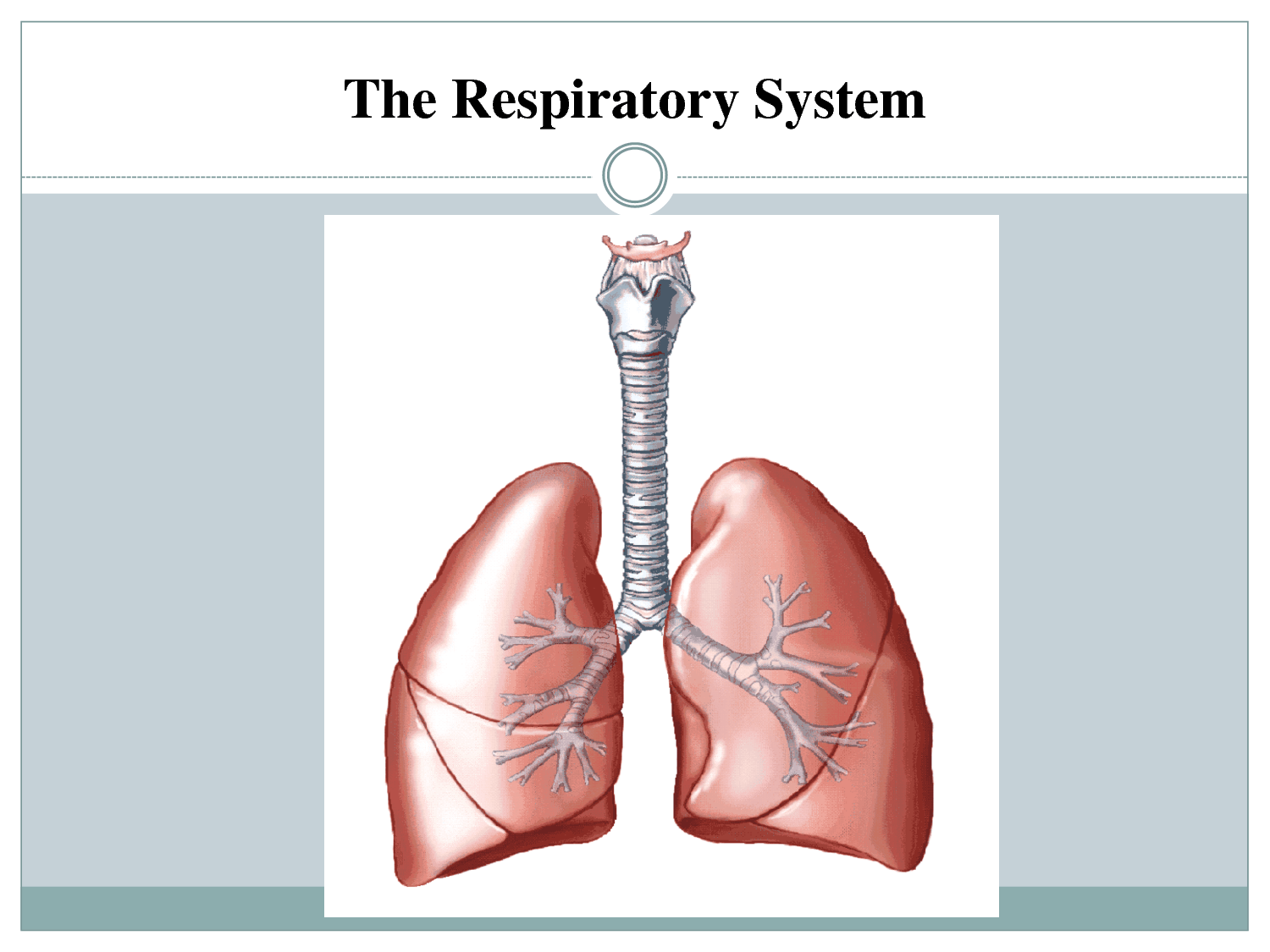


What is the scientific name for the human windpipe?
Trachea
Medulla Oblongata
Adam's Apple
Aorta

What is the function of the system pictured?
Helps pumps your blood through out your body.
Helps move and protect your body.
Helps take in oxygen and remove carbon dioxide.
Helps maintain body your temperature.


You breathe out ________ and breathe in _____________.
oxygen, carbon dioxide
carbon dioxide, oxygen
Oxygenated blood comes from what side of the heart?
Left side
the bottom of the heart
Right side
None of the above

The _______________ have walls that are one cell thick.
arterioles
arteries
veins
venules
capillaries

Which of the following are the strongest blood vessels that function to carry blood away from the heart?
arterioles
arteries
veins
venules
capillaries
Red blood cells (RBCs)
Erythrocytes
Leukocytes
Thrombocytes
White blood cells (WBCs)
Erythrocytes
Leukocytes
Thrombocytes

