
 Worksheets
Imaging II: Minimizing Patient Dose Review
Total questions: 65
Worksheet time: 33mins
Which statement correctly distinguishes exposure from dose in diagnostic radiography?
Exposure is the amount of radiation absorbed by the patient and measured in gray (Gy).
Exposure is the amount of radiation in the air near the patient, used to calculate entrance skin exposure, measured in C/kg.
Dose is the amount of radiation in the air near the patient and formerly measured in roentgens.
Dose is the amount of radiation produced by the x‑ray tube per mAs.
What is the formal historical unit that measured exposure in air near the patient?
Rad
Roentgen
Gray
Sievert
Dose refers to the amount of radiation absorbed by the patient. What is its SI unit?
Coulomb per kilogram (C/kg)
MilliRoentgen (mR)
Gray (Gy)
Milliampere‑seconds (mAs)
Entrance Skin Exposure (ESE) is monitored because the skin receives the highest amount of radiation. When calculating ESE, which Source‑to‑Object Distance (SOD) is used?
Maximum SOD to represent the safest scenario
Average SOD typical for the exam room
Minimum SOD to represent the worst‑case (closest tube) scenario
Any SOD, because distance does not affect skin dose
Why is the minimum SOD used for ESE estimation?
Because a larger distance increases radiation dose to the skin
Because the closer the x‑ray tube is to the patient, the higher the skin dose
Because tube distance only changes image magnification, not dose
Because regulatory guidelines require the farthest possible tube distance
Which quantity and relationship must be known to compute ESE according to the instructional material?
The patient’s body mass index and exposure time
The machine’s output in milliRoentgen per milliampere‑second (mR/mAs) and the selected mAs
The detector sensitivity and kilovolt peak (kVp)
The grid ratio and source‑to‑image distance (SID)
The mR/mAs chart is typically created at which standard Source‑to‑Image Distance (SID)?
30 inches
36 inches
40 inches
48 inches
Which quantity is defined as the amount of radiation in air near the patient and is used for calculating ESE?
Dose
Exposure
Kerma in tissue
Effective dose
Which statement best distinguishes radiation exposure from absorbed dose in medical imaging?
Exposure is the radiation measured in air before it interacts with the patient; absorbed dose is the energy deposited in tissue.
Exposure is the energy deposited in tissue; absorbed dose is the radiation measured in air.
Exposure and absorbed dose are identical measures of patient risk.
Exposure is determined only by patient thickness; absorbed dose is independent of interaction.
Which unit is used to express radiation exposure?
Gray (Gy)
Rad
Coulomb per kilogram (C/kg) or roentgens (R)
Sievert (Sv)
Which unit is appropriate for reporting absorbed dose to tissue?
Roentgen (R)
Coulomb per kilogram (C/kg)
Gray (Gy) or rad
Curie (Ci)
A radiographer notes that the machine’s radiation output is measured in C/kg, but the patient’s absorbed dose is given in Gy. What does this difference indicate?
Output represents exposure in air, while dose indicates energy actually absorbed by the patient’s tissues.
Both values represent the same quantity using different unit systems.
Gy is used only for measuring background radiation exposure, not patient dose.
C/kg measures energy absorbed by tissue, while Gy measures photon fluence in air.
Which equation from the mR/mAs chart is used to find the initial output before applying distance corrections?
Output (mR) = (mR/mAs) × mAs
Output (mR) = kVp × mAs
Output (mR) = mAs ÷ (mR/mAs)
Output (mR) = kVp ÷ mAs
What does SOD represent in ESE calculations?
Source-to-object distance (tube to patient)
Source-to-image distance (tube to receptor)
Object-to-image distance (patient to receptor)
Skin-to-detector distance (patient skin to receptor)
How is SOD computed when SID and OID are known?
SOD = SID − OID
SOD = SID + OID
SOD = OID − SID
SOD = SID × OID
Which statement best describes the Inverse Square Law as applied to patient dose?
Radiation intensity decreases with the square of the distance from the source
Radiation intensity increases linearly with distance from the source
Radiation intensity is independent of distance from the source
Radiation intensity decreases linearly with distance from the source
According to the instructional material, which factor directly lowers dose as it increases?
Distance from the x-ray source
mAs setting
Patient thickness
OID
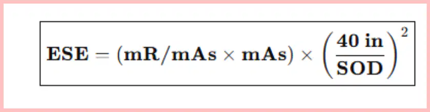
Which formula converts the chart output into Entrance Skin Exposure (ESE) using the reference distance of 40 inches?
ESE = (mR/mAs×mAs)×(40in/SOD)2
ESE = (mR/mAs÷mAs)×(SOD/40in)2
ESE = (kVp×mAs)×(SOD/40in)2
ESE = (mR/mAs × mAs) × (SOD / 40 in)
Which step should be completed first when estimating ESE?
Use the mR/mAs chart to determine output
Apply the Inverse Square Law
Calculate SOD from SID and OID
Choose immobilization devices
Which distances are needed to determine SOD?
SID and OID
SID and SSD
OID and patient thickness
kVp and mAs
Which projection choice is stated to reduce radiation to the ovaries in a female pelvis exam?
PA pelvis compared to AP pelvis
AP pelvis compared to PA pelvis
Lateral pelvis compared to AP pelvis
Oblique pelvis compared to PA pelvis
For skull imaging, which positioning reduces radiation to the eyes compared to the alternative?
PA skull reduces compared to AP skull
AP skull reduces compared to PA skull
Lateral skull reduces compared to PA skull
Oblique skull reduces compared to AP skull
How does effective patient communication help reduce dose according to the material?
It increases patient trust and stillness, reducing repeats
It allows higher mAs to be used safely
It replaces the need for shielding
It permits shorter SID without changing dose
What is a stated benefit of using immobilization to prevent motion during radiography?
Improves image quality and reduces total radiation dose
Allows use of lower kVp without detail loss
Eliminates the need for distance corrections
Increases OID to sharpen the image
In the ESE estimation workflow, what is the purpose of calculating SOD before applying the inverse square law?
To determine the effective distance from x‑ray source to the patient’s skin for dose fall‑off
To select the correct filtration level for image contrast
To choose the focal spot size that minimizes motion blur
To compute milliampere-seconds (mAs) from kVp
Which adjustment to kVp and mAs best reduces patient dose while maintaining image contrast?
Use the lowest kVp and highest mAs possible
Use the highest kVp that still provides acceptable contrast and reduce mAs
Keep both kVp and mAs as low as possible regardless of image quality
Increase both kVp and mAs to shorten exposure time
According to the simplified factor table, how does increasing mAs affect patient dose?
It decreases patient dose by improving penetration
It has no effect on dose but sharpens detail
It increases patient dose because more radiation is emitted
It decreases scatter without changing dose
Which statement about distance (SID/SOD) and patient dose is most accurate?
Decreasing distance reduces dose because the beam spreads out
Increasing distance results in less radiation hitting the patient
Distance does not influence dose in any circumstance
Shorter distance always improves image contrast while lowering dose
What is the primary purpose of filtration in radiography in relation to patient dose?
To sharpen the image by reducing focal spot size
To remove weak x-rays that would only hit the skin, decreasing skin dose
To increase penetration of the beam to reduce exposure time
To eliminate scatter by replacing the need for grids
When should a small focal spot be selected based on the technical factor guidance?
Whenever dose reduction is the highest priority
When possible, to improve image detail without directly changing dose
Only when imaging thick body parts to reduce scatter
Never, because it increases patient dose
What field size choice helps reduce dose and scatter according to the technical factor tips?
Widen the field to include adjacent anatomy
Match the field size to the entire table to avoid cut-off
Make the x-ray beam as small as possible around the area of interest
Use the largest field size when using a grid
Which patients benefit from gonad shielding and when should it be used?
All patients regardless of anatomy, used on every exam
Only pediatric patients, used when the entire torso is imaged
Patients with reproductive organs in or near the beam, used whenever feasible
Only pregnant patients, used during chest imaging
How should kVp be adjusted for body parts of different densities?
Lower kVp for thicker or denser parts; higher kVp for smaller parts
Higher kVp for thicker or denser parts; lower kVp for smaller parts like hand or wrist
Use the same kVp for all parts to standardize dose
Always pair low kVp with high mAs regardless of part thickness
Which statement best explains why digital image receptor systems help reduce patient dose?
They require higher mAs because they are less sensitive
They need less radiation due to higher sensitivity
They produce images only at very high kVp
They require the patient to be closer to the x-ray tube
Which prime exposure factor most directly controls the quantity of x-ray photons and thus patient dose?
mAs
kVp
Focal spot size
Filtration
According to the interrelationship of prime factors, increasing kVp has what general effect on radiation reaching the patient?
Minimal change in radiation output
Greatly increases radiation output
Cuts radiation output in half
Eliminates the need for mAs
What happens to radiation intensity at the patient when source-to-image distance (SID) is doubled?
It doubles
It is reduced to one-half
It is reduced to one-fourth
It remains unchanged
To minimize patient dose while maintaining image quality, which combination is recommended?
Low kVp, high mAs, minimal distance
High enough kVp to penetrate, low mAs, as much distance as possible
High kVp, high mAs, short distance
Low kVp, low mAs, short distance
If mAs is cut in half, what is the expected change in radiation reaching the patient, assuming other factors remain constant?
Radiation doubles
Radiation is unchanged
Radiation cuts in half
Radiation reduces to one-fourth
Why does film/screen imaging typically result in a higher patient dose than digital imaging?
Film is more sensitive and needs lower mAs
Film is less sensitive and requires higher mAs
Digital systems require longer exposure times
Digital systems cannot produce clear images at low mAs
Which statement reflects the connection among mAs, kVp, and distance regarding patient dose?
Changing one factor does not affect the others
All factors influence total dose and are interconnected
Only kVp determines total dose
Distance has no effect on dose
A radiographer increases kVp slightly to improve penetration. What caution does the material emphasize?
Small kVp changes have minimal impact on radiation
Small kVp changes can make big changes in radiation
kVp affects only image contrast, not dose
Increasing kVp always allows reducing distance
Which procedural adjustment most directly reduces patient dose without sacrificing image clarity in digital imaging?
Use higher mAs because digital systems need more photons
Use lower mAs because digital systems can produce clear images at reduced mAs
Decrease distance to increase intensity
Select a lower kVp regardless of anatomy
What is the primary rationale for maximizing distance between the x-ray tube and the patient?
Improves spatial resolution by enlarging focal spot
Reduces radiation intensity at the patient due to inverse square law
Allows the use of lower kVp values
Ensures the detector receives more scatter
Which statement best explains why digital image receptor systems can reduce patient dose compared to film/screen systems?
Digital systems are more sensitive, allowing lower mAs for adequate exposure.
Digital systems require higher mAs to avoid motion blur.
Film/screen systems capture more photons, so they need lower kVp.
Film/screen systems are less sensitive, permitting lower mAs.
According to the instructional text, what is the most direct effect of increasing mAs while keeping other factors constant?
Patient dose decreases because fewer photons are produced
Patient dose increases because more x-ray photons are produced
Image contrast decreases because kVp is lowered
Spatial resolution improves because SID is increased
Which practice aligns with dose reduction guidance related to mAs?
Use the highest mAs to shorten exposure time regardless of image quality
Use the lowest mAs that still provides a clear, diagnostic-quality image
Keep mAs constant and only adjust kVp for every exam
Double mAs whenever SID is increased
Which statement best captures the relationship between mAs and radiation quantity?
mAs primarily changes photon energy without affecting quantity
Higher mAs means more x-ray photons, leading to higher dose
Lower mAs produces higher energy photons, increasing penetration
mAs has no impact on patient dose when kVp is fixed
A technologist increases the source-to-image distance (SID) while maintaining the same exposure settings. Based on the inverse relationship described, what outcome is expected at the patient’s skin?
Greater intensity because the beam narrows with distance
Lower intensity because the radiation spreads over a larger area
No change because distance affects only magnification
Higher intensity due to increased photon energy
If mAs is increased and kVp and distance remain unchanged, which balanced factor combination would reduce patient dose back toward the original level?
Increase kVp and increase SID to maintain image quality with lower mAs
Decrease kVp and decrease SID to compensate for higher mAs
Increase mAs further and keep all other factors the same
Decrease mAs to the lowest value that still yields a diagnostic image
Which prime factor adjustment most directly reduces entrance skin exposure according to the material?
Increasing mAs while keeping distance constant
Decreasing kVp while keeping mAs constant
Increasing SID or SOD with other factors unchanged
Switching from fixed to automatic exposure control
Choose the statement that correctly summarizes the interrelationship of prime factors in radiographic exposure.
mAs controls quantity of radiation; increasing mAs increases dose
kVp alone determines dose regardless of mAs or distance
Distance does not affect radiation intensity at the patient’s skin
Increasing mAs reduces dose because exposure time is shorter
Which setting primarily determines x‑ray beam penetration through tissue, and therefore influences contrast and patient dose via energy?
kVp
mAs
SID
Collimation
Milliamperage‑seconds (mAs) most directly controls which aspect of the exposure, affecting patient dose?
Beam energy (penetration)
Amount of radiation (quantity)
Scatter direction
Field size
According to the 15% Rule, what change to kVp allows you to halve mAs while maintaining similar receptor exposure?
Decrease kVp by 5%
Increase kVp by 15%
Double kVp
Increase mAs by 15%
When you raise kVp and lower mAs appropriately using the 15% Rule, the typical effect on patient dose is what?
Dose increases due to more radiation quantity
Dose decreases because higher kVp with lower mAs reduces entrance skin exposure
Dose is unchanged in all cases
Dose increases unless filtration is added
If image quality requires a minimum mAs to control noise, what is the safest way to reduce dose while preserving that mAs?
Lower kVp slightly and keep mAs the same
Raise kVp and keep mAs the same or slightly lower per 15% Rule
Increase SID to shorten exposure time
Open collimation to include more anatomy
Which statement best describes the interrelationship of kVp and mAs for maintaining similar receptor exposure?
They are independent and should not be changed together
Increasing kVp allows a proportional increase in mAs to keep exposure constant
Increasing kVp can be balanced by decreasing mAs (and vice versa) to keep exposure similar
Decreasing mAs always requires decreasing kVp
A technologist wants to lower patient dose while keeping image noise acceptable. Which change is most appropriate?
Reduce mAs below the optimal level, keeping kVp fixed
Increase kVp by about 15% and reduce mAs by half
Double mAs and lower kVp slightly
Keep both kVp and mAs unchanged
Which statement best explains why accurate use of gonad shielding is recommended in radiography?
It eliminates the need for collimation by narrowing the x‑ray beam automatically.
It decreases patient dose to reproductive organs when positioned correctly.
It increases image sharpness by blocking only scatter radiation.
It allows the use of higher mAs without affecting patient dose.
According to the listed types, which option is NOT one of the major gonad shielding designs?
Flat contact
Shadow
Shaped contact
Focused grid
A radiographer chooses a higher ratio grid. What is the direct consequence described that affects exposure settings?
Less x‑rays are blocked, so mAs can be reduced.
More x‑rays are blocked, so mAs must be increased.
Scatter radiation increases, so kVp must be lowered.
Patient dose decreases automatically, so exposure can stay the same.
What is the relationship between grid strength and patient dose as stated?
Stronger grid requires less radiation, leading to lower patient dose.
Stronger grid has no effect on radiation, keeping patient dose unchanged.
Stronger grid needs more radiation, resulting in higher patient dose.
Stronger grid increases scatter transmission, reducing patient dose.
Which purpose of a grid is identified in the material?
To focus the primary beam and increase magnification.
To clean up scatter radiation and make the image sharper.
To reduce kVp while maintaining image brightness.
To shield gonads during pelvic imaging.
What key caution is emphasized about digital systems’ ability to adjust image brightness?
Because brightness is auto‑adjusted, overexposure is acceptable.
The computer can fix exposure errors and also remove radiation from the patient.
Brightness adjustment does not justify using high exposure; patient dose still matters.
Auto‑adjustment only works when the exposure is too low, not too high.

