
 Worksheets
First trimester pt 4
Total questions: 117
Worksheet time: 2hrs 53mins
The early central nervous system begins as a simple neural plate that folds to form a groove => (a) , open initially at each end
Cranial and caudal ends close at between __ and __ gestational days
24 to 26
26 to 30
20 to 21
27 to 28
The lumen of the neural tube develops into the (a) of the brain and spinal cord
(a) begins at the trilaminar embryo with formation of the notochord and somites, both of which underly the embryonic ectoderm and do not contribute to the nervous system, but are involved with patterning its initial formation
The central portion of the ectoderm then forms the (a) that folds to form the neural tube and neural crest
Epiblast - Ectoderm - Neural Plate - Neural groove and Neural Crest - Neural Tube and Neural Crest
(a)
(a) gives rise to the brain & spinal cord of the central nervous system
(a) develops onto almost all of the structures in the peripheral nervous system
(a) forming from neural plate on upper surface
How many primary brain vesicles are there?
1
2
3
4
develop initially due to the neural plate being broader at the cranial (brain) end that the narrower caudal
(spinal cord) end
(a)
(prosencephalon) – anterior portion of brain
(a)
(mesencephalon)
(a)
(rhombencephalon)
(a)
How many secondary vesicles are there?
1
3
4
5
1) telencephalon
2) diencephalon
3) mesencephalon
4) metencephalon
5) myelencephalon
(a)
Prosencephalon =>
(a)
Mesencephalon =>
(a)
Rhombencephalon =>
(a)
=>Rhinencephalon, Amygdala, Hippocampus, Cerebrum
(Cortex), Hypothalamus, Pituitary | Basal Ganglia, lateral ventricles
(a)
Epithalamus, Thalamus, Subthalamus, Pineal, third ventricle
(a)
Pons, Cerebellum
(a)
Medulla Oblongata
(a)
Embryonic head can be seen by (a) weeks
Cerebral hemisphere grow and the mesenchyme or embryonic mesoderm is caught in the middle => this forms (a)
is a nerve complex in the brain that projects into the side of the lateral ventricles
(a)
cystic structure appearing in the embryonic cranium within the posterior aspect at 6 to 8 weeks that should not be confused with an abnormality
(a)
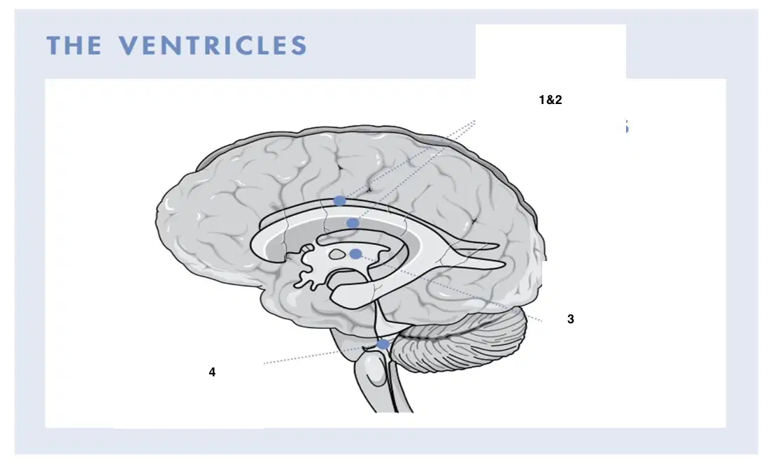
1&2
(a)
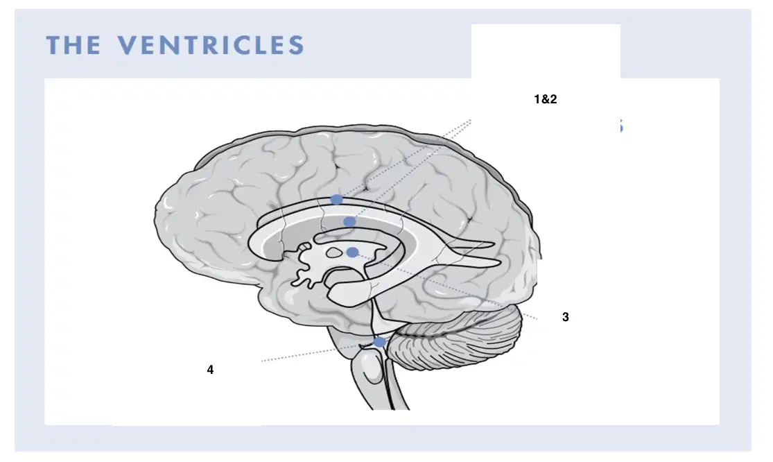
3
(a)
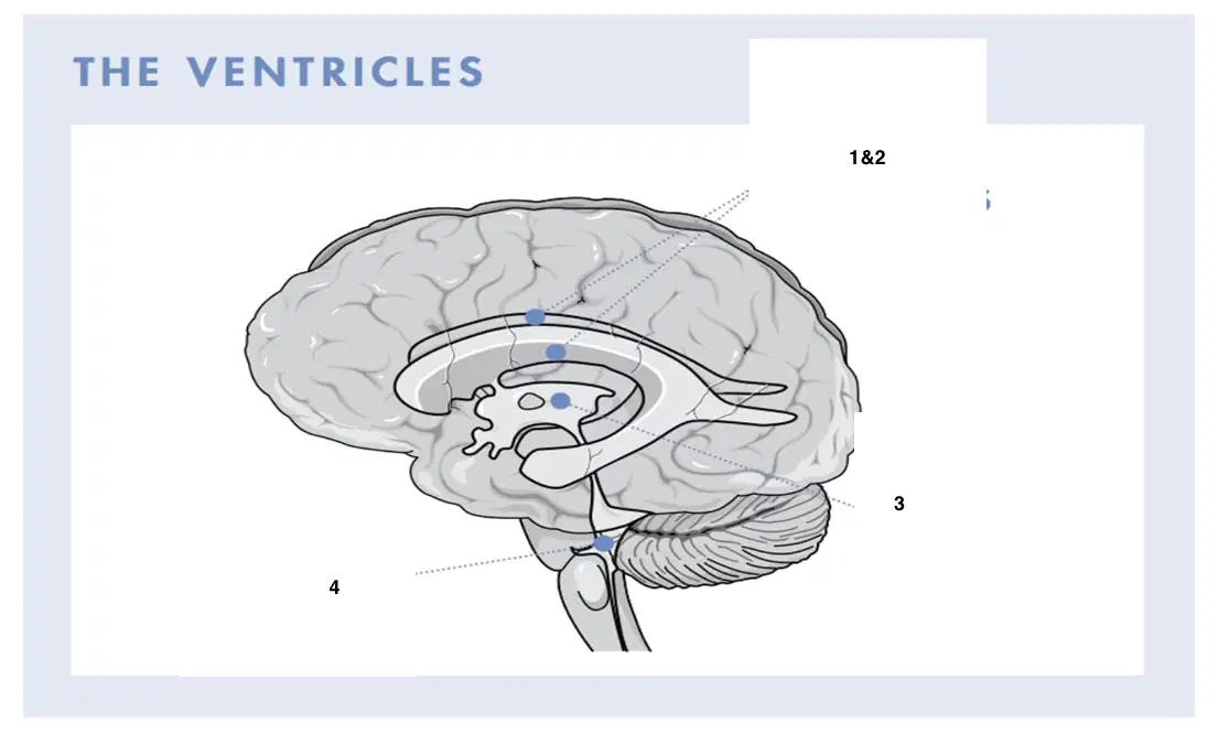
4
(a)
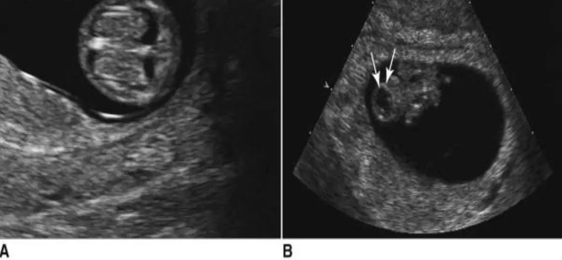
A
(a)
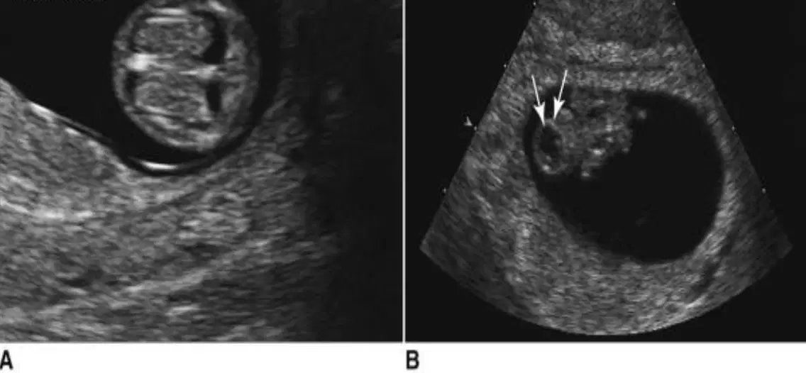
B
(a)
Complete absence of cranium or calvarium
• Abnormal mineralization of bony structures (lack of echogenicity)
• Abnormally shaped “Mickey Mouse” head
(a)
Congenital absence of the brain and cranial vault, with the cerebral hemispheres missing or reduced to small masses
(a)
Midline cranial defect
• Herniation of brain substance and meninges
(a)
Defect in occiput involving the foramen magnum
• Extreme retroflexion of spine
• Open spinal defect
(a)
Dilation of ventricular system without enlargement of the cranium
(a)
Compression of choroid plexus
• Increased cerebrospinal fluid
• Dangling choroid in dilated lateral ventricle
(a)
Failure of prosencephalon to differentiate into cerebral hemispheres and lateral ventricles between 4th and 8th weeks
(a)
Complete to partial failure of cleavage of prosencephalon
• Facial dysmorphism
(a)
Dandy-walker malformation occurs in the ___ week of gestation
6-7
8-9
10-11
11-12
Cystic dilation of fourth ventricle
• Dysgenesis or agenesis of cerebellar vermis and hydrocephaly
Elevated tentorium
(a)
Normally developed skull with fluid replacing the cerebral hemispheres
(a)
Hydranencephaly is a ____ condition
acquired
congenital
loss of all intracranial anatomy, no cerebral hemispheres
(a)
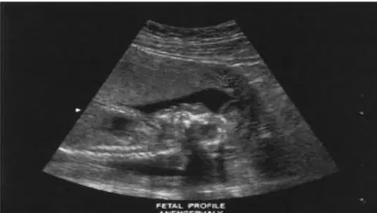
this image shows:
(a)
Ossification of cranium begins after (a) weeks
Acrania has been reported as early as (a) weeks
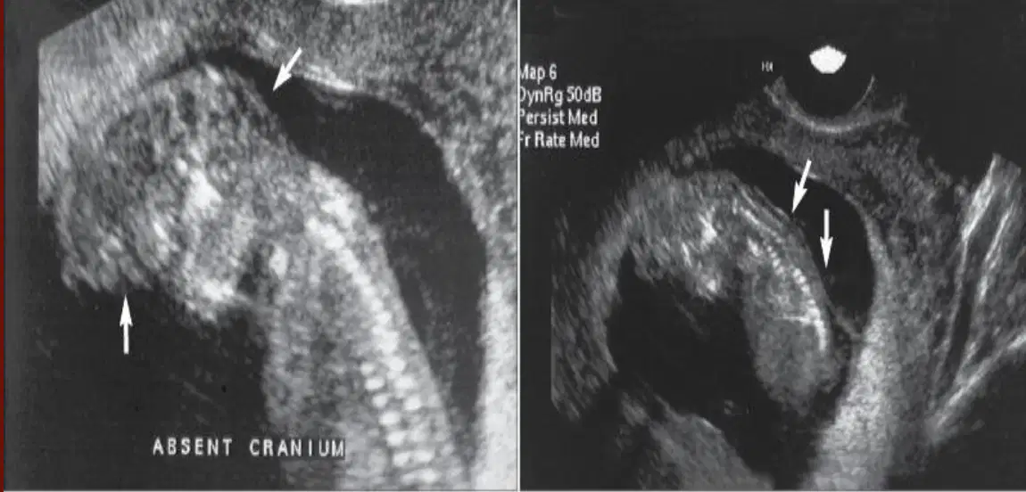
This shows:
(a)
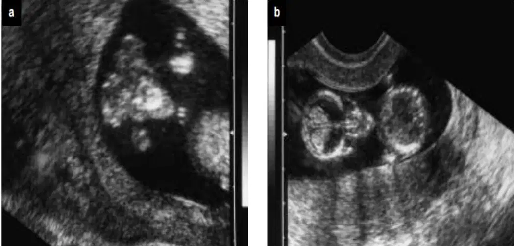
This shows
(a)
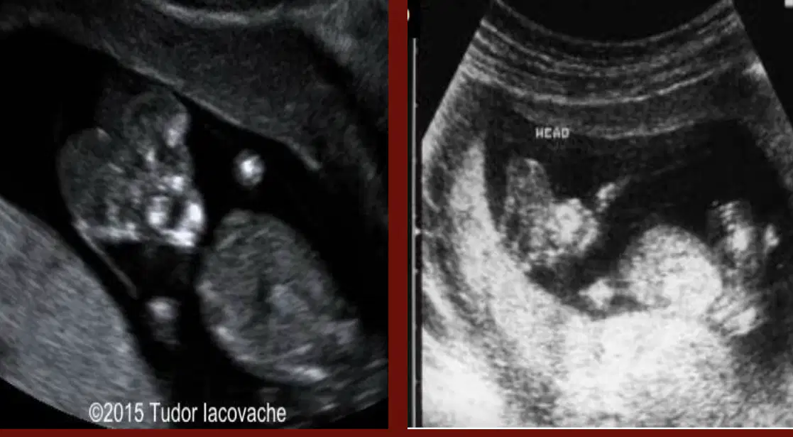
this shows
(a)
Midline cranial defect in which there is herniation of the brain and meninges
(a)
Rare, lethal anomaly of cranial development
(a)
✓ Defect in occiput involving foramen magnum
✓ Retroflexion of spine where fetus looks upward with its occipital
cranium directed toward lumbar spine
✓ Open spinal defects
(a)
Dilation of ventricular system without enlargement of cranium, generally seen after 11 weeks
(a)
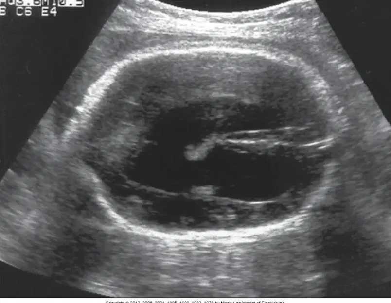
This shows
(a)
Cephalic disorder in which the prosencephalon (the forebrain of the embryo) fails to develop into two hemispheres
(a)
The forebrain and face form in the __ and __ weeks of pregnancy
(a)
Malformation sequence that results from failure of prosencephalon to differentiate into cerebral hemispheres and lateral ventricles between 4th and 8th gestational weeks
(a)
Anomaly ranges from complete to partial failure of cleavage of prosencephalon with variable degrees of facial dysmorphism (dysmorphism = anatomical malformations, facial dysmorphism =
facial malformations)
(a)
Alobar, semi lobar, lobar
(a)
most serious and consists of single ventricle, small cerebrum, fused thalami, agenesis of corpus callous, and fall cerebri
(a)
Important to remember that before 9 weeks, normal fetal brain appears to have “ (a) ” ventricle until falx cerebri develops after 9 weeks
1) large posterior fossa cyst continuous with 4th ventricle
2) absent cerebellum
3) dilated third and lateral ventricles
(a)
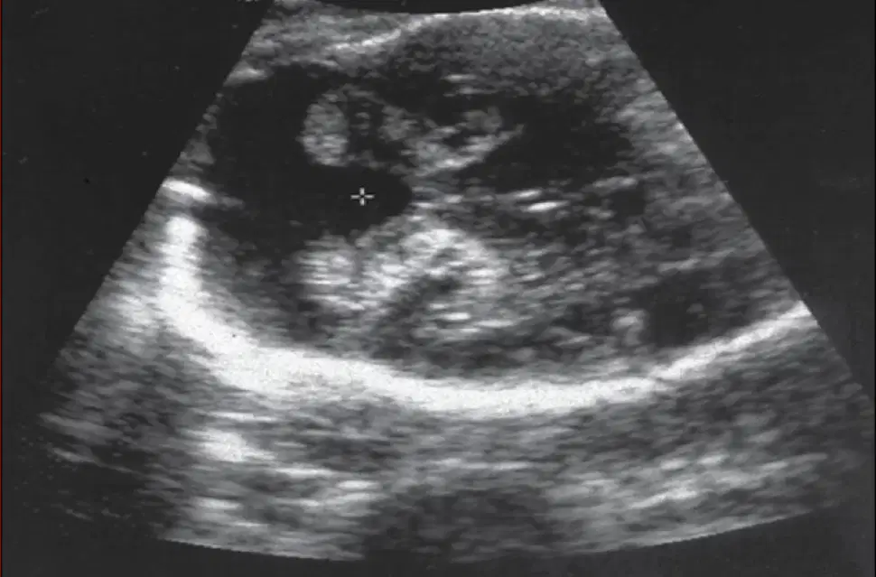
This shows
(a)
Occurs when there is failure of neural tube to close after 6 weeks of gestation
(a)
Can range from small to large defects with protrusion of meninges (Meningocele) or meninges & cord (Meningomyelocele)
(a)
(scalloping of frontal bones)
(a)
(curved appearance of cerebellum)
(a)
The cerebellum as it herniates through the foramen magnum takes up the shape of a
(a)
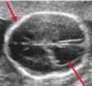
This shows the (a)
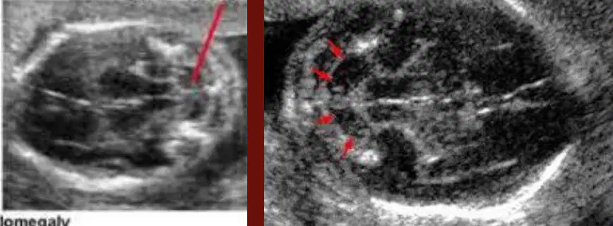
These show
(a)
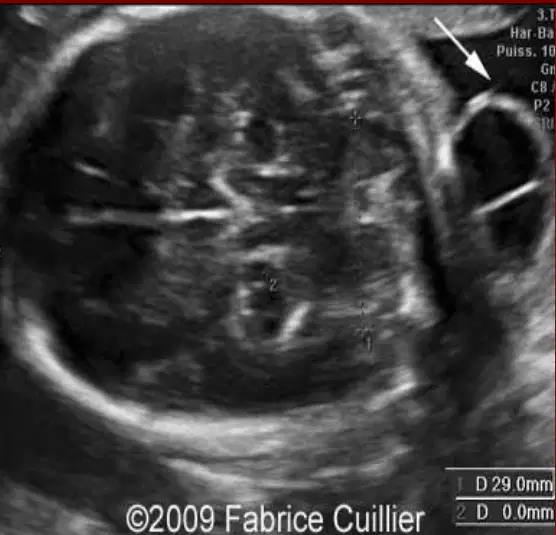
This shows
(a)
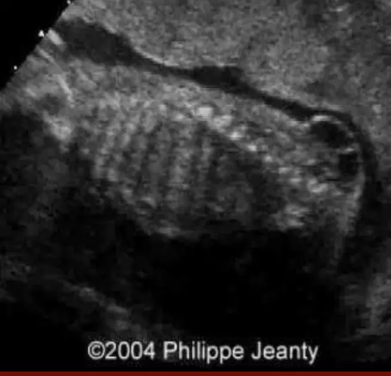
This shows
(a)
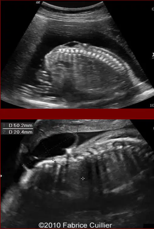
These show
(a)
Omphalocele, Gastroschisis, and Limb–Body Wall complex
(a)
appears sonographically as an echogenic mass at the base of the umbilical cord between 8 and 12 weeks
(a)
Any gut herniation that is ___ is suspicious
>5
>6
<6
<5
Liver is never normally herniated into the base of the umbilical cord => any evidence of the liver outside the anterior abdominal wall=
(a)
Congenital birth defect of the anterior abdominal wall through which the abdominal contents freely protrude into the base of the umbilical cord, which appears to enter the mass
(a)
The infant’s intestines, liver, or other organs stick outside of the belly through
the belly button. The organs are covered in a thin, nearly transparent sac that
hardly ever is open or broken
(a)
Another congenital birth defect of the abdominal wall, just to the
Right of the umbilical cord
(a)
The baby's intestines stick outside of the baby's body, through a hole beside the
belly button (umbilical cord) The hole can be small or large and sometimes other
organs, such as the stomach and liver, can also stick outside of the baby's body
(a)
Rare condition characterized by multiple, severe congenital abnormalities in a fetus
(a)
Results in openings in the anterior body wall (chest and belly) and defects of the limbs (arms and legs).
(a)
facial clefts; a short or missing
umbilical cord; scoliosis; neural tube defects; and abnormalities of
the urogenital organs (i.e. kidney, bladder, and/or genitals
(a)
LBWC is (a) , incompatable with life
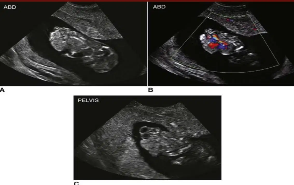
This shows
(a)
is a condition in which the flow of urine is blocked
(a)
This causes the urine to back up and injure one or both kidneys
(a)
Fetal urinary bladder becomes sonographically apparent at __-__ weeks of gestation
(a)
Very large urinary bladder at the level of the urethra
(a)
Bladder may be large enough to extend out of pelvis into fetal abdomen, presenting cystic mass.
(a)
A fluid-filled sac that results from a blockage in the lymphatic system
(a)
Most commonly located in the neck or head area
(a)
One of the most common abnormalities seen in the 1st trim
(a)
If seen early in fetal life => high association with chromosomal
abnormalities (trisomies 13, 18, and 21)
(a)
In fetuses detected with cystic hygroma in 2nd and 3rd trimesters=>
(a) syndrome is most common karyotype abnormality
In 1st trimester may vary in size, but all appear on posterior aspect
of fetal neck and upper thorax
(a)
Any posterior neck thickness > (a) mm, with or without septations, should be
followed
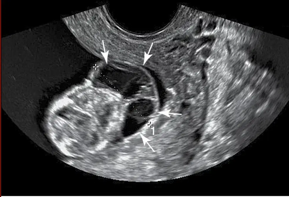
This shows
(a)
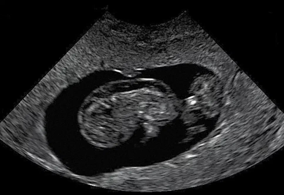
This shows
(a)
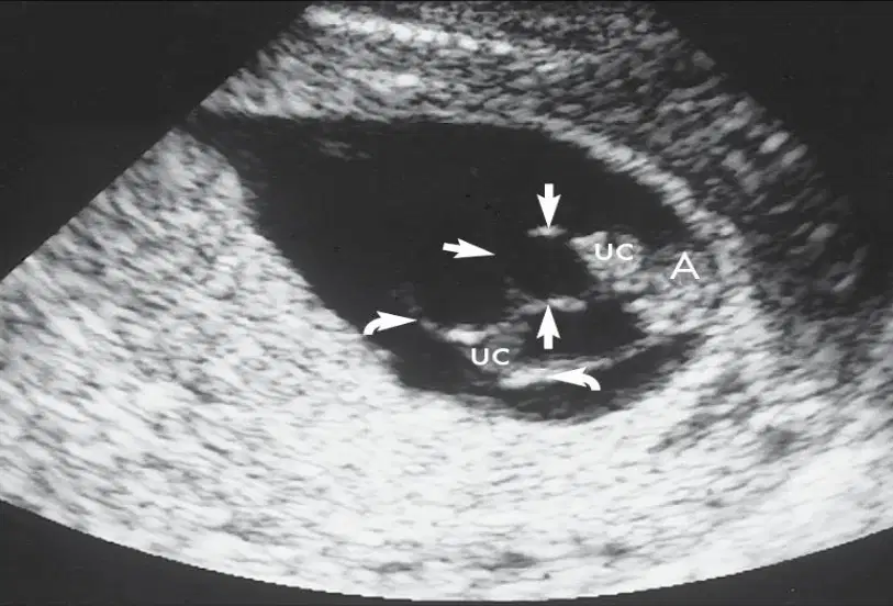
This shows
(a)
Most common ovarian mass seen in the first trim
(a)
which hormone is produced by corpus luteum cysts?
estrogen
progesterone
Color flow - ring of increased vascularity surrounding corpus luteum
• Pulsed Doppler - low-resistance (high-diastolic) waveforms
➢ Such findings similar to decidual flows characterized in ectopic pregnancies but
are intraovarian in location
(a)
Typical corpus luteum cysts are :
<5cm
>10cm
>5cm
<10cm
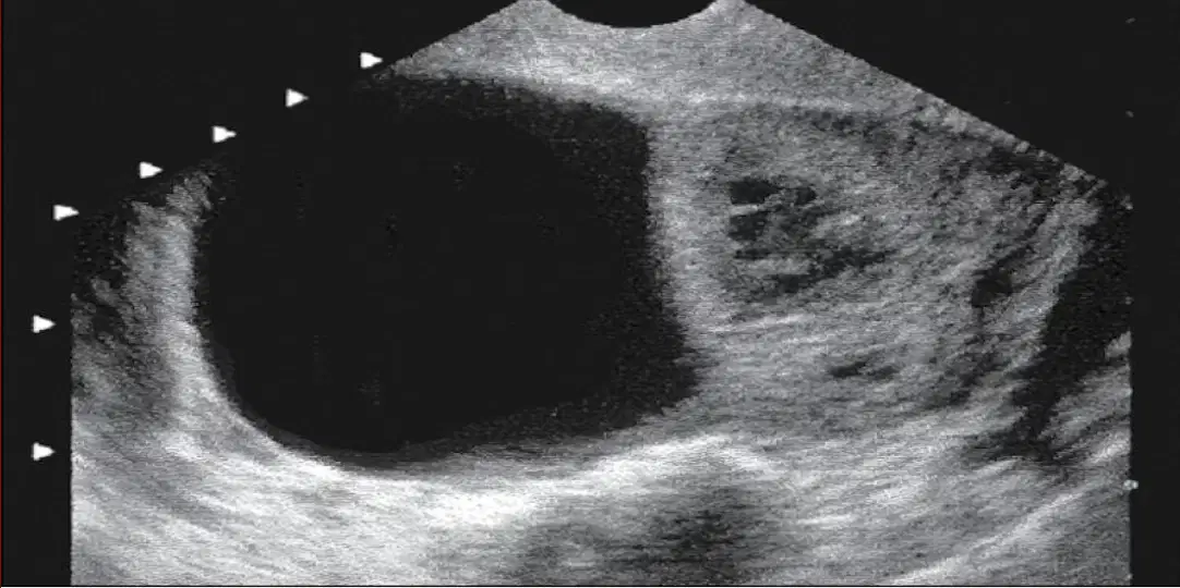
This shows
(a)
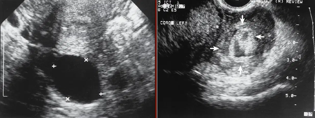
This shows
(a)
If fibroids coexist with 1st trimester => shoul be identified in relation
to
(a)
Uterine leiomyomas increase in size due to (a)
Rapid increase in fibroid size => may lead to (a) of the leiomyoma (degenerating fibroid that may cause significant pain)
Rapidly growing fibroids may compress gestational sac, causing (a)
Myomectomy generally avoided during pregnancy due to increased vasularity of uterus and possibility of marked bleeding
True
False
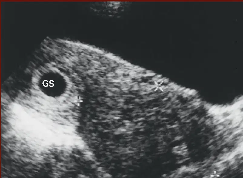
This shows
(a)
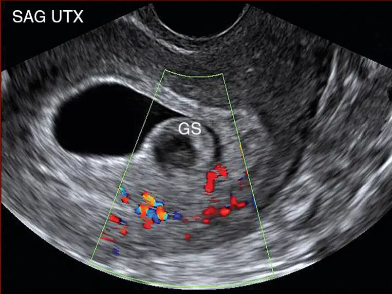
This shows
(a)
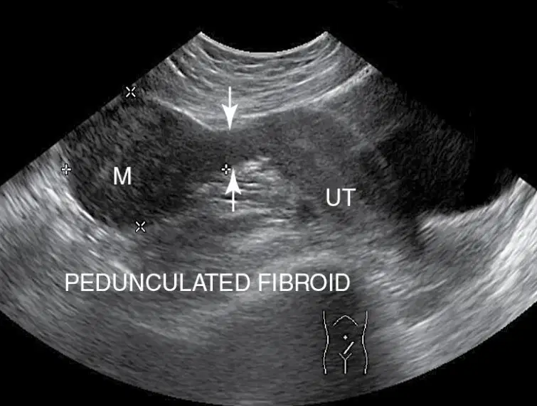
This shows
(a)

