
Font size
 Worksheets
RS 212 Final Review FA25
Total questions: 68
Worksheet time: 35mins
1. Team Member names
(a)
Exposure factors of 350 mA at 1/4 seconds yields:
8.75 mAs
9.0 mAs
87.5 mAs
90 mAs
The body plane that divides the body into front and half parts is:
axial
coronal
sagittal
paranormal
The body plane that divides the body into right and left halves is:
axial
coronal
sagittal
paranormal
The body plane that divides the body into transverse sections is:
axial
coronal
sagittal
paranormal
The body movement that allows for movement of the arm away from the body is known as:
abduction
adduction
flexion
extension
The movement of the arm towards the body is known as:
abduction
adduction
flexion
extension
Contracting the biceps muscle results from _____ of the arm.
abduction
adduction
flexion
extension
The fingers of the hand are located ______ to the forearm.
palmar
dorsal
proximal
distal
The wrist is located ______ to the forearm.
palmar
dorsal
distal
proximal
The elbow is located ______ to the forearm.
palmar
dorsal
distal
proximal
The position that indicates a patient is lying on their back:
lateral recumbent
dorsal recumbent
prone
Fowlers
A position that indicates a patient is laying on his/her right or left side with knees flexed is:
lateral recumbent
dorsal recumbent
prone
supine
Patient laying face down is known as:
supine
prone
Sims'
Trendelenburg
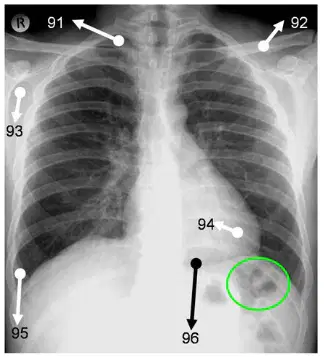
The projection seen here is:
Posterior Anterior
Lateral
Decubitus
None of the above
What anatomical landmark corresponds with T6/7?
Jugular Notch
Inferior angle of the scapula
Xiphoid process
None of the above
All of the following are positioning criteria for a PA chest projection except:
Central Ray to MSP and T6-7
Double inhalation
90 kVp
Shield placed anteriorly
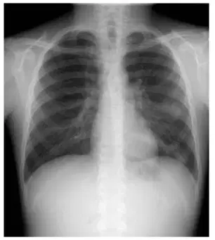
The following is a properly positioned chest radiograph?
True
False
The term that describes a patient who is extremely large is:
Hyposthenic
Hypersthenic
Asthenic
Magnification of the heart is reduced during chest radiography by all of the following except:
Place the patient in the PA position
Use of the left lateral
Seat the patient on a stool
Use a 72" SID
Which projection would best demonstrate a lesion in the right apex?
PA
Lateral
Decubitus
AP Axial
Which projection would demonstrate an air fluid level in the right lung?
PA
Lateral
Decubitus
AP Axial
None of the above
The patient is suspected of having a left sided pneumothorax. He is unable to stand. What projection would be performed to confirm this pathology?
Left Lateral Decubitus
Right Lateral Decubitus
Dorsal Decubitus
Ventral Decubitus
A 90 YR OLD UNSTABLE PATIENT IS IN THE DEPARTMENT FOR DYSPNEA. AFTER COMPLETING THE ROUTINE EXAM THE RADIOLOGIST REQUESTS IMAGES TO DEMONSTRATE RIGHT SIDED FLUID LEVEL. WHAT IMAGE WOULD YOU PERFORM? GIVE THE POSITIONING CRITERIA.
Right lateral Decubitus
Left Lateral Decubitus
Dorsal Decubitus
Ventral Decubitus
A well aerated PA chest will demonstrate lungs filled down to the 9-10th posterior rib.
True
False
How is rotation assessed on a PA chest radiograph?
Symmetry of the Lungs
Symmetry of the ribs
Symmetry of the SC joint
All of the above
Which of the following terms is used to describes "Shortness of Breath"?
Dyspnea
Bronchiectasis
Pleurisy
Atelectasis
What is the positioning error seen here?
Patient is rotated to the left
Patient is rotated to the right
Patient is positioned correctly
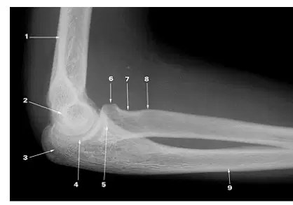
Identify the anatomy labeled #3
Olecranon
Coronoid
Trochlea
Epicondyles

This image best demonstrates the phalanges to identify anterior/posterior displacement of fractures.
True
False
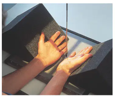
Proper central ray placement for this view would be:
3rd MCP
2nd CMC
5th MCP
5th CMC
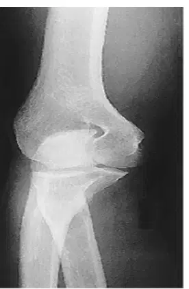
Identify the projection seen here.
AP
AP Oblique with internal rotation
AP Oblique with external rotation
Lateral
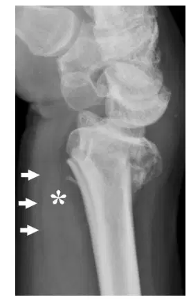
What is the name of the fat stripe seen here?
Supinator
Pronator
Lateral
Scaphoid
What view will best demonstrate the pisiform?
PA
PA Oblique
AP Oblique
Lateral
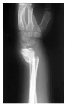
What is the proper name of the fracture seen here?
Barton's
Colle's
Smith's
None of the above
A patient arrives to the Department for a chest exam with a history of pleural effusion. The technique adjustment required is:
Increase exposure factors to penetrate fluid
Decrease exposure factors to reduce dose
Use a lower kVp for better contrast
No adjustment is necessary
A patient with a history of Atelectasis would require an increase in exposure factors for imaging.
True
False
Patient's with history of pneumoperitoneum would require an increase in exposure factors.
True
False
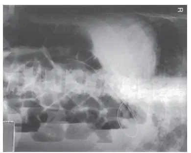
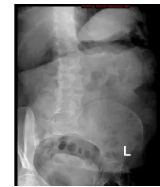
What projection is this?
KUB
LLD
RLD
Erect
What is the arthrology of the metacarpophalangeal joints?
Ginglymus
Ellipsoidal
Saddle
Plane
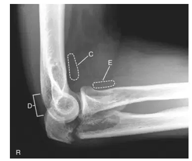
The anatomy labeled "C" is
Posterior fat pad
anterior fat pad
supinator fat pad
pronator fat pad
Rheumatoid Arthritis requires a decrease in technique due to the destructive nature of the pathology.
True
False
This is a properly positioned PA finger image.
True
False
The Olecranon can be well demonstrated on which image(s)? Select all that apply.
AP
Internal Oblique
Lateral
Partial Flexion
Acute Flexion
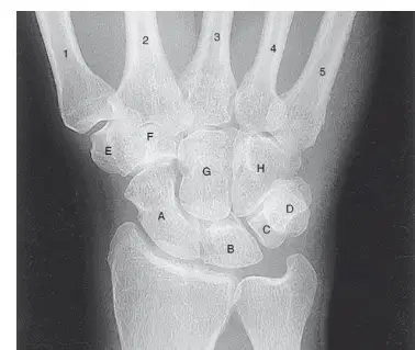
The bone labeled "D" is best demonstrated in what projection?
PA Wrist
AP Wrist
PA Oblique Wrist
AP Oblique Wrist
The anatomy best demonstrated when the upper extremity is rotated 45 degrees medially is:
Pisiform
Radial Head
Lateral Epicondyle
Trapezium
The tube is angled 15 degrees proximal. The pathology best demonstrated would be:
Rolando's Fracture
Carpal Tunnel Syndrome
Radial Head Fracture
Coronoid Process Fracture
Salter Harris classifications are used for what anatomy?
Epiphysis Fractures
Humerus
Radius
Scaphoid
The gap created on an AP wrist with disruption of the scapholunar ligament can be referred to as:
Terry Thomas Sign
Schwarzanager Gap
Wrist crater
Rocky Groove
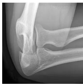
The projection demonstrated here is:
A) Axiolateral Elbow
B) Lateral Oblique Elbow
C) Lateral elbow
D) Medial Oblique Elbow
A radiograph of the PA scaphoid projection reveals extensive superimposition of the scaphoid and adjacent carpals. Which of the following factors can lead to this problem?
A) Elevation of the hand and wrist
B) Insufficient CR angle distally
C) Insufficient ulnar deviation
D) Slight flexion of the phalanges
Which of the following best demonstrates the radial head using the trauma lateral Coyle method routine?
Elbow flexed 80 Degrees, CR angled 45 degrees away from shoulder
Elbow flexed 90 Degrees, CR angled 45 degrees toward shoulder
Elbow flexed 90 Degrees, CR angled 30 degrees toward shoulder
Elbow flexed 90 degrees, CR perpendicular to image receptor
How should the humeral epicondyles be aligned for a lateral projection of the elbow?
Parallel to image receptor
Perpendicular to image receptor
45 degrees to image receptor
40 degrees to image receptor
In which of the 4 quadrants of the abdomen would the cecum be found?
RUQ
RLQ
LUQ
LLQ
The coracoid process is part of the clavicle.
True
False
Which projection best demonstrates the acromioclavicular joints?
AP projection with weights
Lateral scapula
AP shoulder
PA clavicle
The humerus is part of the shoulder girdle.
True
False
The clavicle is the only long bone in the body that lies horizontally.
True
False
Which radiographic view is used to assess the subacromial space?
Grashey
Garth
Lawrence
Gaynor-Hart
The clavicle articulates with the sternum at the sternoclavicular joint.
True
False
Which position is used to best visualize the greater tubercle of the humerus?
External rotation
Internal rotation
Neutral position
Supine position
The humeral head articulates with the glenoid cavity of the scapula.
True
False
What is the purpose of the Y-view in shoulder radiography?
To measure bone density
To evaluate soft tissue
To check for fractures
To assess shoulder dislocation
An AP chest X-ray typically results in greater magnification of the heart compared to a PA view.
True
False
Why is it important to take a patient history before imaging?
To understand patient anxiety levels
To inform technique and identify contraindications
To identify food allergies
To reduce patient wait time
A lateral chest X-ray is performed to assess the retrocardiac space and lower lobes.
True
False
The left lateral decubitus position is used to visualize free air under the right hemidiaphragm.
True
False
What is a primary reason for using an upright position for chest X-rays?
To assess air-fluid levels in the chest
To speed up the procedure
To increase patient comfort
To reduce radiation dose

